


Article Figures & Data
Figures
- Fig. 1.
Structure of physostigmine. CAS number 57-47-6.
- Fig. 2.
Physostigmine directly activates the mouse adult wild-type nAChR. Single-channel currents and the respective open time histograms from patches exposed to 1 μM (A), 10 μM (B), or 100 μM physostigmine (C). The activity consisted of single, isolated openings without the characteristic clustering behavior observed with ACh or many other nicotinic agonists. The open time histograms were fitted to sums of two exponentials (1 and 10 μM physostigmine) or a single exponential (100 μM physostigmine). In the presence of 1 μM physostigmine, the open times for the data in this histogram were 0.22 ms (65%) and 0.74 ms. In the presence of 10 μM physostigmine, the open times were 0.12 ms (51%) and 0.49 ms. In the presence of 100 μM physostigmine, the mean open duration was 0.13 ms. Due to lack of clusters and the uncertainty in the number of active receptors in the patch, the closed times were not analyzed. The data are consistent with physostigmine being a low-potency or a low-efficacy agonist on the wild-type receptor.
- Fig. 3.
Activation of the αS269I mutant receptor by physostigmine. Single-channel currents and the respective open time histograms from patches exposed to 1 μM (A), 10 μM (B), 100 μM physostigmine (C), or 10 μM carbachol (D). The open times were prolonged compared with the wild-type receptor, but no clustering behavior was observed. The open time histograms were fitted to sums of two exponentials (1 and 10 μM physostigmine and 10 μM carbachol) or a single exponential (100 μM physostigmine). The open times for these patches were as follows: 1 μM physostigmine, 0.15 ms (60%) and 1.43 ms; 10 μM physostigmine, 0.19 ms (75%) and 0.96 ms; 100 μM physostigmine, 0.11 ms (100%); and 10 μM carbachol, 0.23 ms (23%) and 2.34 ms. Carbachol produced activity grouped in clusters. Due to lack of clusters of activity elicited by physostigmine and the uncertainty in the number of active receptors in the patch, the closed times were not analyzed. The mutation enhances the channel opening rate constant for nicotinic agonists and the APL galantamine. The lack of clusters in the presence of physostigmine suggests that physostigmine is a low-efficacy agonist of the adult-type muscle nAChR.
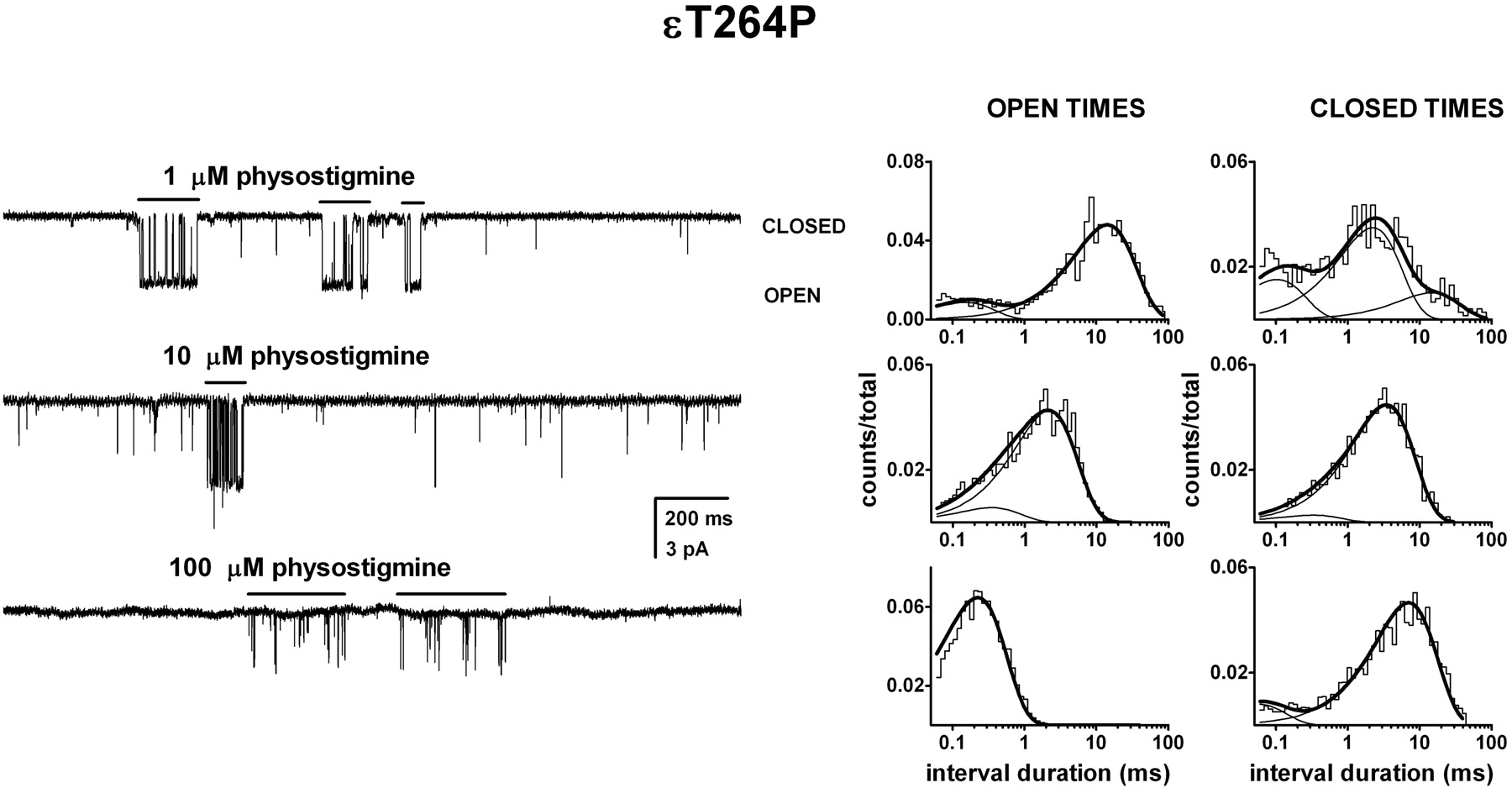
- Fig. 4.
Activation of the ϵT264P mutant receptor by physostigmine. Single-channel currents and the respective open and closed time histograms from patches exposed to 1 μM (A), 10 μM (B), or 100 μM physostigmine (C). The channel activity consisted of episodes or bursts of activity (shown with lines above current traces) intermixed with brief, isolated openings. The bursts were isolated from the recording and analyzed for open and closed time durations. The open times were prolonged compared with the wild-type receptor. In the presence of 1 μM physostigmine, the open times were 0.14 ms (15%) and 13.1 ms, and the closed times were 0.10 ms (25%), 2.1 ms (58%), and 13.6 ms. In the presence of 10 μM physostigmine, the open times were 0.33 ms (12%) and 2.0 ms, and the closed times were 0.30 ms (6%) and 3.2 ms. In the presence of 100 μM physostigmine, the mean open duration was 0.21 ms, and the closed times were 0.05 ms (20%) and 4.9 ms. Due to the potent blocking action of physostigmine, we were unable to identify the activation-related closed time components. It is likely that the more prominent 3- to 5-ms closed time component arises from dwells in the blocked state.
- Fig. 5.
Voltage does not affect channel open durations in the presence of physostigmine. The long open time component from the ϵT264P mutant receptor activated by 1 μM physostigmine (circles) was estimated at -100-, -75-, -50-, -25-, and +50-mV membrane potentials. No voltage sensitivity was observed. For control, we estimated the open interval durations from the ϵT264P mutant receptor activated by 100 μM carbachol (squares) at -50 and +50 mV membrane potentials. Carbachol-elicited openings showed a voltage sensitivity of 93 mV per e-fold change. This value is similar to previous estimates for the wild-type receptor and a number of mutant receptors activated by nicotinic agonists (Auerbach et al., 1996; Akk and Steinbach, 2000). Points show mean ± S.D. for data from three to six patches.
- Fig. 6.
Mutations to the αLys125 site reduce channel open durations in the presence of physostigmine but not carbachol. Single-channel currents and the respective open time histograms for ϵT264P, αK125Q + ϵT264P, and αK125E + ϵT264P mutant receptors activated by 10 μM physostigmine (A), or 100 μM carbachol (B). In the presence of 10 μM physostigmine, the activity consisted of bursts of activity intermixed with brief, isolated openings. All the openings within long sections of the recording were analyzed for open time durations. The open times for the ϵT264P receptor activated by physostigmine were 0.10 ms (80%) and 1.4 ms, weighted average mean open duration of 0.28 ms. The open times for the αK125Q + ϵT264P receptor activated by physostigmine were 0.07 ms (87%) and 1.2 ms, weighted average mean open duration of 0.22 ms. The open times for the ϵT264P + αK125E receptor activated by physostigmine were 0.13 ms (64%) and 1.7 ms, weighted average mean open duration of 0.54 ms. In the presence of 100 μM carbachol, the activity consisted of bursts of activity. The open times for the ϵT264P receptor activated by carbachol were 0.08 ms (25%) and 28.6 ms. The open times for the αK125Q + ϵT264P receptor activated by carbachol were 0.07 ms (30%) and 48 ms. The open times for the ϵT264P + αK125E receptor activated by carbachol were 0.08 ms (45%) and 60 ms.
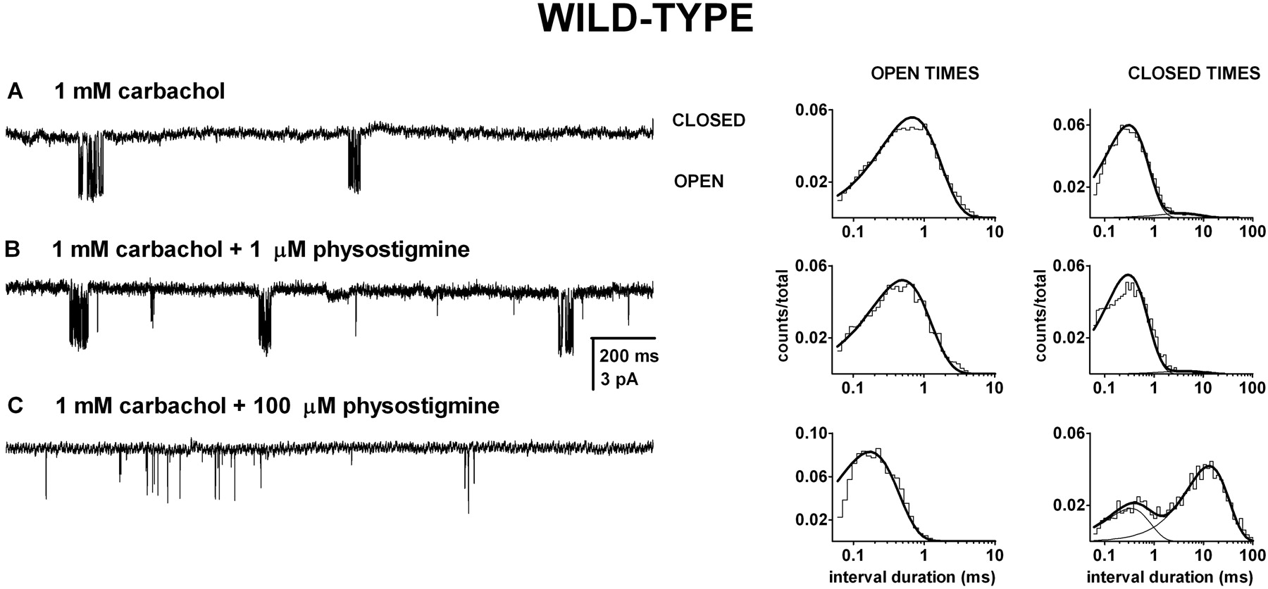
- Fig. 7.
The presence of physostigmine does not interfere with channel activation by carbachol. Single-channel clusters and the respective open and closed time histograms from wild-type receptors exposed to 1 mM carbachol (A), carbachol + 1 μM physostigmine (B), or carbachol + 100 μM physostigmine (C). The activity consisted of easily identified clusters. In the presence of 1 mM carbachol, the mean intracluster open duration was 0.54 ms, and the closed times were 0.25 ms (96%) and 3.4 ms. In the presence of 1 mM carbachol + 1 μM physostigmine, the mean intracluster open duration was 0.39 ms, and the closed times were 0.24 ms (98%) and 9.6 ms. In the presence of 1 mM carbachol + 100 μM physostigmine, the mean intracluster open duration was 0.18 ms, and the mean closed times were 0.27 ms (40%) and 9.6 ms. The presence of 100 μM physostigmine led to a decrease in channel open probability through a decrease in the mean open duration and an increase in the prevalence and duration of the longer lived closed time component. The presence of physostigmine was ineffective at modifying the duration of the shorter-lived closed time component (inverse of the effective opening rate), which is a measure of agonist binding and channel opening. We conclude that physostigmine does not interact with the sites that mediate channel activation by carbachol.
- Fig. 8.
Properties of channel block by physostigmine. The increase in the concentration of physostigmine was found to result in reduced open duration and an increase in the rate of entry into an ∼3- to 7-ms closed state. Both observations are consistent with physostigmine-induced channel block. A, inverse of the open duration (circles and dashed line) and the rate of entry into the putative blocked state (squares and solid line) for ϵT264P mutant receptors are plotted as a function of physostigmine concentration. The lines were fitted to rate = rate at no physostigmine + k+B* × [physostigmine], where k+B* is the apparent blocking rate. The estimates for k+B* were 44 ± 2 μM-1 s-1 when fitting the reduction in the open duration, and 39 ± 2 μM-1 s-1 when fitting the increase in the rate of entry into the putative blocked state. B, relationship between the inverse of the duration of the putative blocked state and physostigmine concentration in the ϵT264P mutant receptor. The increase in the duration of the putative blocked state at higher physostigmine concentrations is consistent with the presence of two (or more) blocking sites per receptor. The line was fitted to 1/τBlocked = k-B (2k-B/(2k-B + k+B × [physostigmine])). This equation assumes the presence of two, equivalent blocking sites per receptor. The fitting results are k+B = 15.6 ± 4.8 μM-1 s-1 and k-B = 458 ± 28 s-1. C, inverse of the open duration (circles) and the rate of entry into the putative blocked state (squares) for the wild-type receptor activated by 1 mM carbachol in the presence of physostigmine are plotted as a function of physostigmine concentration. The estimates for k+B* were 36 ± 2 μM-1 s-1 when fitting the reduction in the open duration and also 36 ± 2 μM-1 s-1 when fitting the increase in the rate of entry into the putative blocked state. D, changes in membrane potential strongly affected the rate of return from the blocked state, but they were ineffective at altering the rate of entry into the blocked state. The values for k+B* (solid lines) and k-B* (dashed lines) in the presence of 100 μM physostigmine were estimated for wild type receptors activated by 1 mM carbachol (circles) or 200 μM ACh (squares), ϵT264P receptors activated by 100 μM carbachol (triangles), and ϵT264P receptors exposed solely to physostigmine (crosses) at -50 mV and +50 mV membrane potential. For the rate of development of block, the H value (change in membrane potential needed for an e-fold change in parameter) was 1141 mV (wild type + carbachol), 268 mV (wild type + ACh), 261 mV (ϵT264P + carbachol), or 227 mV (ϵT264P + physostigmine alone). For the rate of recovery from block, the H value was 44 mV (wild type + carbachol), 45 mV (wild type + ACh), 31 mV (ϵT264P + carbachol), or 32 mV (ϵT264P + physostigmine alone). For A to C, each point shows data from one patch, whereas for D each point shows results from the combined analysis of data from two or three patches.


