
Main menu

  • Home
  • Articles
    • Current Issue
    • Fast Forward
    • Latest Articles
    • Archive
  • Information
    • Instructions to Authors
    • Submit a Manuscript
    • For Subscribers
    • For Advertisers
    • Terms & Conditions of Use
    • Permissions
  • About Us
    • About Molecular Pharmacology
    • Feedback
  • Editorial Board
  • Alerts
    • Alerts
    • RSS Feeds
  • Other Publications
    • Drug Metabolism and Disposition
    • Journal of Pharmacology and Experimental Therapeutics
    • Molecular Pharmacology
    • Pharmacological Reviews
    • Pharmacology Research & Perspectives
    • ASPET

User menu

  • My alerts
  • Log in

Search

  • Advanced search
Molecular Pharmacology
  • Other Publications
    • Drug Metabolism and Disposition
    • Journal of Pharmacology and Experimental Therapeutics
    • Molecular Pharmacology
    • Pharmacological Reviews
    • Pharmacology Research & Perspectives
    • ASPET
  • My alerts
  • Log in
Molecular Pharmacology

Advanced Search

  • Home
  • Articles
    • Current Issue
    • Fast Forward
    • Latest Articles
    • Archive
  • Information
    • Instructions to Authors
    • Submit a Manuscript
    • For Subscribers
    • For Advertisers
    • Terms & Conditions of Use
    • Permissions
  • About Us
    • About Molecular Pharmacology
    • Feedback
  • Editorial Board
  • Alerts
    • Alerts
    • RSS Feeds
  • Visit molpharm on Facebook
  • Follow molpharm on Twitter
  • Follow molpharm on LinkedIn
Research ArticleMinireview—A Latin American Perspective on Ion Channels

Structure-Driven Pharmacology of Transient Receptor Potential Channel Vanilloid 1

Ignacio Díaz-Franulic, Javier Caceres-Molina, Romina V. Sepulveda, Fernando Gonzalez-Nilo and Ramon Latorre
Molecular Pharmacology September 2016, 90 (3) 300-308; DOI: https://doi.org/10.1124/mol.116.104430
Ignacio Díaz-Franulic
Centro Interdisciplinario de Neurociencias de Valparaíso, Facultad de Ciencias, Universidad de Valparaíso, Valparaíso, Chile (I.D.-F., R.L., F.G.-N.); Centro de Bioinformática y Biología Integrativa, Universidad Andrés Bello, Santiago, Chile (I.D.-F., J.C.-M., R.V.S., F.G.-N.); and Fraunhofer Chile Research, Santiago, Chile (I.D.-F.)
  • Find this author on Google Scholar
  • Find this author on PubMed
  • Search for this author on this site
Javier Caceres-Molina
Centro Interdisciplinario de Neurociencias de Valparaíso, Facultad de Ciencias, Universidad de Valparaíso, Valparaíso, Chile (I.D.-F., R.L., F.G.-N.); Centro de Bioinformática y Biología Integrativa, Universidad Andrés Bello, Santiago, Chile (I.D.-F., J.C.-M., R.V.S., F.G.-N.); and Fraunhofer Chile Research, Santiago, Chile (I.D.-F.)
  • Find this author on Google Scholar
  • Find this author on PubMed
  • Search for this author on this site
Romina V. Sepulveda
Centro Interdisciplinario de Neurociencias de Valparaíso, Facultad de Ciencias, Universidad de Valparaíso, Valparaíso, Chile (I.D.-F., R.L., F.G.-N.); Centro de Bioinformática y Biología Integrativa, Universidad Andrés Bello, Santiago, Chile (I.D.-F., J.C.-M., R.V.S., F.G.-N.); and Fraunhofer Chile Research, Santiago, Chile (I.D.-F.)
  • Find this author on Google Scholar
  • Find this author on PubMed
  • Search for this author on this site
Fernando Gonzalez-Nilo
Centro Interdisciplinario de Neurociencias de Valparaíso, Facultad de Ciencias, Universidad de Valparaíso, Valparaíso, Chile (I.D.-F., R.L., F.G.-N.); Centro de Bioinformática y Biología Integrativa, Universidad Andrés Bello, Santiago, Chile (I.D.-F., J.C.-M., R.V.S., F.G.-N.); and Fraunhofer Chile Research, Santiago, Chile (I.D.-F.)
  • Find this author on Google Scholar
  • Find this author on PubMed
  • Search for this author on this site
Ramon Latorre
Centro Interdisciplinario de Neurociencias de Valparaíso, Facultad de Ciencias, Universidad de Valparaíso, Valparaíso, Chile (I.D.-F., R.L., F.G.-N.); Centro de Bioinformática y Biología Integrativa, Universidad Andrés Bello, Santiago, Chile (I.D.-F., J.C.-M., R.V.S., F.G.-N.); and Fraunhofer Chile Research, Santiago, Chile (I.D.-F.)
  • Find this author on Google Scholar
  • Find this author on PubMed
  • Search for this author on this site
  • Article
  • Figures & Data
  • Info & Metrics
  • eLetters
  • PDF
Loading

Visual Overview

Figure1
  • Download figure
  • Open in new tab
  • Download powerpoint

Abstract

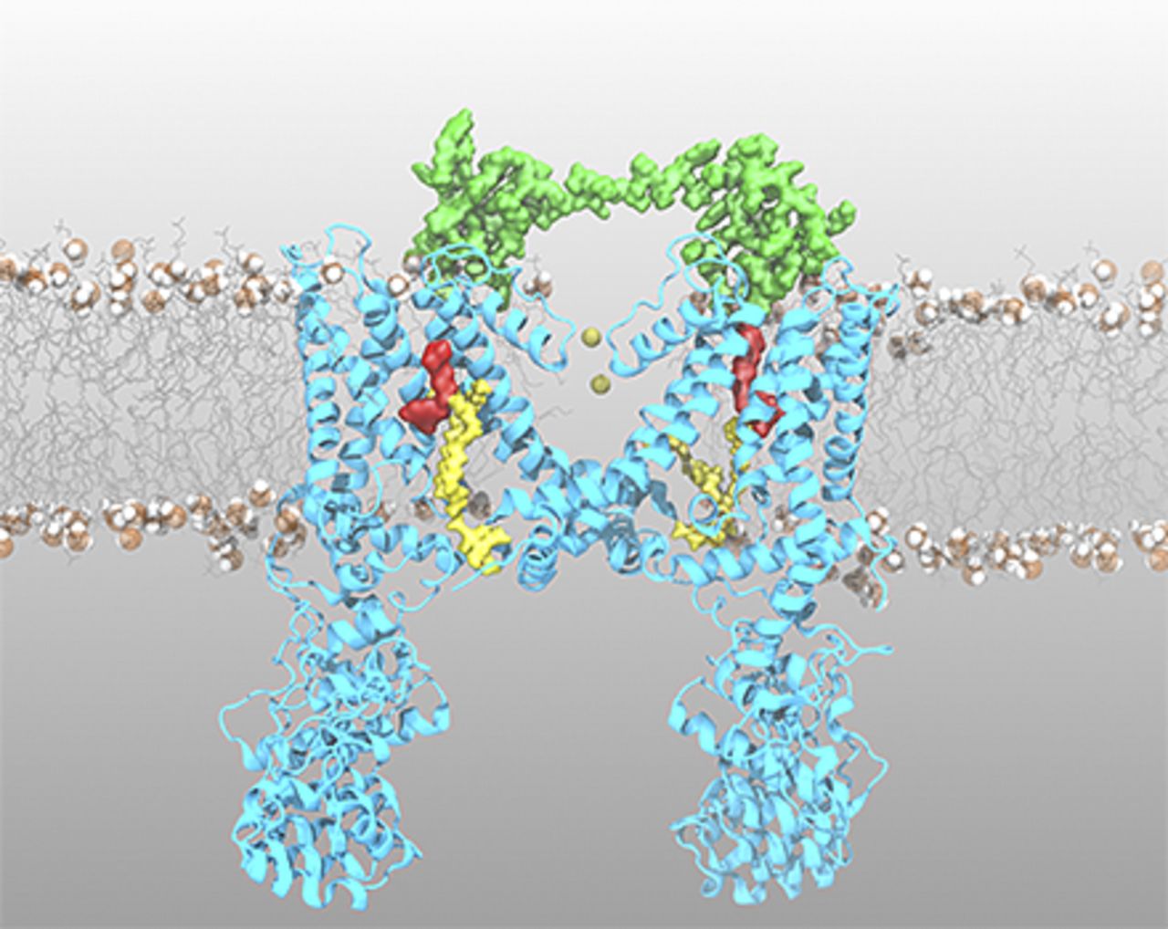
The transient receptor potential vanilloid 1 (TRPV1) ion channel is a polymodal receptor that mediates the flux of cations across the membrane in response to several stimuli, including heat, voltage, and ligands. The best known agonist of TRPV1 channels is capsaicin, the pungent component of “hot” chili peppers. In addition, peptides found in the venom of poisonous animals, along with the lipids phosphatidylinositol 4,5-biphosphate, lysophosphatidic acid, and cholesterol, bind to TRPV1 with high affinity to modulate channel gating. Here, we discuss the functional evidence regarding ligand-dependent activation of TRPV1 channels in light of structural data recently obtained by cryoelectron microscopy. This review focuses on the mechanistic insights into ligand binding and allosteric gating of TRPV1 channels and the relevance of accurate polymodal receptor biophysical characterization for drug design in novel pain therapies.

Introduction

The pain sensation is triggered when the terminals of a specific subset of peripheral neurons called nociceptors are activated by noxious stimuli, such as irritant substances or heat. Specifically, the cationic nonselective transient receptor potential vanilloid 1 (TRPV1) ion channel detects these stimuli and induces the opening of the channel pore and a subsequent increase in membrane permeability (Caterina et al., 1997). The TRPV1 channel is a polymodal receptor originally shown to be activated by capsaicin, heat, protons (Tominaga et al., 1998), lipids (Hernández-García and Rosenbaum, 2014; Morales-Lázaro and Rosenbaum, 2015), and peptide toxins from some venomous animals (Bohlen et al., 2010; Hakim et al., 2015; Yang et al., 2015b). The TRPV1 channel has a tetrameric structure, with each subunit possessing six transmembrane domains, several ankyrin repeat domains (ARDs) at the N terminus, and a large intracellular C terminus containing a conserved amino acidic sequence called the “TRP box”. (Liao et al., 2013). As with many other members of the transient receptor potential (TRP) ion channel superfamily, TRPV1 has been the focus of intense research because of the intriguing nature of its polymodal activation mechanism (Nilius et al., 2005; Latorre et al., 2007; Tominaga, 2007); however, until recently, experimental data lacked a structural framework to interpret functional findings.

A major breakthrough in the field of TRP channels came from David Julius’s laboratory when the structure of the TRPV1 channel in different conformations was resolved using single-particle cryoelectron microscopy (cryo-EM) (Cao et al., 2013b; Liao et al., 2013). This picture showed that the TRPV1 channel shares many structural features with other ion channels of known structures, such as the voltage-gated Kv1.2 channel and the Kv1.2-2.1 paddle chimera (Long et al., 2005a, 2007). The TRPV1 channel structure supported previous studies proposing that the TRPV1 channel is assembled as a tetramer (Kedei et al., 2001; Kuzhikandathil et al., 2001) and confirmed functional studies showing that the channel pore and corresponding intracellular activation gate(s), formed by the S5 to S6 transmembrane segments, exist along the tetramer axis of symmetry (Oseguera et al., 2007; Jara-Oseguera et al., 2008; Salazar et al., 2009). The TRPV1 structure shows that a peripheral domain in each subunit, encompassing S1–S4 segments, is connected to the pore by a short linker, a feature also conserved in voltage-dependent potassium channels (Long et al., 2005a). The short stretch of 25 amino acids at the intracellular C terminus, conserved in several TRP channels and known as the “TRP domain,” adopts an α-helical structure running parallel to the membrane (Liao et al., 2013). This TRP domain is tightly packed with the S4 to S5 linker, a region that has been proposed to couple the voltage sensor domain with pore opening in voltage-gated ion channels (Long et al., 2007). The long ARD characteristic of many TRP channels interacts with both the ARD-S1 linker and the C terminus of the neighboring subunit (Liao et al., 2013). The structure of the TRPV1 channel was obtained under several conditions: in the absence of ligand (so-called APO form), in the presence of capsaicin, and in the presence of resiniferatoxin (RTX) and the double-knot toxin (DkTx) in which both compounds acted as irreversible TRPV1 channel openers (Siemens et al., 2006). Among the several sensing modalities displayed by the TRPV1 channel, a number of unique features of the ligand–channel interaction make for an interesting study. One issue to consider is how the conformational changes triggered by small molecule binding are translated into the mechanical energy necessary to open the channel pore. Second, since the TRPV1 channel is a major component of pain pathways, suppressing this activity may lead to novel strategies that would alleviate pain stemming from several diseases (Julius, 2013).

This review focuses on the structural relationships between three major classes of ligands that are able to modulate the activity of TRPV1 channels: compounds that bind to the vanilloid binding site, peptides derived from poisonous animal venom extract, and lipids.

Ligand Binding at the Vanilloid Binding Pocket

Capsaicin, the pungent active compound of “hot” chili peppers, increases the cation permeability of sensory neuron membranes (Bevan and Szolcsányi, 1990; Oh et al., 1996). However, its molecular target was not identified until the cloning of a functional cDNA encoding a protein of 838 amino acids—the TRPV1 channel—from dorsal root ganglia (DRG) neurons (Caterina et al., 1997). This study showed that TRPV1 channels were activated by capsaicin, noxious heat, and protons, thereby revealing the channel’s role as an integrator of painful stimuli (Caterina et al., 1997). Furthermore, a generation of transgenic mice lacking the TRPV1 channels exhibited impaired nociceptive responses induced by vanilloids (Caterina et al., 2000) and during inflammatory conditions (Davis et al., 2000). Once the target for capsaicin was identified, attention focused on determining the binding site within the TRPV1 channels and the operating mechanism for channel opening. Contrary to what is observed in rodents, DRG neurons from chicks are capsaicin insensitive even though they display heat-induced cationic currents (Wood et al., 1988). Taking advantage of the species-specific differences, Jordt and Julius (2002) showed that transferring transmembrane segments 2–4 from rat to bird TRPV1 channels restored capsaicin sensitivity (Jordt and Julius, 2002). Rabbits, like chicks, are also virtually insensitive to capsaicin; the EC50 of capsaicin-evoked currents of the rat transient receptor potential vanilloid 1 (rTRPV1) channel is 1000-fold lower than that of its rabbit counterpart. However, the rabbit TRPV1 channel becomes capsaicin sensitive if leucine at position 547 or isoleucine at position 550 in segments S3 and S4 is substituted by the residues present at these positions in rats (a methionine and threonine, respectively) (Gavva et al., 2004). These results suggest that residues contained in S3 and S4 constituted the capsaicin binding site.

The structure of the rTRPV1 channel in the presence of capsaicin (Protein Data Bank i 3JR5) did not reveal the precise orientation of the ligand at the vanilloid binding site; thus, specific interactions with residues forming the pocket cannot be inferred (Cao et al., 2013b). Figure 1A shows a capsaicin molecule (red) inside of the vanilloid binding pocket at the S3 to S4 interface of the TRPV1 channel after molecular docking using AutoDock Vina software (Scripps Research Institute, La Jolla, CA) (Trott and Olson, 2010). A recent study (Yang et al., 2015a) addressed this issue by performing a molecular docking of capsaicin at the capsaicin-bound (or so-called the HOLO form) structure of the TRPV1 channel (Cao et al., 2013b) and found that capsaicin adopted a “tail-up, head-down” orientation (Fig. 1, B and C; see also Poblete et al., 2015). Removal of six carbon atoms from the aliphatic tail of capsaicin produced a 6000-fold increase in the EC50, but thermodynamic mutant cycle analysis (Hidalgo and MacKinnon, 1995; Ranganathan et al., 1996) revealed that the capsaicin tail is not energetically coupled (ΔΔG<1.5kT) to any residue of the binding pocket (Yang et al., 2015a). The oxygen of the amide group connecting the capsaicin tail with the vanilloid ring is coupled with T550 in S4 by almost 2.2kT. The vanilloid ring, despite showing extensive contacts with residues at the pocket during the molecular docking, was found to be energetically coupled only to E570 at the S4 to S5 linker (Fig. 1, B and C; Yang et al., 2015a). This segment has been proposed to couple voltage sensor activation with channel opening in Kv channels (Long et al., 2005b) and is likely to play a similar role in coupling ligand binding to the opening of the lower activation gate in TRPV1. In silico characterization of capsaicin binding to TRPV1 channel by free energy methods also suggests that capsaicin adopts the tail-up, head-down orientation (Poblete et al., 2015; Yang et al., 2015a). The atomic detail that molecular dynamics (MD) provides also shows that capsaicin interacts with Y511 and T550, both directly and through water-mediated hydrogen bonds (Fig. 1C; Darré and Domene, 2015).

Fig. 1.
  • Download figure
  • Open in new tab
  • Download powerpoint
Fig. 1.

The vanilloid binding pocket of the TRPV1 channel. (A) Side view of the TRPV1 channel bound to a capsaicin molecule (red) embedded in a POPC lipid bilayer in an 140-Å periodic box containing 100 mM KCl. (B) Chemical structure of capsaicin. (C) The right image shows the lowest energy conformation (∆G = −7.2 kcal⋅mol−1) from 50 independent docking simulations of capsaicin into the vanilloid binding pocket of TRPV1 (Protein Data Bank ID 3J5Q), where residues involved in capsaicin binding (Y511, T550, and E570) are pointed by arrows. (D) Chemical structure of CPZ. (E) CPZ into the vanilloid binding pocket of TRPV1 (Protein Data Bank ID 5IS0), where residues involved in CPZ binding I514 and V518 in S3 and M547 are denoted with arrows. (F) The left image is the chemical structure of RTX. (G) RTX into the vanilloid binding pocket of TRPV1 (Protein Data Bank ID 5IRX). Transmembrane segments of the TRPV1 channel are color coded as follows: S3, light blue; S4, green; S4 to S5 linker, purple; and S6, blue.

Capsazepine (CPZ), a chlorinated compound with a Kd of approximately 220 nM obtained by capsaicin chemical modification (Fig. 1D; Walpole et al., 1994), was originally described to specifically and competitively antagonize the effects of capsaicin in rat DRG neurons (Bevan et al., 1992). Species-specific effects of CPZ have been described in the past; the compound antagonizes capsaicin-mediated responses in TRPV1 from humans, rats, and guinea pigs, but it displays a reduced effectiveness in the inhibition of heat- and pH-dependent activation of rTRPV1 channels (McIntyre et al., 2001; Savidge et al., 2002). The rTRPV1 CPZ sensitivity phenotype is recovered after replacement of three rTRPV1 vanilloid binding pocket residues with the corresponding human residues (I514M and V518L in S3 and M547L in S4) (Phillips et al., 2004). The reasons for such a differential inhibition have not been determined and more functional studies are required.

The structure of the TRPV1 channel in the presence of CPZ was recently obtained by cryo-EM (Gao et al., 2016). Figure 1E (right) shows CPZ into the vanilloid binding site in a head-up, tail-down configuration, with the chlorine atom pointing toward the extracellular side of the membrane. Halogenation has proven to be a successful strategy for transforming a high-affinity agonist into a potent competitive antagonist in RTX-related compounds (Kang et al., 2007). Introduction of halogen atoms at the head of either capsaicin or RTX transform the compounds into competitive antagonists (Wahl et al., 2001; Appendino et al., 2005) and the potency of these substituted competitive antagonists correlates with halogen size (I > Br > Cl > F) (Lim et al., 2011). Even in the presence of the structural details that cryo-EM provides, the mechanism of CPZ-mediated inhibition and its dependence on the size of the halogen in other related TRPV1 inhibitors remains unknown, and functional data, combining site-directed mutagenesis and chemical modification of the ligand, are required.

The structure of the TRPV1 channel obtained by cryo-EM showed that the binding site of RTX (Fig. 1, F and G), a potent capsaicin analog, is located close to Y511 and S512 in S3 and M547 and T550 in S4 (Cao et al., 2013b). The receptor activation response elicited by several TRPV1 agonists, such as capsaicin, piperine, arvanil, olvanil, RTX, and N-4-(2-aminoethoxy)-3-methoxybenzyl)-N′-(4-t-butylbenzyl)urea (SDZ 249665), was strongly correlated with compound hydrophobicity and pungency (Ursu et al., 2010). However, the underlying binding mechanism of these compounds was only recently solved when the diffusional pathway of capsaicin from the extracellular solution to the vanilloid binding site was characterized with MD simulations of the TRPV1 channel structure inserted into a lipid bilayer (Hanson et al., 2015). Results showed that capsaicin flips from the external to the internal leaflets of the lipid bilayer and gains access to its binding site by lateral diffusion (Hanson et al., 2015). Diffusion across the plasma membrane may represent a substantial energy barrier that some hydrophobic ligands must surmount when attempting to reach a buried binding site. Thus, the correlation between potency and hydrophobicity reported by Ursu et al. (2010) may reflect the diffusional coefficient differences for each ligand when traveling from the outer to the inner membrane leaflet. These studies suggest that this energetic cost should be included in drug design in silico studies that attempt to tackle the shortcomings of current approaches.

Single-channel recordings of TRPV1 channels suggest that both partial and full binding of capsaicin are able to open the channel pore (Hui et al., 2003). Furthermore, as shown with concatemers containing variable numbers of defective capsaicin binding sites, maximal activation of TRPV1 by capsaicin is independent of the number of available binding sites (Hazan et al., 2015). In line with the classic Monod–Wyman–Changeux model that has previously been proposed for other ion channels, a fully functional ligand-sensitive TRPV1 channel with a single capsaicin binding site can be satisfactorily explained by allosteric gating (Rothberg and Magleby, 1999; Altomare et al., 2001; Horrigan and Aldrich, 2002; Brauchi et al., 2004; Latorre et al., 2007; Matta and Ahern, 2007; Auerbach, 2012; Raddatz et al., 2014).

The mechanism for ligand binding–mediated channel opening is far from understood, but a comparison of closed and open TRPV1 channel structures obtained by cryo-EM shows a slight rearrangement of Y511 (Cao et al., 2013b; Liao et al., 2013). Comparison of the TRPV1 structures in different conformations suggests that conformational changes at the capsaicin binding pocket are directly transmitted to the lower activation gate through hydrogen bonding between D576 in the S4 to S5 linker and M682 in S6 (Liao et al., 2013). Later analysis of both structures revealed a modest 10% decrease in the solvent accessible area (Elokely et al., 2016) and a small increase in the pore dimensions (approximately 2 to 3 Å) in the lower gate (Cao et al., 2013a). These findings suggest that subtle conformational changes in the ligand binding pocket, such as a single side chain orientation, may be able to spread out toward the activation gates of the channel.

MD simulations of the hTRPV1 channel, using the TRPV1 channel structure obtained by Cao et al. (2013b) as a template, gave some hints on how ligand binding influences the operation of the channel activation gates. Feng et al. (2015) showed that the APO structure, in the presence of RTX, increases the radius of the TRPV1 upper gate, whereas the lower gate remained closed. When simulations were performed in the presence of AMG9819 ((2E)-N-(2,3-Dihydro-1,4-benzodioxin-6-yl)-3-[4-(1,1-dimethylethyl)phenyl]-2-propenamide), a potent TRPV1 channel antagonist, both gates remained in their nonconductive conformation, suggesting that pore opening occurs in a nonsynchronous fashion after ligand binding (Feng et al., 2015). Despite comparisons between the APO and RTX/DkTx structures that showed differences in the interactions between the side chain of E600 and nitrogen atoms of Y653 and D654 (all residues located in the S5 to S6 loop), it is unknown how conformational changes at the vanilloid binding pocket are structurally linked to upper TRPV1 channel gate operation.

Thus, channel-sensing modules and channel gate activation induced by temperature, ligands, voltage, pH, and lipids, along with allosteric crosstalk, require further study. These issues are directly relevant to the design and development of novel TRPV1 modulators and pain inhibition therapies. One of the most physiologically relevant side effects of CPZ and other TRPV1 antagonists is homeostatic deregulation leading to severe hyperthermia (Gavva et al., 2008; Honore et al., 2009), and this has been a major impediment in late-stage clinical trials for TRPV1 inhibitors (De Petrocellis and Moriello, 2013). Dissection of TRPV1 channel structure/function dynamics may lead to the development of a novel class of selective TRPV1 inhibitors that are able to target the specific pain-related TRPV1-sensing modules and avoid the aforementioned metabolic side effects.

Peptide Toxins as TRPV1 Channel Modulators

During evolution, venomous animals have developed a large repertoire of peptide toxins that bind to predators’ ion channels and produce paralysis, pain, and/or death. A subset of these peptides, the family of so-called inhibitor cystine knot (ICK) toxins, consists of peptides of 26–48 amino acids in length containing three disulfide bridges in their folded structure (Craik et al., 2001). The effect of two ICK toxins, charybdotoxin and hanatoxin, in ion channel function has been particularly well characterized from the biophysical point of view. Moreover, these peptide toxins helped to reveal structural details of the Kv channel conduction pathway well before the crystal structure of this channel became available (MacKinnon and Miller, 1988; Hidalgo and MacKinnon, 1995). Charybdotoxin binds to the outer face of the pore domain of potassium channels, preventing ion permeation (Miller et al., 1985; MacKinnon and Miller, 1988; Goldstein et al., 1994; Naranjo and Miller, 1996; Banerjee et al., 2013), and hanatoxin prevents ion conduction through a different mechanism that involves the direct binding to the Kv channel voltage-sensing apparatus (Lee et al., 2003; Phillips et al., 2005). These precedents suggest that a better understanding of the effect of peptide toxins on TRPV1 channel function will also shed light on functional features that are not revealed by the detailed, but static, structural study pictures.

Vanillotoxins (VaTx1–VaTx3) are members of the family of ICK peptides isolated from the venom of the spider Psalmopoeus cambridgei. These compounds activate TRPV1 channels with an EC50 in the range of 0.45–9.9 μM and inhibit voltage-dependent potassium channel Kv2.1 activity with an EC50 of approximately 7.4 μM (Siemens et al., 2006). Hanatoxin, a close relative of VaTx, binds to the S3b to S4 helix in Kv channels, stabilizing the voltage sensor in its resting state and thus preventing channel opening (Phillips et al., 2005; Alabi et al., 2007; Milescu et al., 2007). Although a comparison of Kv and TRPV1 channel crystal structures suggests conserved channel architecture, the binding of VaTx to TRPV1 seems to occur through a different mechanism. Alanine scanning of the entire region encompassed by S1 and S4 failed to produce a VaTx-insensitive rTRPV1 channel (Bohlen et al., 2010). Investigating the species-specific differences in TRPV1 channel behavior has turned out to be a successful strategy for the identification of molecular determinants of ligand binding in TRPV1 channels (Jordt and Julius, 2002; Gavva et al., 2004; Phillips et al., 2004). In the particular case of VaTx, TRPV1 channels from Xenopus laevis are insensitive to the toxin (Bohlen et al., 2010). Bohlen et al. (2010) took advantage of this by generating several chimeric channels between X. laevis TRPV1 and rTRPV1. This experimental strategy helped determine that a single residue mutation at the outer end of S6 (alanine 657 to proline) accounts for VaTx1 sensitivity of the TRPV1 channel in mammals (Bohlen et al., 2010).

DkTx, which was extracted from the venom of the Chinese bird spider Ornithoctonus huwena, is a 70–amino acid, bivalent peptide that targets TRPV1 and acts as an irreversible channel opener (Bohlen et al., 2010). The two separate knotted domains (K1 and K2) display high sequence homology (67%) but were shown to possess a 10-fold difference in their EC50 values after enzymatic cleavage (8.9 μM for K1 and 0.97 μM for K2) (Bohlen et al., 2010; Bae et al., 2012). The EC50 for DkTx is increased 100-fold at TRPV1 channels with the same mutation that accounts for VaTx sensitivity (A657P), compared with the wild type. Other single point mutations such as I599A at the top of S5, F649A in the pore loop, and F659A in S6 disrupt channel activation by DkTx (Bohlen et al., 2010). The advent of membrane protein structural characterization by cryo-EM also revealed mechanistic insights of DkTx binding to TRPV1 channels. Comparison of APO (closed) TRPV1 channel structures (Liao et al., 2013) with channels bound to DkTx and RTX (Cao et al., 2013b) revealed that DkTx binding increases the distance from 4.6 to 7.6 Å between the carbonyl atoms of Gly643, which is the narrowest point of the channel selectivity filter. Additional structural changes to the channel pore that promote ion conduction involve an increase in the distance between methionines at position 644 (from 5.9 to 13 Å) and a shortening of the distance between aspartates 644 (from 15.6 to 13 Å). Although these findings revealed TRPV1 channel modifications promoted by DkTx binding, the moderate resolution of the TRPV1 cryo-EM (approximately 4 Å) structure did not allow for the identification of the interaction surface of the TRPV1/DkTx complex. Recent work by Bae et al. (2016) tackled this issue with a combination of techniques, including nuclear magnetic resonance, MD simulations, in silico refinement of novel cryo-EM structures of the TRPV1/DkTx complex, tryptophan fluorescence, and electrophysiological assays, and this detailed structural characterization revealed that the channel/toxin interaction occurs in a relatively small surface of 556 and 655 A2 for K1 and K2, respectively. The residues in each lobe responsible for DkTx binding were W11 and F27 in K1 and W53 and F67 in K2 (Fig. 2), and the model also suggested direct interaction of the peptide with F649, A657, T650, and Y631 at the channel surface (Bae et al., 2016). In addition, alanine replacement of a cluster of residues in S4, S6, and the pore helix (I599, F649, and F659) strongly influenced the effect of DkTx on the TRPV1 channel (Bohlen et al., 2010). Tryptophan fluorescence experiments showed that K1 partitions more efficiently into the membrane; since K2 displayed a larger affinity for the channel than K1 (Bohlen et al., 2010), it has been proposed that the initial step of DkTx binding is the interaction of K1 with the membrane to increase the local concentration of DkTx and a later step involves the interaction of both lobes with the channel (Bae et al., 2016).

Fig. 2.
  • Download figure
  • Open in new tab
  • Download powerpoint
Fig. 2.

Binding of DkTx to the TRPV1 channel. (A) Side view of the TRPV1 channel bound to DkTx (K1 lobe and linker in green and K2 lobe in light blue) embedded in a POPC lipid bilayer in a 1400-Å periodic box containing 100 mM KCl. (B ) Upper view of the system shown in (A). (C) Closer view of the K1 lobe and the K1 to K2 linker (green), where the key residues in toxin binding are shown in licorice representation and labeled in light blue. Residues at the channel shown to be involved in DkTx-mediated channel opening are colored in red. (D) Closer view of the K2 lobe (light blue), where the key residues in toxin binding are shown in the licorice representation (light blue). Residues at the channel shown to be involved in DkTx-mediated channel opening are colored in red.

The replacement of residues I599 and F659 decreased the effects of DkTx on the TRPV1 channel, but neither of these residues is in direct contact with the peptide, suggesting the existence of a more intricate network of interactions during DkTx-mediated TRPV1 channel gating (Bae et al., 2016). Figure 2A is a snapshot from a 10-ns MD simulation of the TRPV1 channel (Protein Data Bank ID 3J5Q) in the presence of DkTx from the side and upper views (Fig. 2, A and B, respectively). Closer views of K1-lobe and K2-lobe interaction with the TRPV1 channel surface are shown in Fig. 2, B and C, respectively. TRPV1 channel residues involved in DkTx binding are highlighted in red and the key residues at DkTx are displayed in atom representation using Visual Molecular Dynamics (VMD) software (Humphrey et al., 1996).

The coexistence of toxin–lipid and toxin–protein interaction surfaces increases the lifetime of the channel/DkTx complex dramatically; therefore, DkTx induces virtual irreversible opening of the wild-type form of the channel (Bohlen et al., 2010; Bae et al., 2016). However, the modulation exerted by lipids in the binding of DkTx to the TRPv1 channels seems to not be limited to this type of toxin. For example, the voltage-gated NaV1.7 channel inhibitor Hainatoxin I, a 30–amino acid peptide derived from Haemadipsa hainana spider venom, does not bind 1-palmitoyl-2-oleoylglycero-3-phosphoglycerol (POPG) membranes, but the presence of the inhibitor in the lipid phase increases significantly after introducing single and double point mutations at G7W and N24S. This increase in distribution of the lipid phase is accompanied by a 4- to 15-fold decrease in the EC50, which indicates that lipid/peptide interactions play a role in stabilizing the Hainatoxin I/NaV1.7 channel complex (Klint et al., 2015), as was shown to occur for DkTx (Bae et al., 2016).

Work by Andreev et al. (2008) showed that a 56–amino acid peptide termed analgesic peptide Heteractis crispa (APHC1) obtained from the sea anemone H. crispa inhibits capsaicin-meditated rTRPV1 channel activation, with an EC50 of 54 nM and a maximal inhibition of 32% relative to the control response. Despite its moderate apparent affinity and low potency, systemic administration of APHC1 induced analgesic activity without producing hyperthermia in rodents during pain-related behavioral tests (Andreev et al., 2008). These findings were extended to other related peptides found in H. crispa, including APHC2 andAPHC3, both of which display analgesic activity in rodents (Kozlov et al., 2009). Interestingly, even though APHC1 and APHC3 differ in only 4 of 56 amino acids, only APHC3 is able to inhibit low pH-mediated activation of TRPV1 channels (Andreev et al., 2013). In agreement with these results, APHC3 was effective in displaying analgesic activity after intraperitoneal injection of acetic acid, a classic test for evaluating pain responses after tissue acidification (Andreev et al., 2013).

Changes in body temperature have been reported for several inhibitors of TRPV1 channels. AMG9810 inhibits capsaicin-, pH-, and temperature-mediated activation of TRPV1 channels and induces a dose-dependent increase in rat body temperature at a systemic level (Gavva et al., 2007). Overcoming a major problem of almost all TRPV1 inhibitors tested to date, APHC1 and APHC3 induce only a small, nonstatistically significant 0.3°C–0.5°C increase in body temperature. These findings not only make APHC1 and APHC3 excellent probes for biophysical studies that attempt to understand the structural basis of TRPV1 gating, but these compounds also display remarkable potential as drug design templates for pain inhibition.

Modulation of TRPV1 Channel Gating by Lipids

Phosphatidylinositol 4,5-bisphosphate [PI(4,5)P2] represents only a small fraction of lipids at the plasma membrane inner leaflet in eukaryotic cells, but this lipid is a key component of several signaling pathways (McLaughlin and Murray, 2005). PI(4,5)P2 also directly modulates the function of several ion channels and transporters (Suh and Hille, 2005; Robertson, 2007), including members of the TRP channel family (Qin, 2007; Rohacs, 2014). The mode of action of PI(4,5)P2 in TRPV1 channels has been the subject of debate (cf., Cao et al., 2013a; Senning et al., 2014); however, there is apparent consensus that PI(4,5)P2 behaves as a TRPV1 channel agonist (Brauchi et al., 2007; Lukacs et al., 2007; Ufret-Vincenty et al., 2011, 2015; Senning et al., 2014; Poblete et al., 2015). Brauchi et al. (2007) showed that alanine replacement of two positively charged residues (R701 and K710) located at the C-terminal region of the TRPV1 channel shifted the dose-response curve of PI(4,5)P2 to the right. The mutation of these residues caused a 1000-fold increase in the EC50 but had no effect on voltage or temperature gating of TRPV1, which suggests that two separate allosterically coupled sensing modules exist (Brauchi et al., 2007). This study was extended in work by Poblete et al. (2015) utilizing MD simulations of rTRPV1 crystal structures at 3.4-Å resolution (Cao et al., 2013b), site-directed mutagenesis, and patch-clamp electrophysiology; this study found that the neutralization of two positively charged residues at the S4 to S5 linker (R575 and R579) and K694 at the TRP domain decreased the apparent affinity of the TRPV1 channel for PI(4,5)P2. MD simulations also showed that capsaicin and PI(4,5)P2 induce opening of the lower activation gate at I679 in a process that involves Q561, K571, and K694 and causes an increase in the curvature of S6-TRP domain by approximately 20° when moving from the closed to open state (Fig. 3; Poblete et al., 2015). The cryo-EM structure of the TRPV1 channel in its APO form shows an electron density at the interface between the S4 and S5/S6 of adjacent subunits that corresponds to a phosphatidylinositol molecule (Gao et al., 2016). Although there is a rough coincidence in the location of the phospholipid binding site inferred from cryo-EM structure (Gao et al., 2016) and those proposed for PI(4,5)P2 binding after electrophysiological experiments and MD simulations by Poblete et al. (2015), the position of the lipid in both studies is different. However, it is possible that the two observations can be reconciled if we consider that PI(4,5)P2 has two extra phosphate groups in PI(4,5)P2 that can interact with the positively charged residues reported by Poblete et al. (2015) (see Fig. 3).

Fig. 3.
  • Download figure
  • Open in new tab
  • Download powerpoint
Fig. 3.

Atomistic representation of the PI(4,5)P2 binding site in the TRPV1 channel. (A) Side view of the TRPV1–PI(4,5)2 complex, where PI(4,5)2 bound at the interface of two adjacent subunits is colored in yellow. (B) Closer view of the PI(4,5)2 binding pocket in the TRPV1 channel. Key residues involved in TRPV1–PI(4,5)2 interaction are R575, R579, and K694, according to Poblete et al. (2015).

PI(4,5)P2 induces the opening of the inwardly rectifying Kir2.2 potassium channel and regulates the cell’s resting potential (Hilgemann et al., 2001). The crystal structure of Kir2.2 was obtained in the presence of a short chain derivative of PI(4,5)P2 that promotes channel opening (Hansen et al., 2011). Comparisons with the binding site for PI(4,5)P2 proposed by Poblete et al. (2015) showed similarity to the Kir2.2 crystal structure (Hansen et al., 2011) in terms of pocket electrostatics and distance from the channel pore (Poblete et al., 2015). These features make it tempting to speculate that the mechanism for phosphatidylinositol 4,5-biphosphate-dependent gating is conserved in TRPV1 and distantly related ion channels, but additional experimental evidence is required to confirm this.

As stated above, based on structural and functional studies, it has been proposed that the S4 to S5 linker plays a pivotal role in coupling the channel-sensing modules with the pore domain in voltage-dependent potassium channels (Lu et al., 2002; Long et al., 2005b; Chowdhury et al., 2014). For TRPV1, structural data show that the S4 to S5 linker appears to be close to the “TRP domain” located at the proximal C-terminal region. In agreement with functional studies, it has been suggested that these domains may constitute the machinery that couples the channel-sensing modules to the activation gate (Brauchi et al., 2007; Susankova et al., 2007; Boukalova et al., 2010; Cao et al., 2013b; Taberner et al., 2014; Yang et al., 2015a). The binding site of PI(4,5)P2 is therefore nontrivial because it seems that this location may be related to the channel activation gate, although the modulation of channel gating has not been exhaustively dissected as a function of PI(4,5)P2 chemistry. However, this may eventually lead to the discovery of novel compounds from the de novo design of TRPV1 channel modulators.

Lysophosphatidic acid (LPA) plays a role in many cellular processes, including cell migration, apoptosis, cell differentiation, and angiogenesis (Oude Elferink et al., 2015). It has been proposed that LPA is the trigger for neuropathic pain through a signaling cascade involving the LPA receptor and the Rho-Rho kinase pathway (Inoue et al., 2004). As shown in rats, LPA potentiates TRPV1 activity in DRG neurons during bone cancer via an indirect mechanism involving protein kinase Cε (Pan et al., 2010) and this potentiation of TRPV1 channel activity occurs after the blockage of the signaling pathways associated with LPA. Given that the effect of LPA was markedly reduced using extracellular LPA applications, an intracellular LPA binding site was hypothesized to mediate these actions (Nieto-Posadas et al., 2011). Deletion of the channel region comprising residues 777–821 rendered the channel LPA insensitive, suggesting that the LPA binding site is located at the C terminus of the channel. Further charge neutralization of R701 and K710 showed that these residues, which had previously been proposed to stabilize the phosphatidylinositol 4,5-biphosphate binding pocket (Brauchi et al., 2007), are key components in LPA-dependent potentiation of TRPV1 channel activity (Nieto-Posadas et al., 2011; Morales-Lázaro and Rosenbaum, 2015).

Cholesterol, an abundant component of biologic membranes, is involved in membrane mechanical stability, fluidity, and subdomain organization (García-Sáez and Schwille, 2010). Cholesterol directly modulates the function of several ion channels, and at least three cholesterol-binding motifs have been described to date (Levitan et al., 2014). Depletion of cholesterol from membranes of DRG neurons via methyl-β-cyclodextrin application decreases both capsaicin-induced responses and TRPV1 immunoreactivity. These effects were specific for TRPV1 and not the purinergic receptor P2X3 suggesting that cholesterol is involved in the stability of lipid rafts where TRPV1 channels are located (Liu et al., 2006). Consistent with this hypothesis, pharmacological disruption of lipid rafts in DRG neurons by the enzyme sphingomyelinase decreases the calcium influx in response to capsaicin (Szoke et al., 2010). To avoid trafficking effects that had previously been reported by Liu et al. (2006), Picazo-Juárez et al. (2011) assessed the effects of cholesterol levels on TRPV1 channel function in excised membrane patches. Data from excised membrane patches suggested that cholesterol specifically binds to rTRPV1 channels; an increase in cholesterol levels decreased capsaicin-evoked currents, whereas the increase in cholesterol’s stereoisomer epicholesterol did not. The sensitivity of rTRPV1 channels to cholesterol was affected by mutations at isoleucine 585 in S5, a region wherein the sequence contains a cholesterol-binding motif between residues 579 and 586 (Picazo-Juárez et al., 2011). These findings suggest that cholesterol levels are critical for TRPV1 channel function in several ways and a decrease in its levels may produce structural disruption of membrane elements that are key for ion channel function/assembly. On the other hand, increases in cholesterol levels inhibit channel activity possibly by trapping the channel in its closed state (Picazo-Juárez et al., 2011).

Concluding Remarks

The TRPV1 channel is a polymodal receptor whose activation is driven by ligands, heat, voltage, and lipids (Nilius and Voets, 2004; Baez-Nieto et al., 2011). This channel has become an attractive target for developing novel pain inhibitors because of its role in nociceptive and inflammatory responses. The structure of the TRPV1 channel has been resolved by cryo-EM (Cao et al., 2013b; Liao et al., 2013), and this work has provided most of the structural details for protein–ligand docking algorithms that rapidly evaluate the binding of thousands of compounds from virtual libraries (Sousa et al., 2013). However, the search of novel TRPV1 modulators can be a hard task because of the polymodal nature of this ion channel, which becomes a double-edged sword. While dissection of channel activation modes and their allosteric crosstalk results is fascinating from a biophysical point of view, this represents a major challenge for pharmacologists who look for an unique, well defined response after ligand binding. This may represent a major shortcoming during the transit from in vitro assays to clinical trials. Several molecules acting as potent TRPV1 channel inhibitors displayed analgesic activity during in vivo assays but were also found to produce hyperthermia or impaired heat sensing, rendering them unsafe for human use (Lee et al., 2015). Thus, future work will require function-oriented pharmacology that includes the suppression of activation of the module that is sensitive to decreases in external pH, a widespread phenomenon during inflammatory processes (White et al., 2011). To achieve this goal, a detailed biophysical understanding of ion channel function will be necessary. Future efforts should also include a search for the structural determinants of each sensing module and aim to understand how these domains are functionally connected.

Acknowledgments

ASPET thanks Dr. Katie Strong for copyediting of this article.

Authorship Contributions

Performed data analysis: Caceres-Molina, Sepulveda.

Wrote or contributed to the writing of the manuscript: Díaz-Franulic, Gonzalez-Nilo, Latorre.

Footnotes

    • Received March 26, 2016.
    • Accepted June 16, 2016.
  • This research was supported by the National Fund of Science and Technology [Grants 1150273, 1131003, and RI-130006]. The Centro Interdisciplinario de Neurociencia de Valparaíso is a Millennium Institute supported by the Millennium Scientific Initiative of the Ministerio de Economía, Fomento y Turismo.

  • dx.doi.org/10.1124/mol.116.104430.

Abbreviations

AMG9810
(2E)-N-(2,3-dihydro-1,4-benzodioxin-6-yl)-3-[4-(1,1-dimethylethyl)phenyl]-2-propenamide
APHC
analgesic peptide Heteractis crispa
APO
apoenzime
ARD
ankyrin repeat domain
CPZ
capsazepine
cryo-EM
single-particle cryoelectron microscopy
DkTx
double-knot toxin
DRG
dorsal root ganglia
HOLO
holoenzime
ICK
inhibitor cystine knot
LPA
lysophosphatidic acid
MD
molecular dynamics
PI(4,5)P2
phosphatidylinositol 4,5-bisphosphate
rTRPV1
rat transient receptor potential vanilloid 1
RTX
resiniferatoxin
SDZ 249665
N-4-(2-aminoethoxy)-3-methoxybenzyl)-N′-(4-t-butylbenzyl)urea
TRP
transient receptor potential
TRPV1
transient receptor potential vanilloid 1
VaTx
vanillotoxin
  • Copyright © 2016 by The American Society for Pharmacology and Experimental Therapeutics

References

  1. ↵
    1. Alabi AA,
    2. Bahamonde MI,
    3. Jung HJ,
    4. Kim JI, and
    5. Swartz KJ
    (2007) Portability of paddle motif function and pharmacology in voltage sensors. Nature 450:370–375.
    OpenUrlCrossRefPubMed
  2. ↵
    1. Altomare C,
    2. Bucchi A,
    3. Camatini E,
    4. Baruscotti M,
    5. Viscomi C,
    6. Moroni A, and
    7. DiFrancesco D
    (2001) Integrated allosteric model of voltage gating of HCN channels. J Gen Physiol 117:519–532.
    OpenUrlAbstract/FREE Full Text
  3. ↵
    1. Andreev YA,
    2. Kozlov SA,
    3. Korolkova YV,
    4. Dyachenko IA,
    5. Bondarenko DA,
    6. Skobtsov DI,
    7. Murashev AN,
    8. Kotova PD,
    9. Rogachevskaja OA,
    10. Kabanova NV,
    11. et al.
    (2013) Polypeptide modulators of TRPV1 produce analgesia without hyperthermia. Mar Drugs 11:5100–5115.
    OpenUrlCrossRefPubMed
  4. ↵
    1. Andreev YA,
    2. Kozlov SA,
    3. Koshelev SG,
    4. Ivanova EA,
    5. Monastyrnaya MM,
    6. Kozlovskaya EP, and
    7. Grishin EV
    (2008) Analgesic compound from sea anemone Heteractis crispa is the first polypeptide inhibitor of vanilloid receptor 1 (TRPV1). J Biol Chem 283:23914–23921.
    OpenUrlAbstract/FREE Full Text
  5. ↵
    1. Appendino G,
    2. Daddario N,
    3. Minassi A,
    4. Moriello AS,
    5. De Petrocellis L, and
    6. Di Marzo V
    (2005) The taming of capsaicin. Reversal of the vanilloid activity of N-acylvanillamines by aromatic iodination. J Med Chem 48:4663–4669.
    OpenUrlCrossRefPubMed
  6. ↵
    1. Auerbach A
    (2012) Thinking in cycles: MWC is a good model for acetylcholine receptor-channels. J Physiol 590:93–98.
    OpenUrlCrossRefPubMed
  7. ↵
    1. Bae C,
    2. Anselmi C,
    3. Kalia J,
    4. Jara-Oseguera A,
    5. Schwieters CD,
    6. Krepkiy D,
    7. Won Lee C,
    8. Kim EH,
    9. Kim JI,
    10. Faraldo-Gómez JD,
    11. et al.
    (2016) Structural insights into the mechanism of activation of the TRPV1 channel by a membrane-bound tarantula toxin. eLife 5:e11273.
    OpenUrlAbstract/FREE Full Text
  8. ↵
    1. Bae C,
    2. Kalia J,
    3. Song I,
    4. Yu J,
    5. Kim HH,
    6. Swartz KJ, and
    7. Kim JI
    (2012) High yield production and refolding of the double-knot toxin, an activator of TRPV1 channels. PLoS One 7:e51516.
    OpenUrlCrossRefPubMed
  9. ↵
    1. Baez-Nieto D,
    2. Castillo JP,
    3. Dragicevic C,
    4. Alvarez O, and
    5. Latorre R
    (2011) Thermo-TRP channels: biophysics of polymodal receptors. Adv Exp Med Biol 704:469–490.
    OpenUrlCrossRefPubMed
  10. ↵
    1. Banerjee A,
    2. Lee A,
    3. Campbell E, and
    4. Mackinnon R
    (2013) Structure of a pore-blocking toxin in complex with a eukaryotic voltage-dependent K(+) channel. eLife 2:e00594.
    OpenUrlAbstract/FREE Full Text
  11. ↵
    1. Bevan S,
    2. Hothi S,
    3. Hughes G,
    4. James IF,
    5. Rang HP,
    6. Shah K,
    7. Walpole CS, and
    8. Yeats JC
    (1992) Capsazepine: a competitive antagonist of the sensory neurone excitant capsaicin. Br J Pharmacol 107:544–552.
    OpenUrlCrossRefPubMed
  12. ↵
    1. Bevan S and
    2. Szolcsányi J
    (1990) Sensory neuron-specific actions of capsaicin: mechanisms and applications. Trends Pharmacol Sci 11:330–333.
    OpenUrlPubMed
  13. ↵
    1. Bohlen CJ,
    2. Priel A,
    3. Zhou S,
    4. King D,
    5. Siemens J, and
    6. Julius D
    (2010) A bivalent tarantula toxin activates the capsaicin receptor, TRPV1, by targeting the outer pore domain. Cell 141:834–845.
    OpenUrlCrossRefPubMed
  14. ↵
    1. Boukalova S,
    2. Marsakova L,
    3. Teisinger J, and
    4. Vlachova V
    (2010) Conserved residues within the putative S4-S5 region serve distinct functions among thermosensitive vanilloid transient receptor potential (TRPV) channels. J Biol Chem 285:41455–41462.
    OpenUrlAbstract/FREE Full Text
  15. ↵
    1. Brauchi S,
    2. Orio P, and
    3. Latorre R
    (2004) Clues to understanding cold sensation: thermodynamics and electrophysiological analysis of the cold receptor TRPM8. Proc Natl Acad Sci USA 101:15494–15499.
    OpenUrlAbstract/FREE Full Text
  16. ↵
    1. Brauchi S,
    2. Orta G,
    3. Mascayano C,
    4. Salazar M,
    5. Raddatz N,
    6. Urbina H,
    7. Rosenmann E,
    8. Gonzalez-Nilo F, and
    9. Latorre R
    (2007) Dissection of the components for PIP2 activation and thermosensation in TRP channels. Proc Natl Acad Sci USA 104:10246–10251.
    OpenUrlAbstract/FREE Full Text
  17. ↵
    1. Cao E,
    2. Cordero-Morales JF,
    3. Liu B,
    4. Qin F, and
    5. Julius D
    (2013a) TRPV1 channels are intrinsically heat sensitive and negatively regulated by phosphoinositide lipids. Neuron 77:667–679.
    OpenUrlCrossRefPubMed
  18. ↵
    1. Cao E,
    2. Liao M,
    3. Cheng Y, and
    4. Julius D
    (2013b) TRPV1 structures in distinct conformations reveal activation mechanisms. Nature 504:113–118.
    OpenUrlCrossRefPubMed
  19. ↵
    1. Caterina MJ,
    2. Leffler A,
    3. Malmberg AB,
    4. Martin WJ,
    5. Trafton J,
    6. Petersen-Zeitz KR,
    7. Koltzenburg M,
    8. Basbaum AI, and
    9. Julius D
    (2000) Impaired nociception and pain sensation in mice lacking the capsaicin receptor. Science 288:306–313.
    OpenUrlAbstract/FREE Full Text
  20. ↵
    1. Caterina MJ,
    2. Schumacher MA,
    3. Tominaga M,
    4. Rosen TA,
    5. Levine JD, and
    6. Julius D
    (1997) The capsaicin receptor: a heat-activated ion channel in the pain pathway. Nature 389:816–824.
    OpenUrlCrossRefPubMed
  21. ↵
    1. Chowdhury S,
    2. Haehnel BM, and
    3. Chanda B
    (2014) Interfacial gating triad is crucial for electromechanical transduction in voltage-activated potassium channels. J Gen Physiol 144:457–467.
    OpenUrlAbstract/FREE Full Text
  22. ↵
    1. Craik DJ,
    2. Daly NL, and
    3. Waine C
    (2001) The cystine knot motif in toxins and implications for drug design. Toxicon 39:43–60.
    OpenUrlCrossRefPubMed
  23. ↵
    1. Darré L and
    2. Domene C
    (2015) Binding of capsaicin to the TRPV1 ion channel. Mol Pharm 12:4454–4465.
    OpenUrlCrossRef
  24. ↵
    1. Davis JB,
    2. Gray J,
    3. Gunthorpe MJ,
    4. Hatcher JP,
    5. Davey PT,
    6. Overend P,
    7. Harries MH,
    8. Latcham J,
    9. Clapham C,
    10. Atkinson K,
    11. et al.
    (2000) Vanilloid receptor-1 is essential for inflammatory thermal hyperalgesia. Nature 405:183–187.
    OpenUrlCrossRefPubMed
  25. ↵
    1. De Petrocellis L and
    2. Moriello AS
    (2013) Modulation of the TRPV1 channel: current clinical trials and recent patents with focus on neurological conditions. Recent Patents CNS Drug Discov 8:180–204.
    OpenUrl
  26. ↵
    1. Elokely K,
    2. Velisetty P,
    3. Delemotte L,
    4. Palovcak E,
    5. Klein ML,
    6. Rohacs T, and
    7. Carnevale V
    (2016) Understanding TRPV1 activation by ligands: insights from the binding modes of capsaicin and resiniferatoxin. Proc Natl Acad Sci USA 113:E137–E145.
    OpenUrlAbstract/FREE Full Text
  27. ↵
    1. Feng Z,
    2. Pearce LV,
    3. Xu X,
    4. Yang X,
    5. Yang P,
    6. Blumberg PM, and
    7. Xie XQ
    (2015) Structural insight into tetrameric hTRPV1 from homology modeling, molecular docking, molecular dynamics simulation, virtual screening, and bioassay validations. J Chem Inf Model 55:572–588.
    OpenUrlCrossRefPubMed
  28. ↵
    1. Gao Y,
    2. Cao E,
    3. Julius D, and
    4. Cheng Y
    (2016) TRPV1 structures in nanodiscs reveal mechanisms of ligand and lipid action. Nature 534:347–351.
    OpenUrlCrossRefPubMed
  29. ↵
    1. García-Sáez AJ and
    2. Schwille P
    (2010) Stability of lipid domains. FEBS Lett 584:1653–1658.
    OpenUrlCrossRefPubMed
  30. ↵
    1. Gavva NR,
    2. Bannon AW,
    3. Surapaneni S,
    4. Hovland DN Jr.,
    5. Lehto SG,
    6. Gore A,
    7. Juan T,
    8. Deng H,
    9. Han B,
    10. Klionsky L,
    11. et al.
    (2007) The vanilloid receptor TRPV1 is tonically activated in vivo and involved in body temperature regulation. J Neurosci 27:3366–3374.
    OpenUrlAbstract/FREE Full Text
  31. ↵
    1. Gavva NR,
    2. Klionsky L,
    3. Qu Y,
    4. Shi L,
    5. Tamir R,
    6. Edenson S,
    7. Zhang TJ,
    8. Viswanadhan VN,
    9. Toth A,
    10. Pearce LV,
    11. et al.
    (2004) Molecular determinants of vanilloid sensitivity in TRPV1. J Biol Chem 279:20283–20295.
    OpenUrlAbstract/FREE Full Text
  32. ↵
    1. Gavva NR,
    2. Treanor JJ,
    3. Garami A,
    4. Fang L,
    5. Surapaneni S,
    6. Akrami A,
    7. Alvarez F,
    8. Bak A,
    9. Darling M,
    10. Gore A,
    11. et al.
    (2008) Pharmacological blockade of the vanilloid receptor TRPV1 elicits marked hyperthermia in humans. Pain 136:202–210.
    OpenUrlCrossRefPubMed
  33. ↵
    1. Goldstein SA,
    2. Pheasant DJ, and
    3. Miller C
    (1994) The charybdotoxin receptor of a Shaker K+ channel: peptide and channel residues mediating molecular recognition. Neuron 12:1377–1388.
    OpenUrlCrossRefPubMed
  34. ↵
    1. Hakim MA,
    2. Jiang W,
    3. Luo L,
    4. Li B,
    5. Yang S,
    6. Song Y, and
    7. Lai R
    (2015) Scorpion toxin, BmP01, induces pain by targeting TRPV1 channel. Toxins (Basel) 7:3671–3687.
    OpenUrlCrossRefPubMed
  35. ↵
    1. Hansen SB,
    2. Tao X, and
    3. MacKinnon R
    (2011) Structural basis of PIP2 activation of the classical inward rectifier K+ channel Kir2.2. Nature 477:495–498.
    OpenUrlCrossRefPubMed
  36. ↵
    1. Hanson SM,
    2. Newstead S,
    3. Swartz KJ, and
    4. Sansom MS
    (2015) Capsaicin interaction with TRPV1 channels in a lipid bilayer: molecular dynamics simulation. Biophys J 108:1425–1434.
    OpenUrlCrossRefPubMed
  37. ↵
    1. Hazan A,
    2. Kumar R,
    3. Matzner H, and
    4. Priel A
    (2015) The pain receptor TRPV1 displays agonist-dependent activation stoichiometry. Sci Rep 5:12278.
    OpenUrlCrossRefPubMed
  38. ↵
    1. Hernández-García E and
    2. Rosenbaum T
    (2014) Lipid modulation of thermal transient receptor potential channels. Curr Top Membr 74:135–180.
    OpenUrlCrossRefPubMed
  39. ↵
    1. Hidalgo P and
    2. MacKinnon R
    (1995) Revealing the architecture of a K+ channel pore through mutant cycles with a peptide inhibitor. Science 268:307–310.
    OpenUrlAbstract/FREE Full Text
  40. ↵
    1. Hilgemann DW,
    2. Feng S, and
    3. Nasuhoglu C
    (2001) The complex and intriguing lives of PIP2 with ion channels and transporters. Sci STKE 2001:re19.
    OpenUrlAbstract/FREE Full Text
  41. ↵
    1. Honore P,
    2. Chandran P,
    3. Hernandez G,
    4. Gauvin DM,
    5. Mikusa JP,
    6. Zhong C,
    7. Joshi SK,
    8. Ghilardi JR,
    9. Sevcik MA,
    10. Fryer RM,
    11. et al.
    (2009) Repeated dosing of ABT-102, a potent and selective TRPV1 antagonist, enhances TRPV1-mediated analgesic activity in rodents, but attenuates antagonist-induced hyperthermia. Pain 142:27–35.
    OpenUrlCrossRefPubMed
  42. ↵
    1. Horrigan FT and
    2. Aldrich RW
    (2002) Coupling between voltage sensor activation, Ca2+ binding and channel opening in large conductance (BK) potassium channels. J Gen Physiol 120:267–305.
    OpenUrlAbstract/FREE Full Text
  43. ↵
    1. Hui K,
    2. Liu B, and
    3. Qin F
    (2003) Capsaicin activation of the pain receptor, VR1: multiple open states from both partial and full binding. Biophys J 84:2957–2968.
    OpenUrlCrossRefPubMed
  44. ↵
    1. Humphrey W,
    2. Dalke A, and
    3. Schulten K
    (1996) VMD: visual molecular dynamics. J Mol Graph 14:27–38.
    OpenUrl
  45. ↵
    1. Inoue M,
    2. Rashid MH,
    3. Fujita R,
    4. Contos JJ,
    5. Chun J, and
    6. Ueda H
    (2004) Initiation of neuropathic pain requires lysophosphatidic acid receptor signaling. Nat Med 10:712–718.
    OpenUrlCrossRefPubMed
  46. ↵
    1. Jara-Oseguera A,
    2. Llorente I,
    3. Rosenbaum T, and
    4. Islas LD
    (2008) Properties of the inner pore region of TRPV1 channels revealed by block with quaternary ammoniums. J Gen Physiol 132:547–562.
    OpenUrlAbstract/FREE Full Text
  47. ↵
    1. Jordt SE and
    2. Julius D
    (2002) Molecular basis for species-specific sensitivity to “hot” chili peppers. Cell 108:421–430.
    OpenUrlCrossRefPubMed
  48. ↵
    1. Julius D
    (2013) TRP channels and pain. Annu Rev Cell Dev Biol 29:355–384.
    OpenUrlCrossRefPubMed
  49. ↵
    1. Kang DW,
    2. Ryu H,
    3. Lee J,
    4. Lang KA,
    5. Pavlyukovets VA,
    6. Pearce LV,
    7. Ikeda T,
    8. Lazar J, and
    9. Blumberg PM
    (2007) Halogenation of 4-hydroxy-3-methoxybenzyl thiourea TRPV1 agonists showed enhanced antagonism to capsaicin. Bioorg Med Chem Lett 17:214–219.
    OpenUrlCrossRefPubMed
  50. ↵
    1. Kedei N,
    2. Szabo T,
    3. Lile JD,
    4. Treanor JJ,
    5. Olah Z,
    6. Iadarola MJ, and
    7. Blumberg PM
    (2001) Analysis of the native quaternary structure of vanilloid receptor 1. J Biol Chem 276:28613–28619.
    OpenUrlAbstract/FREE Full Text
  51. ↵
    1. Klint JK,
    2. Chin YK, and
    3. Mobli M
    (2015) Rational engineering defines a molecular switch that is essential for activity of spider-venom peptides against the analgesics target NaV1.7. Mol Pharmacol 88:1002–1010.
    OpenUrlAbstract/FREE Full Text
  52. ↵
    1. Kozlov SA,
    2. Andreev IaA,
    3. Murashev AN,
    4. Skobtsov DI,
    5. D’iachenko IA, and
    6. Grishin EV
    (2009) [New polypeptide components from the Heteractis crispa sea anemone with analgesic activity]. Bioorg Khim 35:789–798.
    OpenUrlPubMed
  53. ↵
    1. Kuzhikandathil EV,
    2. Wang H,
    3. Szabo T,
    4. Morozova N,
    5. Blumberg PM, and
    6. Oxford GS
    (2001) Functional analysis of capsaicin receptor (vanilloid receptor subtype 1) multimerization and agonist responsiveness using a dominant negative mutation. J Neurosci 21:8697–8706.
    OpenUrlAbstract/FREE Full Text
  54. ↵
    1. Liedtke WB and
    2. Heller S
    1. Latorre R,
    2. Vargas G,
    3. Orta G, and
    4. Brauchi S
    (2007) Voltage and temperature gating of thermoTRP channels, in TRP Ion Channel Function in Sensory Transduction and Cellular Signaling Cascades (Liedtke WB and Heller S eds) pp 287–302, CRC Press, Boca Raton, FL.
  55. ↵
    1. Lee HC,
    2. Wang JM, and
    3. Swartz KJ
    (2003) Interaction between extracellular Hanatoxin and the resting conformation of the voltage-sensor paddle in Kv channels. Neuron 40:527–536.
    OpenUrlCrossRefPubMed
  56. ↵
    1. Lee Y,
    2. Hong S,
    3. Cui M,
    4. Sharma PK,
    5. Lee J, and
    6. Choi S
    (2015) Transient receptor potential vanilloid type 1 antagonists: a patent review (2011 - 2014). Expert Opin Ther Pat 25:291–318.
    OpenUrlCrossRefPubMed
  57. ↵
    1. Levitan I,
    2. Singh DK, and
    3. Rosenhouse-Dantsker A
    (2014) Cholesterol binding to ion channels. Front Physiol 5:65.
    OpenUrlPubMed
  58. ↵
    1. Liao M,
    2. Cao E,
    3. Julius D, and
    4. Cheng Y
    (2013) Structure of the TRPV1 ion channel determined by electron cryo-microscopy. Nature 504:107–112.
    OpenUrlCrossRefPubMed
  59. ↵
    1. Lim KS,
    2. Kang DW,
    3. Kim YS,
    4. Kim MS,
    5. Park SG,
    6. Choi S,
    7. Pearce LV,
    8. Blumberg PM, and
    9. Lee J
    (2011) Receptor activity and conformational analysis of 5′-halogenated resiniferatoxin analogs as TRPV1 ligands. Bioorg Med Chem Lett 1;21(1):299–302.
    OpenUrl
  60. ↵
    1. Liu M,
    2. Huang W,
    3. Wu D, and
    4. Priestley JV
    (2006) TRPV1, but not P2X, requires cholesterol for its function and membrane expression in rat nociceptors. Eur J Neurosci 24:1–6.
    OpenUrlCrossRefPubMed
  61. ↵
    1. Long SB,
    2. Campbell EB, and
    3. MacKinnon R
    (2005a) Crystal structure of a mammalian voltage-dependent Shaker family K+ channel. Science 309:897–903.
    OpenUrlAbstract/FREE Full Text
  62. ↵
    1. Long SB,
    2. Campbell EB, and
    3. MacKinnon R
    (2005b) Voltage sensor of Kv1.2: structural basis of electromechanical coupling. Science 309:903–908.
    OpenUrlAbstract/FREE Full Text
  63. ↵
    1. Long SB,
    2. Tao X,
    3. Campbell EB, and
    4. MacKinnon R
    (2007) Atomic structure of a voltage-dependent K+ channel in a lipid membrane-like environment. Nature 450:376–382.
    OpenUrlCrossRefPubMed
  64. ↵
    1. Lu Z,
    2. Klem AM, and
    3. Ramu Y
    (2002) Coupling between voltage sensors and activation gate in voltage-gated K+ channels. J Gen Physiol 120:663–676.
    OpenUrlAbstract/FREE Full Text
  65. ↵
    1. Lukacs V,
    2. Thyagarajan B,
    3. Varnai P,
    4. Balla A,
    5. Balla T, and
    6. Rohacs T
    (2007) Dual regulation of TRPV1 by phosphoinositides. J Neurosci 27:7070–7080.
    OpenUrlAbstract/FREE Full Text
  66. ↵
    1. MacKinnon R and
    2. Miller C
    (1988) Mechanism of charybdotoxin block of the high-conductance, Ca2+-activated K+ channel. J Gen Physiol 91:335–349.
    OpenUrlAbstract/FREE Full Text
  67. ↵
    1. Matta JA and
    2. Ahern GP
    (2007) Voltage is a partial activator of rat thermosensitive TRP channels. J Physiol 585:469–482.
    OpenUrlCrossRefPubMed
  68. ↵
    1. McIntyre P,
    2. McLatchie LM,
    3. Chambers A,
    4. Phillips E,
    5. Clarke M,
    6. Savidge J,
    7. Toms C,
    8. Peacock M,
    9. Shah K,
    10. Winter J,
    11. et al.
    (2001) Pharmacological differences between the human and rat vanilloid receptor 1 (VR1). Br J Pharmacol 132:1084–1094.
    OpenUrlCrossRefPubMed
  69. ↵
    1. McLaughlin S and
    2. Murray D
    (2005) Plasma membrane phosphoinositide organization by protein electrostatics. Nature 438:605–611.
    OpenUrlCrossRefPubMed
  70. ↵
    1. Milescu M,
    2. Vobecky J,
    3. Roh SH,
    4. Kim SH,
    5. Jung HJ,
    6. Kim JI, and
    7. Swartz KJ
    (2007) Tarantula toxins interact with voltage sensors within lipid membranes. J Gen Physiol 130:497–511.
    OpenUrlAbstract/FREE Full Text
  71. ↵
    1. Miller C,
    2. Moczydlowski E,
    3. Latorre R, and
    4. Phillips M
    (1985) Charybdotoxin, a protein inhibitor of single Ca2+-activated K+ channels from mammalian skeletal muscle. Nature 313:316–318.
    OpenUrlCrossRefPubMed
  72. ↵
    1. Morales-Lázaro SL and
    2. Rosenbaum T
    (2015) A painful link between the TRPV1 channel and lysophosphatidic acid. Life Sci 125:15–24.
    OpenUrlCrossRefPubMed
  73. ↵
    1. Naranjo D and
    2. Miller C
    (1996) A strongly interacting pair of residues on the contact surface of charybdotoxin and a Shaker K+ channel. Neuron 16:123–130.
    OpenUrlCrossRefPubMed
  74. ↵
    1. Nieto-Posadas A,
    2. Picazo-Juárez G,
    3. Llorente I,
    4. Jara-Oseguera A,
    5. Morales-Lázaro S,
    6. Escalante-Alcalde D,
    7. Islas LD, and
    8. Rosenbaum T
    (2011) Lysophosphatidic acid directly activates TRPV1 through a C-terminal binding site. Nat Chem Biol 8:78–85.
    OpenUrlCrossRefPubMed
  75. ↵
    1. Nilius B,
    2. Talavera K,
    3. Owsianik G,
    4. Prenen J,
    5. Droogmans G, and
    6. Voets T
    (2005) Gating of TRP channels: a voltage connection? J Physiol 567:35–44.
    OpenUrlCrossRefPubMed
  76. ↵
    1. Nilius B and
    2. Voets T
    (2004) Diversity of TRP channel activation. Novartis Found Symp 258:140–149; discussion 149–159, 263–266.
    OpenUrlCrossRefPubMed
  77. ↵
    1. Oh U,
    2. Hwang SW, and
    3. Kim D
    (1996) Capsaicin activates a nonselective cation channel in cultured neonatal rat dorsal root ganglion neurons. J Neurosci 16:1659–1667.
    OpenUrlAbstract/FREE Full Text
  78. ↵
    1. Oseguera AJ,
    2. Islas LD,
    3. García-Villegas R, and
    4. Rosenbaum T
    (2007) On the mechanism of TBA block of the TRPV1 channel. Biophys J 92:3901–3914.
    OpenUrlCrossRefPubMed
  79. ↵
    1. Oude Elferink RP,
    2. Bolier R, and
    3. Beuers UH
    (2015) Lysophosphatidic acid and signaling in sensory neurons. Biochim Biophys Acta 1851:61–65.
    OpenUrl
  80. ↵
    1. Pan HL,
    2. Zhang YQ, and
    3. Zhao ZQ
    (2010) Involvement of lysophosphatidic acid in bone cancer pain by potentiation of TRPV1 via PKCε pathway in dorsal root ganglion neurons. Mol Pain 6:85.
    OpenUrlCrossRefPubMed
  81. ↵
    1. Phillips E,
    2. Reeve A,
    3. Bevan S, and
    4. McIntyre P
    (2004) Identification of species-specific determinants of the action of the antagonist capsazepine and the agonist PPAHV on TRPV1. J Biol Chem 279:17165–17172.
    OpenUrlAbstract/FREE Full Text
  82. ↵
    1. Phillips LR,
    2. Milescu M,
    3. Li-Smerin Y,
    4. Mindell JA,
    5. Kim JI, and
    6. Swartz KJ
    (2005) Voltage-sensor activation with a tarantula toxin as cargo. Nature 436:857–860.
    OpenUrlCrossRefPubMed
  83. ↵
    1. Picazo-Juárez G,
    2. Romero-Suárez S,
    3. Nieto-Posadas A,
    4. Llorente I,
    5. Jara-Oseguera A,
    6. Briggs M,
    7. McIntosh TJ,
    8. Simon SA,
    9. Ladrón-de-Guevara E,
    10. Islas LD,
    11. et al.
    (2011) Identification of a binding motif in the S5 helix that confers cholesterol sensitivity to the TRPV1 ion channel. J Biol Chem 286:24966–24976.
    OpenUrlAbstract/FREE Full Text
  84. ↵
    1. Poblete H,
    2. Oyarzún I,
    3. Olivero P,
    4. Comer J,
    5. Zuñiga M,
    6. Sepulveda RV,
    7. Báez-Nieto D,
    8. González Leon C,
    9. González-Nilo F, and
    10. Latorre R
    (2015) Molecular determinants of phosphatidylinositol 4,5-bisphosphate (PI(4,5)P2) binding to transient receptor potential V1 (TRPV1) channels. J Biol Chem 290:2086–2098.
    OpenUrlAbstract/FREE Full Text
  85. ↵
    1. Qin F
    (2007) Regulation of TRP ion channels by phosphatidylinositol-4,5-bisphosphate. Handbook Exp Pharmacol 179:509–525.
    OpenUrlCrossRef
  86. ↵
    1. Raddatz N,
    2. Castillo JP,
    3. Gonzalez C,
    4. Alvarez O, and
    5. Latorre R
    (2014) Temperature and voltage coupling to channel opening in transient receptor potential melastatin 8 (TRPM8). J Biol Chem 289:35438–35454.
    OpenUrlAbstract/FREE Full Text
  87. ↵
    1. Ranganathan R,
    2. Lewis JH, and
    3. MacKinnon R
    (1996) Spatial localization of the K+ channel selectivity filter by mutant cycle-based structure analysis. Neuron 16:131–139.
    OpenUrlCrossRefPubMed
  88. ↵
    1. Robertson B
    (2007) Regulation of ion channels and transporters by phosphatidylinositol 4,5-bisphosphate. J Physiol 582:901–902.
    OpenUrlCrossRefPubMed
  89. ↵
    1. Rohacs T
    (2014) Phosphoinositide regulation of TRP channels. Handbook Exp Pharmacol 223:1143–1176.
    OpenUrlCrossRef
  90. ↵
    1. Rothberg BS and
    2. Magleby KL
    (1999) Gating kinetics of single large-conductance Ca2+-activated K+ channels in high Ca2+ suggest a two-tiered allosteric gating mechanism. J Gen Physiol 114:93–124.
    OpenUrlAbstract/FREE Full Text
  91. ↵
    1. Salazar H,
    2. Jara-Oseguera A,
    3. Hernández-García E,
    4. Llorente I,
    5. Arias-Olguín 2nd.,
    6. Soriano-García M,
    7. Islas LD, and
    8. Rosenbaum T
    (2009) Structural determinants of gating in the TRPV1 channel. Nat Struct Mol Biol 16:704–710.
    OpenUrlCrossRefPubMed
  92. ↵
    1. Savidge J,
    2. Davis C,
    3. Shah K,
    4. Colley S,
    5. Phillips E,
    6. Ranasinghe S,
    7. Winter J,
    8. Kotsonis P,
    9. Rang H, and
    10. McIntyre P
    (2002) Cloning and functional characterization of the guinea pig vanilloid receptor 1. Neuropharmacology 43:450–456.
    OpenUrlCrossRefPubMed
  93. ↵
    1. Senning EN,
    2. Collins MD,
    3. Stratiievska A,
    4. Ufret-Vincenty CA, and
    5. Gordon SE
    (2014) Regulation of TRPV1 ion channel by phosphoinositide (4,5)-bisphosphate: the role of membrane asymmetry. J Biol Chem 289:10999–11006.
    OpenUrlAbstract/FREE Full Text
  94. ↵
    1. Siemens J,
    2. Zhou S,
    3. Piskorowski R,
    4. Nikai T,
    5. Lumpkin EA,
    6. Basbaum AI,
    7. King D, and
    8. Julius D
    (2006) Spider toxins activate the capsaicin receptor to produce inflammatory pain. Nature 444:208–212.
    OpenUrlCrossRefPubMed
  95. ↵
    1. Sousa SF,
    2. Ribeiro AJ,
    3. Coimbra JT,
    4. Neves RP,
    5. Martins SA,
    6. Moorthy NS,
    7. Fernandes PA, and
    8. Ramos MJ
    (2013) Protein-ligand docking in the new millennium--a retrospective of 10 years in the field. Curr Med Chem 20:2296–2314.
    OpenUrlCrossRefPubMed
  96. ↵
    1. Suh BC and
    2. Hille B
    (2005) Regulation of ion channels by phosphatidylinositol 4,5-bisphosphate. Curr Opin Neurobiol 15:370–378.
    OpenUrlCrossRefPubMed
  97. ↵
    1. Susankova K,
    2. Ettrich R,
    3. Vyklicky L,
    4. Teisinger J, and
    5. Vlachova V
    (2007) Contribution of the putative inner-pore region to the gating of the transient receptor potential vanilloid subtype 1 channel (TRPV1). J Neurosci 27:7578–7585.
    OpenUrlAbstract/FREE Full Text
  98. ↵
    1. Szoke E,
    2. Börzsei R,
    3. Tóth DM,
    4. Lengl O,
    5. Helyes Z,
    6. Sándor Z, and
    7. Szolcsányi J
    (2010) Effect of lipid raft disruption on TRPV1 receptor activation of trigeminal sensory neurons and transfected cell line. Eur J Pharmacol 628:67–74.
    OpenUrlCrossRefPubMed
  99. ↵
    1. Taberner FJ,
    2. López-Córdoba A,
    3. Fernández-Ballester G,
    4. Korchev Y, and
    5. Ferrer-Montiel A
    (2014) The region adjacent to the C-end of the inner gate in transient receptor potential melastatin 8 (TRPM8) channels plays a central role in allosteric channel activation. J Biol Chem 289:28579–28594.
    OpenUrlAbstract/FREE Full Text
  100. ↵
    1. Liedtke WB and
    2. Heller S
    1. Tominaga M
    (2007) The role of TRP channels in thermosensation, in TRP Ion Channel Function in Sensory Transduction and Cellular Signaling Cascades (Liedtke WB and Heller S eds) pp 271–286, CRC Press, Boca Raton, FL.
  101. ↵
    1. Tominaga M,
    2. Caterina MJ,
    3. Malmberg AB,
    4. Rosen TA,
    5. Gilbert H,
    6. Skinner K,
    7. Raumann BE,
    8. Basbaum AI, and
    9. Julius D
    (1998) The cloned capsaicin receptor integrates multiple pain-producing stimuli. Neuron 21:531–543.
    OpenUrlCrossRefPubMed
  102. ↵
    1. Trott O and
    2. Olson AJ
    (2010) AutoDock Vina: improving the speed and accuracy of docking with a new scoring function, efficient optimization, and multithreading. J Comput Chem 31:455–461.
    OpenUrlCrossRefPubMed
  103. ↵
    1. Ufret-Vincenty CA,
    2. Klein RM,
    3. Collins MD,
    4. Rosasco MG,
    5. Martinez GQ, and
    6. Gordon SE
    (2015) Mechanism for phosphoinositide selectivity and activation of TRPV1 ion channels. J Gen Physiol 145:431–442.
    OpenUrlAbstract/FREE Full Text
  104. ↵
    1. Ufret-Vincenty CA,
    2. Klein RM,
    3. Hua L,
    4. Angueyra J, and
    5. Gordon SE
    (2011) Localization of the PIP2 sensor of TRPV1 ion channels. J Biol Chem 286:9688–9698.
    OpenUrlAbstract/FREE Full Text
  105. ↵
    1. Ursu D,
    2. Knopp K,
    3. Beattie RE,
    4. Liu B, and
    5. Sher E
    (2010) Pungency of TRPV1 agonists is directly correlated with kinetics of receptor activation and lipophilicity. Eur J Pharmacol 641:114–122.
    OpenUrlCrossRefPubMed
  106. ↵
    1. Wahl P,
    2. Foged C,
    3. Tullin S, and
    4. Thomsen C
    (2001) Iodo-resiniferatoxin, a new potent vanilloid receptor antagonist. Mol Pharmacol 59:9–15.
    OpenUrlAbstract/FREE Full Text
  107. ↵
    1. Walpole CS,
    2. Bevan S,
    3. Bovermann G,
    4. Boelsterli JJ,
    5. Breckenridge R,
    6. Davies JW,
    7. Hughes GA,
    8. James I,
    9. Oberer L,
    10. Winter J,
    11. et al.
    (1994) The discovery of capsazepine, the first competitive antagonist of the sensory neuron excitants capsaicin and resiniferatoxin. J Med Chem 37:1942–1954.
    OpenUrlCrossRefPubMed
  108. ↵
    1. White JP,
    2. Urban L, and
    3. Nagy I
    (2011) TRPV1 function in health and disease. Curr Pharm Biotechnol 12:130–144.
    OpenUrlCrossRefPubMed
  109. ↵
    1. Wood JN,
    2. Winter J,
    3. James IF,
    4. Rang HP,
    5. Yeats J, and
    6. Bevan S
    (1988) Capsaicin-induced ion fluxes in dorsal root ganglion cells in culture. J Neurosci 8:3208–3220.
    OpenUrlAbstract
  110. ↵
    1. Yang F,
    2. Xiao X,
    3. Cheng W,
    4. Yang W,
    5. Yu P,
    6. Song Z,
    7. Yarov-Yarovoy V, and
    8. Zheng J
    (2015a) Structural mechanism underlying capsaicin binding and activation of the TRPV1 ion channel. Nat Chem Biol 11:518–524.
    OpenUrlCrossRefPubMed
  111. ↵
    1. Yang S,
    2. Yang F,
    3. Wei N,
    4. Hong J,
    5. Li B,
    6. Luo L,
    7. Rong M,
    8. Yarov-Yarovoy V,
    9. Zheng J,
    10. Wang K,
    11. et al.
    (2015b) A pain-inducing centipede toxin targets the heat activation machinery of nociceptor TRPV1. Nat Commun 6:8297.
    OpenUrlCrossRefPubMed
View Abstract
PreviousNext
Back to top

In this issue

Molecular Pharmacology: 90 (3)
Molecular Pharmacology
Vol. 90, Issue 3
1 Sep 2016
  • Table of Contents
  • Table of Contents (PDF)
  • About the Cover
  • Index by author
  • Editorial Board (PDF)
  • Front Matter (PDF)
Download PDF
Article Alerts
Sign In to Email Alerts with your Email Address
Email Article

Thank you for sharing this Molecular Pharmacology article.

NOTE: We request your email address only to inform the recipient that it was you who recommended this article, and that it is not junk mail. We do not retain these email addresses.

Enter multiple addresses on separate lines or separate them with commas.
Structure-Driven Pharmacology of Transient Receptor Potential Channel Vanilloid 1
(Your Name) has forwarded a page to you from Molecular Pharmacology
(Your Name) thought you would be interested in this article in Molecular Pharmacology.
Citation Tools
Research ArticleMinireview—A Latin American Perspective on Ion Channels

Ligand Binding on TRPV1 Channels

Ignacio Díaz-Franulic, Javier Caceres-Molina, Romina V. Sepulveda, Fernando Gonzalez-Nilo and Ramon Latorre
Molecular Pharmacology September 1, 2016, 90 (3) 300-308; DOI: https://doi.org/10.1124/mol.116.104430

Citation Manager Formats

  • BibTeX
  • Bookends
  • EasyBib
  • EndNote (tagged)
  • EndNote 8 (xml)
  • Medlars
  • Mendeley
  • Papers
  • RefWorks Tagged
  • Ref Manager
  • RIS
  • Zotero

Share
Research ArticleMinireview—A Latin American Perspective on Ion Channels

Ligand Binding on TRPV1 Channels

Ignacio Díaz-Franulic, Javier Caceres-Molina, Romina V. Sepulveda, Fernando Gonzalez-Nilo and Ramon Latorre
Molecular Pharmacology September 1, 2016, 90 (3) 300-308; DOI: https://doi.org/10.1124/mol.116.104430
del.icio.us logo Digg logo Reddit logo Twitter logo CiteULike logo Facebook logo Google logo Mendeley logo
  • Tweet Widget
  • Facebook Like
  • Google Plus One

Jump to section

  • Article
    • Visual Overview
    • Abstract
    • Introduction
    • Ligand Binding at the Vanilloid Binding Pocket
    • Peptide Toxins as TRPV1 Channel Modulators
    • Modulation of TRPV1 Channel Gating by Lipids
    • Concluding Remarks
    • Acknowledgments
    • Authorship Contributions
    • Footnotes
    • Abbreviations
    • References
  • Figures & Data
  • Info & Metrics
  • eLetters
  • PDF

Related Articles

Cited By...

More in this TOC Section

  • Autophagy and Ca2+ in Human Disease
  • Redox Modulation of GABAA Receptors
Show more Minireview—A Latin American Perspective on Ion Channels

Similar Articles

  • Home
  • Alerts
Facebook   Twitter   LinkedIn   RSS

Navigate

  • Current Issue
  • Fast Forward by date
  • Fast Forward by section
  • Latest Articles
  • Archive
  • Search for Articles
  • Feedback
  • ASPET

More Information

  • About Molecular Pharmacology
  • Editorial Board
  • Instructions to Authors
  • Submit a Manuscript
  • Customized Alerts
  • RSS Feeds
  • Subscriptions
  • Permissions
  • Terms & Conditions of Use

ASPET's Other Journals

  • Drug Metabolism and Disposition
  • Journal of Pharmacology and Experimental Therapeutics
  • Pharmacological Reviews
  • Pharmacology Research & Perspectives
ISSN 1521-0111 (Online)

Copyright © 2018 by the American Society for Pharmacology and Experimental Therapeutics