


Article Figures & Data
Figures
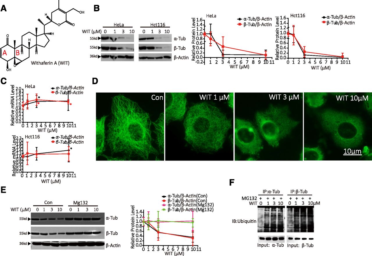
- Fig. 1.
WIT promotes tubulin degradation in a post-transcriptional manner. (A) Chemical structure of WIT. (B) HeLa and Hct116 cells were treated with increasing concentrations of WIT for 16 hours and protein levels of α- and β-tubulin detected via Western blot. The right image presents quantitative data on α- and β-tubulin proteins. Values are presented as mean ± 95% CI of three independent experiments. (C) HeLa and Hct116 cells were treated with increasing concentrations of WIT for 16 hours, and mRNA levels of α- and β-tubulin were detected via quantitative PCR. Values are presented as mean ± 95% CI of three independent experiments. *P < 0.05. (D) Cells were treated with different concentrations of WIT (1 μM, 3 μM, and 10 μM) for 16 hours, and microtubules were imaged using an Olympus fluorescence microscope. (E) HeLa cells were pretreated with or without 10 µM MG132 for 1 hour before treatment with increasing concentrations of WIT for 16 hours and protein levels of α- and β-tubulin detected via Western blot. The right image presents quantitative data on α- and β-tubulin proteins. Values are presented as mean ± 95% CI of three independent experiments. (F) HeLa cells were incubated with MG132(10 μM) for 1 hour before treatment with 0, 1, 3, or 10 μM WIT for 16 hours, α- or β-tubulin were immunoprecipitated from the lysates, and ubiquitinylated α- or β-tubulin were detected with anti-ubiquitin antibodies. The results are representative of three independent experiments. α-Tub, α-tubulin; β-Tub, β-tubulin; Con, Control.
- Fig. 2.
WIT binds to the colchicine site of β-tubulin. (A) Purified tubulin was incubated with the indicated compounds at 4°C and optical density values at 340 nm measured once a minute. (B) Cells were treated with high concentrations of the specified compounds (10 μM paclitaxel, 10 μM colchicine, 10 μM vinblastine, or 100 μM WIT) for 1 hour, and microtubules were imaged using an Olympus fluorescence microscope. (C) Purified tubulin (1 μM) was incubated with the specified compounds for 2 hours before incubation with 100 μM EBI for another 2 hours and subjected to Western blot for detection of β-tubulin protein. The lower image depicts the EBI-β-tubulin protein level. Values are presented as mean ± 95% CI of three independent experiments. (D) HeLa cells were pretreated with 3 µM colchicine or 3 µM vinblastine for 1 hour before treatment with 3 µM WIT for 16 hours. Protein levels of α- and β-tubulin were detected via Western blot. The lower image represents quantitative data on α- and β-tubulin proteins. Values are presented as mean ± 95% CI of three independent experiments. (E) HeLa cells were pretreated with 3 µM nocodazole, 3 µM plinabulin, or 3 µM combretastatin A4 for 1 hour before treatment with 3 µM WIT for 16 hours. Protein levels of α- and β-tubulin were detected via Western blot. The lower image represents quantitative data on α- and β-tubulin proteins. Values are presented as mean ± 95% CI of three independent experiments. Con, control; Col, colchicine; PTX, paclitaxel; Vin, vinblastine; Noc, nocodazole; Pil, plinabulin; CA4, combretastatin A4; α-Tub, α-tubulin; β-Tub, β-tubulin.
- Fig. 3.
WIT exerts irreversible cellular effects. (A) HeLa cells were treated with 10 μM colchicine, 10 μM colcemid, or 100 μM WIT for 8 hours. After the compounds were completely washed off, the cells were cultured for an additional 24 hours, and their microtubule morphology was examined via immunofluorescence at the 0-, 8-, 16-, 24-, and 32-hour time points. (B) Western blot analysis of p-H3 expression was further examined at 0, 8, 16, 24, and 32 hours. The right image depicts quantitative data on p-H3 expression. Values are presented as mean ± 95% CI of three independent experiments.
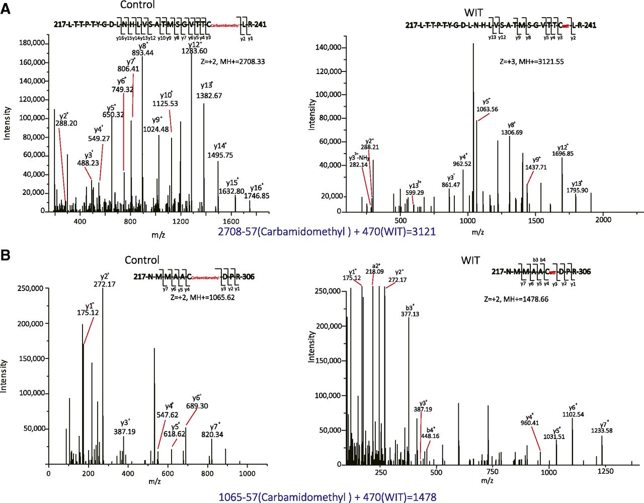
- Fig. 4.
WIT forms covalent bonds with Cys303 and Cys239 of β-tubulin. (A) Left: MS/MS fragmentation patterns of the untreated peptide 217LTTPTYGDLNHLVSATMSGVTTCLR241. Right: MS/MS fragmentation patterns of the covalently bound peptide 217LTTPTYGDLNHLVSATMSGVTTCLR241 indicate that WIT binds to Cys239. (B) Left: MS/MS fragmentation pattern of the untreated peptide 298NMMAACDPR306. Right: MS/MS fragmentation pattern of the covalently bound peptide 298NMMAACDPR306 indicate that WIT binds to Cys303.
- Fig. 5.
Covalent binding between WIT and Cys239 accounts for WIT-induced tubulin degradation. (A) Selected amino acid sequences of β2-, β3-, β4-, β5 (β)-, and β6-tubulin isoforms. (B) HeLa cells were treated with increasing concentrations of WIT for 16 hours, and β2-, β3-, β4- and β6-tubulin protein levels were detected via Western blot. The right image depicts quantitative data on β2-, β3-, β4-, and β6-tubulin protein expression. Values are presented as mean ± 95% CI of three independent experiments. (C) HeLa cells were transiently transfected with MSCV-IRES-GFP vectors coexpressing GFP and WT-FLAG-β-Tubulin, C239S-FLAG-β-Tubulin, or C303S-FLAG-β-Tubulin for 24 hours. Cells were treated with or without 3 µM WIT for 16 hours, and GFP and FLAG-tubulin detected via Western blot. The right image depicts quantitative data on GFP and FLAG-tubulin. Values are presented as mean ± 95% CI of three independent experiments. (D) Chemical structure of dihydrowithaferin A (DWIT). (E) HeLa cells were treated with increasing concentrations of WIT or DWIT for 16 hours, and β-tubulin protein was detected via Western blot. The right image depicts quantitative data on β-tubulin protein. Values are presented as mean ± 95% CI of three independent experiments. (F) HeLa cells were treated with increasing concentrations of WIT or DWIT for 48 hours, and cell viability was assessed with the trypan blue exclusion assay.Values are presented as mean ± 95% CI of three independent experiments. β-Tub, β-tubulin; β2-Tub, β2-tubulin; β3-Tub, β-3tubulin; β4-Tub, β4-tubulin; β6-Tub, β6-tubulin.


