


Abstract
The IκB kinase complex (IKK) mediates activation of the transcription factor nuclear factor-κB (NF-κB). We previously showed that green tea polyphenols inhibited endotoxin-mediated tumor necrosis factor-α (TNFα) production by blocking NF-κB activation. In this study, we evaluated whether green tea polyphenols inhibit NF-κB by blocking IKK activity. We assessed IKK activity by detecting changes in phosphorylation of an IκBα-glutathioneS-transferase (GST) fusion protein. IEC-6 cells pretreated with an extract of green tea polyphenols (GrTPs; 0–0.4 mg/ml) had diminished TNFα-induced IKK and NF-κB activity. Of the various GrTPs, (−)-epigallocatechin-3-gallate (EGCG) was the most potent inhibitor. We next examined whether EGCG inhibited activated IKK. In cytosolic extracts of TNFα-stimulated cells, EGCG inhibited phosphorylation of IκBα-GST (IC50 > 18 μM) consistent with inhibition of IKK activity. Using other polyphenols, we showed that the gallate group was essential for inhibition, and antioxidants were ineffective in blocking activated IKK. Importantly, EGCG decreased IKK activity in cytosolic extracts of NIK transiently transfected cells. This latter finding showed that our findings were not related to nonspecific kinase activity. In conclusion, EGCG is an effective inhibitor of IKK activity. This may explain, at least in part, some of the reported anti-inflammatory and anticancer effects of green tea.
There is increasing interest in the role of tea (Camellia sinensis) in maintaining health and treating disease. Although tea consists of several components, research has focused on polyphenols, especially those found in green tea. The green tea polyphenols include (−)-epicatechin (EC), (−)-epigallocatechin (EGC), (−)-epicatechin-3-gallate (ECG), and (−)-epigallocatechin-3-gallate (EGCG). Of these, EGCG generally accounts for greater than 40% of the total (Hara, 1997). Green tea polyphenols are potent antioxidants (Salah et al., 1995). EGCG usually has the greatest antioxidant activity, and it is the most widely studied polyphenol for disease prevention (for review, see Bagchi, 1999). Many of the putative health benefits of tea are presumed to be caused by its antioxidant effects. One benefit may be cancer prevention. Epidemiological studies suggest that regular consumption of tea reduces the risk of cancer (Katiyar and Mukhtar, 1996). In support of this contention, tea, or, more specifically, the polyphenol fraction, has been reported to decrease the incidence of carcinogen-induced malignancies in animal models (Stoner and Mukhtar, 1995). One proposed mechanism of action is the finding that polyphenols induce apoptosis more readily in cancer cells than their natural counterparts (Chen et al., 1998; Yang et al., 1998b;Suganuma et al., 1999; Ahmad et al., 2000).
Another potential benefit of tea is as an anti-inflammatory agent. Studies in animal models show that green tea polyphenols decrease inflammation. We reported that mice fed an extract of green tea polyphenols had decreased tumor necrosis factor-α (TNFα) production in response to an injection of lipopolysaccharide (LPS) and prevented death after administration of an otherwise lethal dose of LPS (Yang et al., 1998a). Haqqi et al. (1999) reported that the ingestion of a green tea polyphenol extract reduced joint disease in mice with adjuvant-induced arthritis. We recently reported that green tea polyphenols reduced disease activity in the autoimmune disease model, interleukin-2 deficient mice (Varilek et al., 1999). These studies suggest that green tea may have benefit in treating inflammatory disorders.
Several studies have focused on potential mechanisms responsible for the anti-inflammatory and anticancer effects. One potential mechanism of action is the inhibition of nuclear factor-κB activation. Nuclear factor-κB is an oxidative stress sensitive transcription factor that regulates the expression of a variety of genes important in cellular responses, including inflammation, innate immunity, and growth. We showed that EGCG decreased LPS-induced TNFα production in the macrophage cell line RAW264.7 and peritoneal macrophages by blocking NF-κB activation (Yang et al., 1998a). Lin and Lin (1997) reported that EGCG inhibited LPS-induced inducible nitric-oxide synthase gene expression in mouse peritoneal macrophages by decreasing the expression of the transcription factor, NF-κB. Ahmad et al. (2000)recently showed that green tea polyphenols modulate NF-κB in several cancer cell lines, rendering them susceptible to apoptosis.
In unstimulated cells, NF-κB predominantly exists as a heterodimer, composed of p65 and p50 subunits, that resides in the cytoplasm in an inactive state bound to a member of the IκB family of inhibitory proteins (Baldwin, 1996; Barnes and Karin, 1997). IκB masks the nuclear localization sequence of NF-κB, sequestering it in the cytoplasm. NF-κB activity can be induced in most cell types upon exposure to stimuli including cytokines (TNFα, interleukin-1), endotoxin, and oxidative stress (Baldwin, 1996). NF-κB activators induce degradation of IκB by the 26S proteasome. This requires the phosphorylation of two serines (Ser-32 and Ser-36 in IκBα) in the N-terminal regulatory domain of IκB, followed by polyubiquitination. The liberated NF-κB then translocates to the nucleus, binds to specific sequences in the promoter region, and induces gene expression (Karin, 1999). Several anti-inflammatory agents, including aspirin and sodium salicylate, inhibit the activity of IKK (Yin et al., 1998). Antioxidants such as pyrrolidinedithiocarbamate (PDTC), N-acetyl cysteine (NAC), and vitamin E also suppress NF-κB activation (Suzuki and Packer 1993;Beauparlant and Hiscott, 1996). Recently, Pan et al. (2000) showed that the black tea derivative theaflavin-3,3′-digallate and EGCG block the phosphorylation of IκB; however, the study did not determine whether these polyphenols blocked the activation of IKK or inhibited the activity of IKK. Both mechanisms would lead to a reduction in IκB phosphorylation.
In this study, we used IEC-6 intestinal cells, because intestinal epithelial cells are the early targets for consumed green tea polyphenols. We identified EGCG as the most potent inhibitor of IKK among green tea polyphenols. Importantly, this function of EGCG appeared to be independent of its antioxidant property. Our data suggest that most anti-inflammatory effects of green tea are mediated by EGCG rather than other polyphenol extracts. Through comparison among structurally similar compounds, we conclude that the gallate group plays a crucial role for inhibiting IKK activates; however, the catechin structure is also required. This information may be used in design of more potent IKK inhibitors in the future.
Experimental Procedures
Materials.
Extracted green tea polyphenols (GrTP; >95% polyphenols) were purchased from LKT Laboratories, Inc (St. Paul, MN). High-performance liquid chromatography analysis of the green tea extracts revealed the percentage composition of the four polyphenols of interest: 35% EC, 4% EGC, 15% EGC, and 38% EGCG. Cell culture supplies were obtained from Invitrogen (Carlsbad, CA). [γ-32P]ATP (6000 mCi/mmol) was obtained from PerkinElmer Life Science Products (Boston, MA). The fusion protein (70 kDa) containing the full-length IκB-α of human origin and glutathione S-transferase (GST), rabbit polyclonal IgG antibody against GST, and protein-A-agarose were purchased from Santa Cruz Biotechnology (Santa Cruz, CA). EGCG (>98% pure), ECG (>98% pure), and remaining chemicals were purchased from Sigma (St. Louis, MO).
Cell Culture.
The fetal rat intestinal epithelial cell line, IEC-6 was obtained from the American Type Culture Collection (Manassas, VA). The cells were grown in Dulbecco's modified Eagle's medium supplemented with 10% (v/v) endotoxin-free fetal calf serum, 2 mM glutamine, and 1 × 10⁵ U/liter of penicillin/streptomycin at 37°C in an atmosphere of 10% CO2 and 95% relative humidity. Passage consisted of incubating cells in sterile trypsin/EDTA followed by resuspending the cells in fresh medium. The cells were studied between passages 20 and 35.
Cytosolic and Nuclear Extraction.
Cytosolic and nuclear extracts were isolated by the method initially described by Dignam et al. (1983) with the addition of protease inhibitors (1 μg/l leupeptin, 1 μg/l pepstatin, and 1 μg/l leucine thiol) to the lysis and nuclear extraction buffers (Yang et al., 1998a). IEC-6 cells (5 × 10⁶/plate) were seeded onto 60-mm culture plates and cultured to 90% confluence. After various treatments, the cells were washed twice with ice-cold PBS and incubated on ice for 15 min with 0.2 ml of ice-cold lysis buffer. The cells were collected into 1.5-ml polypropylene tubes, incubated on ice for 30 min, and then underwent centrifugation at 1,200g for 10 min at 4°C. For cytosolic extracts, supernatants were collected and underwent a second centrifugation at 13,000g for 10 min at 4°C. The resulting supernatants (cytosolic extracts) were stored at −70°C. For nuclear extracts, pellets from the initial centrifugation were incubated in nuclear extraction buffer on ice for 1 h followed by centrifugation at 110,000g for 15 min at 4°C. The supernatants were stored at −70°C. The protein concentration of the cytosolic and nuclear extracts was measured using the Dc Protein Assay kit (Bio-Rad Laboratories, Hercules, CA).
IKK Assay.
IκB kinase activity was assessed by detecting the phosphorylation of IκBα-GST fusion protein. Ten micrograms of cytosolic extract and 2.5 μg of IκBα-GST fusion protein were incubated for 30 min at 37°C in 20 mM HEPES (pH 7.5) containing 20 mM β-glycerophosphate, 10 mM MgCl2, 10 mMp-nitrophenyl phosphate, 0.1 mM Na3VO4, 2 mM dithiothreitol, 10 μg/ml aprotinin, 20 μM ATP, 0.1 mM NaCl, 0.4 mM phenylmethylsulfonyl fluoride, and 15 μCi γ-32P-ATP (Regnier et al., 1997). The reaction was stopped by adding ten volumes of an immunoprecipitation (IP) buffer consisting of 0.1 M Tris-HCl (pH = 8.0) with 1% (v/v) Triton X-100, 1% (w/v) deoxycholate, 0.5% (w/v) SDS, and 2 mM phenylmethylsulfonyl fluoride. The IκBα-GST proteins were immunoprecipitated by incubating the samples with a rabbit polyclonal IgG antibody against GST (1:500) and protein-A-agarose (20 μl) followed by extensive washes with IP buffer. The phosphorylated IκBα-GST proteins then were visualized by 5 to 15% SDS-polyacrylamide gel electrophoresis and autoradiography. Densitometry was performed on the resulting bands using a Personal Densitometer SI (Molecular Dynamics, Sunnyvale, CA) with ImageQuant V1.1 (Molecular Dynamics).
Electrophoretic Mobility Shift Assay (EMSA).
The DNA binding activity of NF-κB was detected in nuclear protein extracts by an electrophoretic mobility shift assay (Yang et al., 1998a).32P-End-labeled oligonucleotide probes containing the κB enhancer DNA element (Santa Cruz Biotechnology, Inc.) were prepared according to protocol (Sambrook et al., 1989). Incubation of 8 μg of the nuclear extract with the end-labeled probe for 20 min at room temperature was carried out in the presence of sonicated salmon sperm DNA, which served as a nonspecific competitor. Complexed and uncomplexed DNA were then resolved by electrophoresis on a 6% low-ionic-strength nondenaturing polyacrylamide gel and visualized by autoradiography. To identify the specific subunits of NF-κB, supershift experiments were performed by incubating nuclear extracts with 1 μl of antibodies against p65 or p50 (Santa Cruz Biotechnology, Inc.) for 30 min before adding the end-labeled probe. To confirm the binding specificity, competition experiments were performed by adding 100-fold of unlabeled NF-κB consensus oligonucleotides as specific competitors.
Cytokine-Induced Neutrophil Chemoattractant (CINC) Immunoassay.
IEC-6 cells (1 × 10⁵) were plated onto 24-well cluster plate. At 80% confluence, the cells were pretreated with various concentrations of EGCG or EGC (0–100 μM) for 2 h followed by stimulation with TNFα (500 U/ml). The cells were incubated overnight and the supernatants collected at 18 h. The concentrations of CINC in the culture supernatants were determined by sandwich enzyme-linked immunosorbent assay using the method described by Wittwer et al. (1993). The rabbit polyclonal IgG antibody against CINC and CINC (standards) were purchased from Peptides International (Louisville, KY). The goat polyclonal IgG antibody against CINC was kindly provided by Dr. John Zagorski (Laboratory of Immunology, National Institute of Dental Research, National Institutes of Health, Bethesda MD).
NF-κB-Inducing Kinase (NIK) transient transfection.
PRK-Myc-NIK, a full-length NIK expression vector was kindly provided by Dr. Warner Greene (Gladstone Institute of Virology and Immunology, University of California) (Lin et al., 1998). IEC-6 cells (5 × 10⁵) were seeded onto six-well (35 mm diameter) cluster plates and transfected the following day with 1.2 μg of pRK-Myc-NIK using the LipofectAMINE PLUS Reagent kit (Invitrogen) according to the methods provided by the manufacturer. Control plates were treated similarly except they did not receive pRK-Myc-NIK. The cells were harvested at 24 h and cell extracts were prepared for the IKK assay as described above.
Results
Extracted GrTPs Inhibited the Phosphorylation of IκB.
We initially examined the effect of TNFα on IKK activity in IEC-6 cells by monitoring the in vitro phosphorylation of an IκBα-GST fusion protein. At baseline (time 0), there was detectable IKK-like activity in cytosolic extracts (Fig. 1A). After treatment with TNFα (500 U/ml), cytosolic IKK activity increased within 5 min and remained elevated for at least 30 min (Fig. 1A). This was associated with increased nuclear NF-κB binding activity that peaked 30 min after stimulation (Fig. 1B). Supershift experiments confirmed that the observed NF-κB band was the active heterodimer, p65/p50 (data not shown).

TNFα-activated IKK and NF-κB in IEC-6 cells. IEC-6 cells were treated with TNFα (500 U/ml) for the indicated periods. Then cytosolic and nuclear extracts were prepared as described under Experimental Procedures. A, a representative gel showing the time course of IKK activity in cytosolic extracts following stimulation with TNF α. Relative densitometry scores are noted in these and appropriate subsequent figures. IKK activity was detected by the in vitro phosphorylation of IκBα-GST. B, a representative EMSA showing the time course of NF-κB activity in nuclear extracts from IEC-6 cells treated with TNFα.
We examined the effect of extracted GrTPs on TNFα-mediated phosphorylation of IκB. The cells were pretreated with GrTP (0–0.4 mg/ml) for 2 h and then exposed to TNFα (500 U/ml) for 5 min. The cytosolic extracts were then assessed for their ability to phosphorylate IκBα-GST. GrTP decreased TNFα-induced IκBα phosphorylation in a dose-dependent fashion (Fig.2A). This corresponded to a similar reduction in nuclear NF-κB binding activity (Fig. 2B). These data clearly showed that the GrTP extract inhibited the phosphorylation of IκBα resulting in a reduction in NF-κB activation

GrTPs blocked TNFα -mediated activation of IKK and NF-κB in IEC-6 cells. IEC-6 cells were pretreated with the indicated concentrations of GrTP for 2 h and then stimulated with TNFα (500 U/ml) for either 5 min for cytosolic IKK activity or 30 min for nuclear NF-κB activity. IKK activity was determined by in vitro phosphorylation of IκBα-GST and nuclear NF-κB -DNA binding was detected by EMSA. A, a representative gel showing the effect of GrTP on TNFα-stimulated IKK activity. B, a representative EMSA showing the effect of GrTP on TNFα-stimulated NF-κB activation.
The Effects of the Various Green Tea Polyphenols on IκBα Phosphorylation.
We next examined the various green tea polyphenols (EGCG, EGC, EC, ECG) to determine whether differences in efficacy exist. EGCG has been shown to affect the activity of several enzymes (Nakane and Ono, 1990; Stoner and Mukhtar, 1995; Aucamp et al., 1997). Therefore, we initially examined EGCG. IEC-6 cells were pretreated with EGCG (0–200 μM) for 2 h followed by stimulation with TNFα (500 U/ml) for 5 min. The cytosolic extracts were assessed for their ability to phosphorylate IκBα. EGCG decreased IκBα phosphorylation in a dose-dependent fashion (Fig.3A). We then compared EGCG to the other green tea polyphenols (EGC, ECG, EC). The cells were pretreated with equimolar concentrations (200 μM) of EGC, ECG, EC, or EGCG. EGCG reduced the phosphorylation of IκBα to a much greater extent than the other polyphenols. The other polyphenols had little or no effects at the concentration studied. These studies suggested that EGCG primarily was responsible for the observed inhibition of IκBα phosphorylation. We then investigated whether this also was reflected in differences in downstream events. We compared EGCG to EGC, because they differ only in that EGCG has a gallate group. We examined their ability to inhibit TNFα-mediated cytokine production. The cytokine studied was CINC, a NF-κB-responsive chemokine. IEC-6 cells were pretreated with EGCG or EGC (0–100 μM) for 2 h followed by stimulation with TNFα (500 U/ml). The supernatants were collected at 18 h and assayed for CINC (Fig. 4). EGCG decreased CINC release to a much greater extent than EGC. These results were consistent with our prior observations.

EGCG inhibited IKK activity more effectively than other green tea polyphenols. IKK activity was determined in cytosolic extracts by in vitro phosphorylation of IκBα-GST. A, IEC-6 cells pretreated with EGCG (0–200 μM) for 2 h were then stimulated with TNFα (500 U/ml) for 5 min. IKK activity in the cytosolic extracts were determined by in vitro phosphorylation of IκBα-GST proteins. A representative gel is presented. B, IEC-6 cells were pretreated with equimolar concentrations (200 μM) of EGCG, EGG, ECG, or EC for 2 h and then stimulated with TNFα (500 U/ml) for 5 min. A representative gel is presented. Lanes: 1, control; 2, TNFα; 3, TNFα + EGCG; 4, TNFα + EGC; 5, TNFα + ECG; and 6, TNFα + EC.

Comparison of the effects of EGCG and EGC on TNF-mediated release of CINC. IEC-6 cells were pretreated with EGCG or EGC (0–100 μM) for 2 h followed by stimulation with TNFα (500 U/ml). The supernatants were collected at 18 h and assayed for CINC by sandwich enzyme-linked immunosorbent assay. Data expressed as mean ± S.D., n = 6.

EGCG inhibited IKK activity in a cell-free system. Cytosolic extracts from TNFα-stimulated IEC-6 cells (500 U/ml, 5 min) were treated with the indicated concentrations of EGCG on ice for 20 min followed by measurement of IKK activity. A, a representative gel. B, relative densitometry of gel.
EGCG Inhibited TNFα-Induced IKK Activity in a Cell-Free System.
Our data clearly showed that EGCG inhibited TNFα-induced phosphorylation of IκBα. However, these experiments did not determine whether EGCG blocks the activation of IKK or inhibits the activity of IKK. Therefore, we focused on the effects of EGCG on activated IKK. The source of activated IKK was cytosolic extracts from IEC-6 cells stimulated for 5 min with TNFα (500 U/ml). We incubated the TNFα-stimulated cytosolic extracts with EGCG (0–200 μM) on ice for 20 min and detected changes in IKK activity. EGCG decreased IKK activity in a dose-response fashion. Fifty-percent inhibition was calculated to be 18 μM (IC50 = 18 μM) (Figure. 5). We next examined whether or not any of the other green tea polyphenols inhibited activated IKK. The TNFα-stimulated cytosolic extracts were incubated with 200 μM EGCG, EGC, ECG, EC, or catechin (backbone of the polyphenols) and IKK activity was detected. Other than EGCG, only ECG inhibited IKK activity and was about 5-fold less effective (Fig.6, A and B). EGCG and ECG both contain gallate groups. Therefore, we examined gallic acid (200 μM) and its methyl ester (200 μM) and found that they were very weak inhibitors (20–30% inhibition versus 95% inhibition; Fig. 6, A and B). These data showed that EGCG and (to a much lesser extent) ECG inhibited IKK activity. Importantly, the gallate group was functionally necessary for inhibition of IKK activity, and the presence of the catechin structure dramatically enhanced this effect.

EGCG and ECG specifically inhibited IKK activity in vitro. Cytosolic extracts from TNFα-stimulated IEC-6 cells (500 U/ml, 5 min) were preincubated with H2O (control), 0.18 mg/ml of extracted GrTPs, or 200 μM various polyphenols or related compounds on ice for 20 min followed by measuring IKK activity. A, a representative gel; B, relative densitometry of the presented gel. Lanes: 1, control; 2, GrTP; 3, EGCG; 4, EGC; 5, ECG; 6, (+)-EC; 7, (−)-EC; 8, (±)-catechin; 9, gallic acid; and 10, methyl ester gallic acid.
EGCG, a Specific Inhibitor of IKK.
There are several molecules that inhibit NF-κB activation by blocking IκB-α phosphorylation (Beauparlant and Hiscott, 1996). Among these are antioxidants, such as PDTC and NAC. NAC has been shown to inhibit IKK activation in endothelial cells (Spiecker et al., 1998). We examined whether antioxidants could inhibit activated IKK in vitro. The experiments were performed as described above. TNFα-stimulated cytosolic extracts of IEC-6 cells were incubated with various antioxidants [PDTC, α-tocopheryl acetate (vitamin E), NAC, butylated hydroxytolvene, dimethyl sulfoxide] and IKK activity was detected. As shown in Fig. 7, none of these antioxidants inhibited IKK activity. These results suggested that the inhibitory effect of EGCG was not mediated by an antioxidant mechanism, but rather was structure-related. As expected,N-tosyl-l-phenylalanine chloromethylketone (TPCK), a serine protease inhibitor that blocks IκB degradation downstream (Zandi et al., 1998), did not inhibit IKK activity (Fig. 7).

In vitro effects of various antioxidants on IKK activity. Cytosolic extracts from TNFα-stimulated IEC-6 cells (500 U/ml, 5 min) were treated with various agents on ice for 20 min followed by detection of IKK activity. A representative gel showing the effects on IKK activity. Lanes: 1, control; 2, 0.5% dimethyl sulfoxide (DMSO); 3, 200 μM N-tosyl-l-phenylalanine chloromethylketone (TPCK); 4, 200 μM butylated chloromethyl ketone (BHT); 5, 200 μM α-tocopheryl acetate; 6, 200 μM PDTC; and 7, 200 μM NAC.
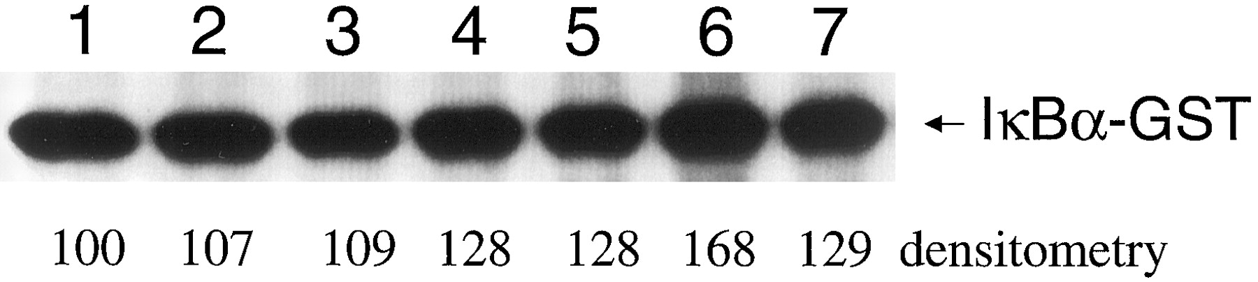
These data strongly suggested that EGCG inhibited the activity of IKK. However, a full-length IκBα protein was used as the substrate for detecting IKK activity. IKK phosphorylates IκBα at Ser-32 and Ser-36. IκBα has other potential phosphorylation sites that could be phosphorylated by other kinases activated in response to TNFα stimulation. To date, only ultraviolet radiation and anoxia are known to phosphorylate IκB without activation of IKK (Karin, 1999). To address this issue, we transiently transfected IEC-6 cells with a full-length NIK expression vector (pRK-Myc-NIK). NIK is an upstream kinase in the activation pathway of IKK. In overexpression experiments, NIK acts as a potent IKK and NF-κB activator (Regnier et al., 1997). We isolated the cytosolic extracts from IEC-6 cells transiently transfected with pRK-Myc-NIK and assessed IKK activity in the presence of various concentrations of EGCG. Similar to the TNFα experiments, EGCG decreased IKK activity in the NIK transiently transfected cells in a dose-dependent fashion (Fig. 8). These data confirmed that EGCG inhibited IKK activity and that our findings were not related to changes in nonspecific kinase activity.

EGCG inhibits IKK activity in IEC-6 cells transiently transfected with pRK-Myc-NIK. We transiently transfected IEC-6 cells with a full-length NIK expression vector (pRK-Myc-NIK) and assessed the effects of EGCG (0–100 μM) on IKK activity. A representative gel is presented.
Discussion
We have identified two structurally related compounds, EGCG and ECG that inhibit IKK phosphorylation of IκBα. These two compounds, derived from green tea, are catechins with gallate groups. The gallate group is essential for this inhibitory effect, because polyphenols lacking the gallate group did not inhibit IKK activity. Gallic acid and its methyl ester are very weak inhibitors in a cell-free system. The catechin backbone confers greater efficacy. Of the two, EGCG was much more potent than ECG. EGCG differs only in that it contains one more hydroxyl group. Importantly, this phenomenon was not related to an antioxidant effect, because antioxidants and polyphenols lacking the gallate group did not inhibit IKK activity. Our data clearly shows that EGCG inhibits IKK phosphorylation of IκBα. This may occur as a direct effect on IKK or by interfering with the interaction of IKK with IκB.
The phosphorylation of IκB is a key regulatory step that dictates NF-κB activation. In nearly all instances, IKK controls IκB phosphorylation (Karin, 1999). IKK is a large 500- to 900-kDa protein complex with three subunits. Two of the subunits, IKKα and IKKβ, are serine kinases (Mercurio et al., 1997; Regnier et al., 1997;Woronicz et al., 1997; Zandi et al., 1997). A third subunit, IKKγ, interacts preferentially with IKKβ and seems necessary for activation (Rothwarf et al., 1998). IKK directly phosphorylates two serine residues at the NH2-termini of IκBα (Ser-32, Ser-36) and IκBβ (Ser-19, Ser-23) (Salah et al., 1995). This phosphorylation event triggers the polyubiquitination of IκB and subsequent degradation by the 26S proteasome (Zandi et al., 1998). Of the three subunits, the phosphorylation of IKKβ is necessary for IKK activation and IKKβ seems responsible for phosphorylation of IκB (Delhase et al., 1999).
EGCG and ECG also inhibit the activity of other enzymes. For instance, EGCG and ECG reduced the activities of reverse transcriptase and cellular DNA and RNA polymerases (Nakane and Ono, 1990). EGCG competitively inhibited xanthine oxidase, although the remaining polyphenols were less potent and showed a mixed pattern of inhibition (Aucamp et al., 1997). EGCG also inhibited epidermal growth factor-stimulated tyrosine phosphorylation of the epidermal growth factor receptor (Liang et al., 1997). Using molecular modeling techniques, EGCG should bind to urokinase and interfere with the ability of urokinase to recognize its substrate (Jankun et al., 1997). These studies suggest that EGCG may have other cellular targets that could alter function at various levels.
A number of antioxidants, such as PDTC, NAC, and vitamin E, can suppress NF-κB activation (Suzuki and Packer, 1993; Beauparlant and Hiscott, 1996), and NAC was reported to inhibit activation of IKK (Spiecker et al., 1998). Our findings indicated that these antioxidants did not affect the activity of IKK. These data support the premise that antioxidants block activation of IKK and that reactive oxygen species play an intermediary role upstream to IKK. The green tea polyphenols are potent antioxidants; as such, they may also inhibit activation of IKK. Therefore, EGCG may block NF-κB activation at two potential steps in the pathway. First, as an antioxidant, it may inhibit signaling events upstream to IKK that result in decreased IKK activation. Secondly, its unique structure inhibits IKK activity. Both mechanisms would lead to inhibition of NF-κB activation.
We chose to study IEC-6 cells because of our research focus in inflammatory bowel disease, because these cells are potent producers of proinflammatory cytokines/chemokines and because the intestinal epithelium is an initial target for polyphenol therapeutic intervention. Thus, the intestinal epithelium may derive antiinflammatory effect from green tea polyphenols even if the polyphenols have limited systemic absorption. The activation of NF-κB may play a critical role in the development and perpetuation of many inflammatory disorders such as inflammatory bowel disease and rheumatoid arthritis (Barnes and Karin, 1997; Baeuerle and Baichwal, 1997). Indeed, several groups have reported increased intestinal NF- κB activation in patients with inflammatory bowel disease and in animal models of inflammatory bowel disease. NF-κB, therefore, is a potential target for anti-inflammatory therapies. Many of the currently accepted medical therapies for these diseases inhibit NF-κB (e.g., glucocorticoids, sulfasalazine). The identification of new more selective inhibitors of NF-κB may lead to more effective treatment of inflammatory disorders. IKK represents a key control point for NF-κB activation and may be a suitable target for modulating NF-κB-mediated cellular responses. The identification of EGCG as an inhibitor of IKK should provide insight into the IKK complex and potentially lead to the development of new IKK inhibitors that are more potent and hopefully more selective. Our findings also suggest that green tea polyphenols have a potential therapeutic utility in the treatment of inflammatory diseases.
Acknowledgment
We thank K. Westberry and D. Schweder for their technical assistance.
Footnotes
-
This work was supported by National Institutes of Health Grants KO8-DK02401-01A, MO1-RR02602, IPO1-NS31220-01A1, and RO1-AA010762 and by the Veterans Administration.
- Abbreviations:
- EC
- epicatechin
- EGC
- (−)-epigallocatechin
- ECG
- (−)-epicatechin-3-gallate
- EGCG
- (−)-epigallocatechin-3-gallate
- TNF
- tumor necrosis factor
- LPS
- lipopolysaccharide
- NF-κB
- nuclear factor-κB
- IKK
- IκB kinase
- PDTC
- pyrrolidinedithiocarbamate
- NAC
- N-acetyl cysteine
- GrTP
- green tea polyphenols
- GST
- glutathione S-transferase
- EMSA
- electrophoretic mobility shift assay
- CINC
- cytokine-induced neutrophil chemoattractant
- NIK
- NF-κB-inducing kinase
- Received May 10, 2001.
- Accepted May 21, 2001.
- The American Society for Pharmacology and Experimental Therapeutics


