


Abstract
This study focused on the question of how the DNA mismatch repair (MMR) system and p53 interact to maintain genomic integrity in the presence of the mutagenic stress induced by hydrogen peroxide (H2O2). The cytotoxic and mutagenic effects of H2O2 were compared in four colon carcinoma sublines: HCT116, HCT116/E6, HCT116+ch3, and HCT116+ch3/E6, representing MMR−/p53+, MMR−/p53−, MMR+/p53+, and MMR+/p53− phenotypes, respectively. Loss of p53 in MMR-proficient cells did not significantly alter cellular sensitivity to H2O2, but disruption of p53 in MMR-deficient cells resulted in substantial resistance to H2O2 (IC50 values of 203.8 and 66.2 μM for MMR−/p53− and MMR−/p53+ cells, respectively). The effect of loss of p53 and MMR function on sensitivity to the mutagenic effect of H2O2 paralleled the effects on cytotoxic sensitivity. In MMR-deficient cells, loss of p53 resulted in a 3.5- and 2.2-fold increase in the generation of 6-thiogunaine and ouabain-resistant clones, respectively. Loss of MMR in combination with loss of p53 synergistically increased the frequency of frameshift mutations in the CA repeat tracts of the out-of-frame shuttle vector pZCA29 and further promoted instability of microsatellite sequences under H2O2 stress. Flow cytometric analysis showed that H2O2 treatment produced a Gl and G2/M phase arrest in MMR+/p53+ cells. Loss of MMR did not alter the ability of H2O2 to activate either checkpoint; loss of p53 in either the MMR-proficient or deficient cells resulted in impairment of the Gl arrest and a more pronounced G2/M arrest. H2O2 caused a greater and more longed increase in p53 protein levels in MMR-proficient than in the MMR-deficient cells. The results demonstrate that the effect of disabling p53 function is modulated by the proficiency of the MMR system (and vice versa) and that there is an overlap between the functions of p53 and the MMR system with respect to the activation of apoptosis and mutagenesis after an oxidative stress.
Cells engage multiple repair mechanisms after DNA damage, reflecting the importance of DNA repair in maintaining both cell viability and genomic stability (ap Rhys and Bohr, 1996). Among these, the postreplicative DNA mismatch repair (MMR) system is of particular importance to the integrity of the genome. In addition to its role in correcting the misincorporation errors that occur during replication, MMR is also implicated in the recognition of several types of DNA damage. We and others have demonstrated that loss of MMR causes resistance to the cytotoxic effects of some of the platinum-containing drugs (Anthoney et al., 1996; Drummond et al., 1996; Fink et al., 1996) and renders cells hypersensitive to the mutagenic effects of cisplatin (Lin and Howell, 1999). One hypothesis is that MMR acts as a sensor for genomic damage that, upon recognition of certain types of DNA lesions, initiates a sequence of events that facilitate repair or promote apoptosis (Kat et al., 1993; Hawn et al., 1995; Nehmé et al., 1997).
The p53 protein plays a central role in the cellular injury response after DNA damage (Lane, 1992). After DNA damage, wild-type p53 activates several genes that lead to cell-cycle arrest, DNA repair, or apoptosis. Some of these genes include p21WAF1/CIP1, GADD45, and MDM2 (El-Deiry et al., 1993; Chen et al., 1994; Zhan et al., 1994). Current evidence is consistent with the hypothesis that loss of p53 contributes to genomic instability by permitting the inappropriate survival of cells that would normally have undergone apoptosis in response to DNA damage.
Reactive oxygen species are formed continuously in living cells as byproducts of normal cellular metabolism, as well as through the action of exogenous compounds and radiation. Adducts produced by oxidative damage may constitute the single most common type of DNA damage, and it has been estimated that each human cell must repair approximately 10,000 oxidatively damaged sites in its genome each day (Ames, 1989). It is a generally accepted that DNA damage derived from molecular oxygen occurs largely through the generation of H2O2 (DiGiuseppi and Fridovich, 1984). H2O2 is clearly mutagenic in bacteria (Levin et al., 1982), and there is substantial evidence that endogenously generated H2O2 is mutagenic in mammalian cells (Hsie et al., 1986; Ziegler-Skylakakis and Andrae, 1987; Oller and Thilly, 1992; Gille et al., 1994). The induction of mutations after oxidative stress represents a failure of the available repair systems to remove the DNA damage, and such mutations are believed to contribute to malignant transformation.
Heterozygosity with respect to germline mutations that disable the function of either p53 or any of several genes whose products play central roles in MMR predisposes to malignancy. It is likely that genomic instability caused by loss of either p53 or MMR plays a role in the phenomenon of tumor progression and the acquisition of drug resistance after the cell has become fully malignant. Mutations in p53 are found in more than 50% of all human tumors (Hollstein et al., 1991). Loss of MMR is characteristic of tumors arising in patients with hereditary nonpolyposis colon cancer and is also found in some cases of sporadic cancer of many types. Loss of p53 or MMR function in at least a subset of the cells in a tumor because of somatic mutation is even more likely. In the present work, we have used sublines of the human colon carcinoma cell line HCT116 that have been engineered to differ in MMR capacity and p53 function to investigate the interaction of p53 with MMR in modulating the cellular sensitivity to the cytotoxic and mutagenic effects of oxidative stress imposed by H2O2.
Experimental Procedures
Cell Lines.
The hMLH1-deficient human colorectal adenocarcinoma cell line HCT116 was obtained from the American Type Culture Collection (Manassas, VA); HCT116 contains a hemizygous mutation in hMLH1 resulting in a truncated, nonfunctional protein (Boyer et al., 1995). A subline complemented with chromosome 3 (HCT116+ch3) was obtained from Drs. C.R. Boland and M. Koi. The chromosome 3-complemented cells are competent in DNA mismatch repair (Koi et al., 1994). HCT116 and HCT116+ch3 sublines expressing papillomavirus E6 (identified here as HCT116/E6 and HCT116+ch3/E6) were obtained from Drs. D.A. Boothman and M. Meyers. p53 function was disrupted in these cell lines by constitutive high-level expression of the human papillomavirus type-16 E6 gene, which stimulates the degradation of p53 through a ubiquitin pathway (Davis et al., 1998). All the four cell lines were maintained in Iscove's modified Dulbecco's medium (Irvine Scientific, Irvine, CA) supplemented with 2 mM l-glutamine and 10% heat-inactivated fetal bovine serum. The chromosome-complemented lines were maintained in medium containing 400 μg/ml geneticin (Life Technologies, Inc., Gaithersburg, MD), and the cell lines expressing papillomavirus E6 were cultured in medium supplemented with 80 μg/ml hygromycin B (Boehringer Mannheim, Indianapolis, IN).
Materials.
In these studies, oxidative stress was generated by exposing cells to exogenously added hydrogen peroxide. Hydrogen peroxide was obtained as a 30% solution from Sigma Chemical Co. (St. Louis, MO) and diluted freshly with serum-free medium to form a stock solution of 50 mM before each experiment. The H2O2 solution was filter sterilized before addition to cell cultures.
Clonogenic Assay.
Clonogenic assays were performed by seeding 250 cells into 60-mm plastic dishes in 5 ml of complete media. After a 24-h incubation at 37°C, the cultures were replaced with serum-free medium and appropriate amounts of hydrogen peroxide were added to the dishes and incubated for 1 h. After treatment with H2O2, cells were rinsed twice at 37°C in PBS to remove any remaining H2O2 and fresh medium was added. Colonies of at least 50 cells were scored visually after 8 to 10 days. Each experiment was performed a minimum of three times using triplicate cultures for each drug concentration. IC50 values were determined using log-linear interpolation.
Mutant Frequency Assay.
HCT116, HCT116/E6, HCT116+ch3, and HCT116+ch3/E6 cells were grown in HAT medium containing 0.4 μM aminopterin, 16 μM thymidine, and 100 μM hypoxanthine for a minimum of 14 days and were then exposed for 1 h to increasing concentrations of H2O2 in serum-free medium. Thereafter, the cells were washed twice and recultured in regular medium for 8 days during which the cultures were split 2:1 as needed to keep them from becoming confluent. All the cells were then trypsinized and seeded into each of 10 100-mm tissue culture dishes at 100,000 cells/dish in the presence of 20 μM 6-thioguanine. At the same time, aliquots of 250 cells were seeded into each of three 60-mm dishes in drug-free medium for determination of cloning efficiency. After 14 days, colonies were counted after staining with 0.1% crystal violet. The procedure for determination of the frequency of mutation to ouabain was the same except that the cells were not grown in HAT medium before the start of the experiments, and the ouabain concentration was 1.0 μM. Mutation frequency was calculated as follows: mutation frequency = a/(b × 106) where “a” is the number of colonies present in the 10 drug-treated dishes and “b” is the cloning efficiency. Each experiment was performed a minimum of three times and the data are presented as mean ± S.D.
Host Cell Microsatellite Instability Assay.
The pZCA29 vector (Diem and Runger, 1998) was obtained from Dr. Runger. Four million HCT116, HCT116/E6, HCT116+ch3, HCT116+ch3/E6 cells were transfected with 2 μg of pZCA29 by electroporation on day 1. Replicated pZCA29 was recovered from the transfected cells on days 3, 5, 7, 9, and 11 by a rapid alkali lysis procedure. For the H2O2 treatment experiment, 2 days after transfection, the cells were treated with 100 μM H2O2 for 1 h, and the vector harvested on days 3, 5, 7, 9, and 11. Unreplicated input plasmid DNA was removed by digestion with DpnI, which cleaves the methylated DNA from bacteria. Escherichia coli XL1- Blue MRF′ (Stratagene) was transformed with recovered pZCA29 and then selected on LB agar plates containing 5-bromo-4-chloro-3-indolyl-β-galactosidase, isopropyl-β-d-thiogalactoside, and ampicillin. Bacterial transformations were performed in triplicate for each of two to three independent samples of pZCA29 recovered at each time point. The total number of white and blue colonies were counted. The mutation frequency was calculated as the mean of the total number of blue colonies divided by the mean of the total number of colonies. Student's t test was used to test for the differences.
Flow Cytometry.
Subconfluent cultures growing in 10-cm tissue culture dishes were exposed to 100 μM H2O2 for 1 h. At 1, 2, and 3 days after H2O2treatment, cells were harvested by trypsinization, washed twice with ice-cold PBS, fixed in ice-cold 70% ethanol, treated with RNase (Sigma) at 37°C for 30 min, and stained with 50 μg/ml propidium iodide (Sigma). After a 30-min incubation on ice, the cells were analyzed on a FACScan flow cytometer (Becton-Dickinson, San Jose, CA) using the FlowJo cell cycle analysis software (Tree Star, Inc., San Carlos, CA) and the “Watson Pragmatic” model.
Western Blotting.
Cells were collected at times from 1 to 7 days after a 1-h treatment with 100 μM H2O2, and lysed in 100 μl of lysis buffer [10 mM Tris·HCl, pH7.4, 150 mM NaCl, 5 mM EDTA, 1% Triton X-100, 5 mM dithiothreitol, 1 mM sodium vanadate, 0.1 mM phenylmethylsulfonyl fluoride, and 5 mM aminocaproic acid] for 30 min on ice. The insoluble material was removed by centrifugation at 14,000 g for 20 min at 4°C. Ten micrograms of protein from each sample were electrophoresed through 10 to 20% SDS-polyacrylamide gel electrophoresis and transferred to a polyvinylidene difluoride membrane (Immobilon P; Millipore, Bedford, MA). The membranes were blotted with p53 monoclonal antibody (Santa Cruz Biotechnology, Santa Cruz, CA). After application of a horseradish peroxidase-coupled secondary antibody, reactive proteins were visualized with enhanced chemiluminescence (Amersham Pharmacia Biotech, Piscataway, NJ). The protein bands that reacted with the antibodies were detected on radiographic film (Fuji Medical X-ray Film; Fuji Medical Systems USA, Stamford, CT) 5 to 60 s after exposure. The bands of p53 on radiographic films were scanned and analyzed densitometrically by a ChemiImager (Alpha Innotech Corporation, San Leandro, CA).
Results
Effect of Loss of p53 and/or MMR Function on the Cytotoxicity of H2O2.
These studies were conducted using a panel of four human colon carcinoma HCT116 sublines engineered to have the following combinations of proficient and deficient phenotypes: p53+/MMR+, p53−/MMR+, p53+/MMR−, and p53−/MMR−. Clonogenic assays were used to determine the effect of loss of p53, MMR, or both on the sensitivity to the cytotoxic effects of H2O2. The H2O2 concentration-survival curves for the 4 HCT116 sublines are shown in Fig.1, and the IC50values and statistical analyses are presented in Table1. The p53+/MMR+ cells were the most sensitive. Loss of p53 function because of the expression of E6 reduced sensitivity by a factor of 1.3-fold, whereas loss of MMR in these cells reduced sensitivity by 1.5-fold. However, loss of both p53 and MMR function rendered the cells 4.5-fold resistant. Thus, loss of p53 function in MMR-proficient cells had relatively little effect on sensitivity to H2O2, whereas loss of p53 function in MMR-deficient cells had a substantially larger effect (3.1-fold).

Effect of the loss of p53 and/or MMR function on the cytotoxicity of H2O2. The graphs depict the cytotoxic effects of H2O2 on the four HCT116 sublines determined by clonogenic assay (●, p53+/MMR+; ▪, p53−/MMR+, ♦, p53+/MMR; ▴, p53−/MMR−). Each data point represents the mean of three to five experiments each performed with triplicate cultures. Bars, S.D.
Effect of the loss of MMR function, p53 function, or both on sensitivity to the cytotoxic and mutagenic effects of H2O2
Effect of Loss of p53 and/or MMR Function on the Ability of H2O2 to Generate Drug-Resistant Variants.
As one measure of mutagenesis, cells were exposed to H2O2 for 1 h, and then the number of 6-thioguanine and ouabain-resistant clones in the surviving population was measured 10 days later. Figure2 shows that the number of 6-thioguanine and ouabain-resistant colonies increased linearly as a function of H2O2 concentration in all four HCT116 sublines over the range tested. Sensitivity to the mutagenic effect of H2O2was determined from the slope of the plot of the number of resistant colonies per 106 clonogenic cells and H2O2 concentration. Based on the ratio of the slopes, as shown in Table 1, loss of p53 function alone increased the generation of 6-thioguanine-and ouabain-resistant variants by 1.6- and 1.3-fold, respectively. Loss of MMR function alone increased the ability of H2O2 to generate 6-thioguanine and ouabain-resistant variants by only 1.4- and 1.3-fold, respectively. Loss of both p53 and MMR resulted in a 4.9-fold increase in the generation of 6-thioguanine-resistant variants and a 3.0-fold increase in the generation of ouabain-resistant variants. Thus, whereas loss of p53 in the MMR-proficient cells had relatively little effect, loss of p53 in the MMR-deficient cells resulted in a large increase in slope of 3.5- and 2.2-fold for 6-thioguanine and ouabain, respectively. Thus, the impact of the loss of one type of genome stabilizing function was modulated by whether the cell was proficient or deficient with respect to the other function. The effect of loss of p53 and MMR function on sensitivity to the mutagenic effect of H2O2 closely paralleled the effects on cytotoxic sensitivity.

Effect of the loss of p53 and/or MMR function on sensitivity to the mutagenic effect of H2O2. The graphs show the number of 6-thioguanine (A) and ouabain-resistant (B) colonies per 106 clonogenic cells as a function of H2O2 concentration for the four HCT116 sublines (●, p53+/MMR+; ▪, p53−/MMR+; ♦, p53+/MMR; ▴, p53−/MMR−). Each point is the mean (± S.D.) of three experiments for each concentration of H2O2.
Effect of Loss of p53 and/or MMR Function on the Ability of H2O2 to Generate Insertion/Deletion Mutants.
Another way to assess the effect of loss of p53 or MMR function on sensitivity to the mutagenic potential of H2O2 is to measure its ability to produce insertion/deletion mutations in a defined DNA sequence. This was approached by comparing the stability of a microsatellite sequence introduced into the four HCT116 sublines in the form of an episomally replicating shuttle vector. The pZCA29 vector contains a 94-base pair (bp) insertion, that includes within it a 28-bp CA repeat tract and a 30-bp GT repeat tract arranged palindromically that renders the coding sequences of a β-galactosidase reporter gene out of frame. The vector also contains the simian virus 40 T-antigen, origin, and enhancer to allow episomal replication in the human cells. In the absence of a frameshift mutation generated during replication of the vector in the tumor cell, β-galactosidase is not expressed when the plasmid is rescued from the mammalian cell and introduced into an appropriate bacterial strain. However, insertions or deletions introduced during replication of the vector in the tumor cells that correct the reading frame permit expression of β-galactosidase when the vector is transferred to the bacteria. The fraction of the plasmids containing such a mutation after passage through the tumor cells can be quantified as the fraction of blue versus white bacterial colonies.
Fig. 3A shows the spontaneous mutant frequencies observed after passage of the pZCA29 vector through the MMR+/p53+, MMR+/p53−, MMR−/p53+, and MMR−/p53− cell lines for various periods of time. These frequencies reflect the ability of the cell to faithfully replicate the out-of-frame vector under basal conditions in the absence of any exogenous oxidative insult. For each cell line, the number of mutations increased as a function of the time during which the vector was allowed to replicate in the tumor cells. Differences between the cell lines were apparent after just 3 days of vector replication. Based on the slope of the plot of mutant frequency versus time, generation of mutants was lowest for the MMR and p53-proficient cell line. Loss of p53 function alone increased the mutant frequency by 1.6-fold, whereas loss of MMR alone increased it by 1.2-fold relative to the MMR+/p53+ cells. However, the greatest increase, 2.4-fold, was observed in the cells that had lost both p53 and MMR function. This increase was statistically significant for comparison with either the MMR+/p53+ cells or cells that had lost either just p53 or MMR function alone. Thus, the pZCA29 vector detected the type of genomic instability produced by both loss of p53 and loss of MMR; these results indicate that both types of loss produced instability in the microsatellite sequence contained in this vector.

Microsatellite instability in the four HCT116 sublines (●, p53+/MMR+; ▪, p53−/MMR+; ♦, p53+/MMR; ▴, p53−/MMR−). The graph shows the frequency of blue bacterial colonies obtained after passage of pZCA29 through the untreated cells (A) and H2O2-treated cells (B), and subsequent transduction into permissive bacteria. Plasmid DNA was isolated from the four cell lines at the indicated times after transfection. Each point represents the mean (± S.D.) of three experiments.
Treatment of cells with 100 μM H2O2 for 1 h starting 48 h after introduction of the vector into the tumor cells increased the number of mutants produced in all the four cell lines as shown in Fig. 3B. Based on the slopes of the curves, loss of p53 permitted H2O2 exposure to generate an average of 1.7-fold more mutants, whereas loss of MMR function permitted it to generate an average of 1.8-fold more mutants. Loss of both p53 and MMR function resulted in an average of 4.1-fold more mutants. Loss of p53 function had a larger effect in MMR-deficient (2.3-fold) than in MMR-proficient cells (1.7-fold). Likewise, loss of MMR had a larger effect in p53-deficient (2.4-fold) than in p53-proficient cells (1.8-fold). Thus, H2O2 produced frameshift mutations in the pZCA29 sequence even in p53 and MMR-proficient cells, and loss of either function, but particularly loss of both, rendered the cells markedly more susceptible to the type of microsatellite instability reported on by this episomal vector.
H2O2-Induced Cell Cycle Arrest.
As a first step toward elucidation of the mechanisms responsible for the differences in sensitivity to the cytotoxic and mutagenic effects of H2O2, we sought to determine how loss of p53 alone, MMR alone, or both functions together altered the ability of H2O2to activate the G1 and G2/M cell cycle control checkpoints. As shown in Fig.4, H2O2 induced a marked and sustained G2/M phase arrest in the MMR+/p53+ cells; 54.7% of these cells were arrested in G2/M at 24 h, 53.4% at 48 h, and 53.0% at 72 h. Only a gradual decay in the fraction of cells in G1 was seen over this time range. In contrast, there was no change in the fraction of control untreated cells in G2/M over this time frame with only 23.5% in this phase of the cell cycle at 72 h (data not shown). Loss of p53 alone resulted in an increase in the fraction of cells arrested in G2/M after H2O2 treatment to a level further above that observed in the MMR+/p53+ cells. It also resulted in a rapid loss of cells from G1consistent with inactivation of the Glcheckpoint. Loss of MMR function alone did not reduce the ability of H2O2 to engage the G2/M arrest mechanism, although the arrest was not as sustained as in the MMR+/p53+ cells. Loss of MMR alone also did not affect the fraction of cells in G1 at various time points compared with the MMR+/p53+ cells. Loss of both p53 and MMR function together produced a profile very similar to that associated with the loss of p53 alone, except that there was an even greater accumulation of cells in G2/M and a more pronounced loss of cells from G1. Thus, the loss of p53 function due to the expression of E6 produced largely similar effects in both MMR-proficient and deficient cells.

Effect of loss of p53 and/or MMR function on cell-cycle phase distribution after H2O2exposure. Each data point represents the mean of duplicate flow cytometric measurements.
Effect of H2O2 on p53 Protein Level.
Fig. 5 demonstrates that the 1 h exposure to 100 μM H2O2induced an increase in p53 protein level in the p53- and MMR-proficient cells that was sustained for at least 6 days. In cells that had lost MMR alone, there was a smaller increase in p53 level than was observed in the fully proficient subline, and this was sustained for a shorter period of time. Expression of E6 reduced the level of p53 and completely abolished the H2O2-induced increase in both the P53−/MMR+ and p53−/MMR− sublines (data not shown). As a control, the cells were also examined for the ability of cisplatin to induce an increase in p53 protein level. The same type of deficit was observed for cisplatin as well. Thus, loss of MMR function partially impaired activation of the signal transduction pathway that mediates the change in p53 protein level after two different types of DNA damage, both of which are detectable by the MMR system.
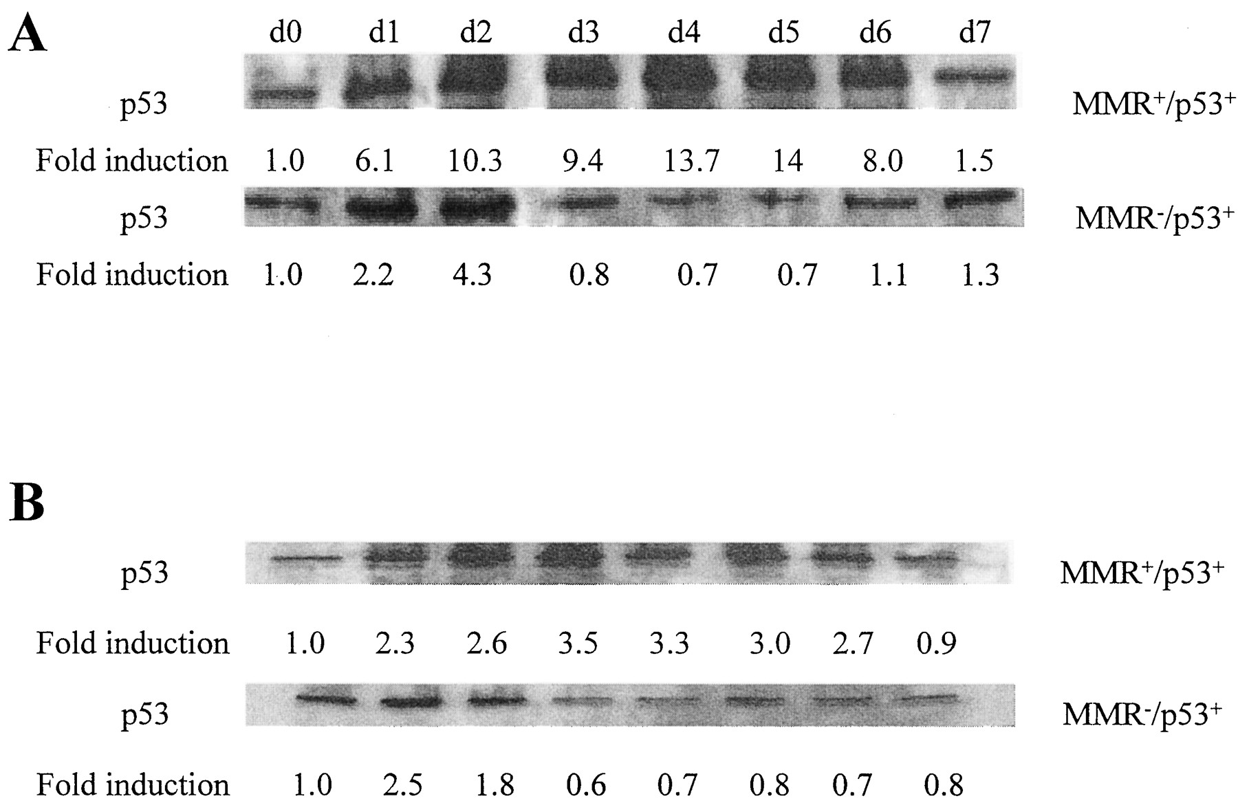
Western blot analysis of p53 levels following exposure to H2O2 (top) or cisplatin (bottom). Cells were exposed to either 100 μM H2O2 or 25 μM cisplatin for 1 h and aliquots were harvested at the indicated times.
Discussion
Mutations that disable p53 are frequently found in human cancers (Hollstein et al., 1991), often in association with tumor progression or high grade malignancy (Carder et al., 1993). Loss of MMR function is a less common but well-described phenomenon, particularly in colon and endometrial cancer (Fishel et al., 1993; Leach et al., 1993; Nicolaides et al., 1994; Papadopoulos et al., 1994). The fact that many malignant cells have defects in genomic stability is of concern with respect to continued accumulation of somatic mutations that result in tumor progression and drug resistance.
H2O2 is generated as a byproduct of normal cellular metabolism. Although it is a potent mutagen, detoxification and DNA repair mechanisms normally prevent the accumulation of oxidatively damaged bases in DNA. The results of this study demonstrate that, in human colon carcinoma cells, loss of either p53 or MMR function alone reduces the cytotoxicity and increases the mutagenicity of H2O2; most importantly, however, there is an interaction between loss of these two functions. Loss of either p53 or MMR function alone produced unequivocal but relatively modest changes in sensitivity to the cytotoxic and mutagenic effect of H2O2, but loss of both together produced much larger effects on both parameters. As depicted schematically in Fig. 6, the net result is that a given exposure to H2O2 produced more mutations, and the mutated cells had a higher probability of surviving to replicate again. Thus, the risk of the generation and persistence of clones carrying mutations capable of contributing to tumor progression or the emergence of drug resistance is increased.

Schematic diagram of potential H2O2 injury response pathways. MMR and p53 seem to function in independent but partially redundant pathways that activate apoptosis and processes that serve to protect genomic stability.
The extent to which endogenously generated H2O2 actually drives the generation of somatic mutations in tumor cells is uncertain. However, the results of this study indicate that the risk of such somatic mutations is increased when both p53 and MMR function are lost. This has already been documented for an exogenous mutagen. Previous studies from this laboratory demonstrated that p53 and MMR-deficient cells are hypersensitive to the ability of cisplatin to generate variants in the population that are resistant to several different classes of other chemotherapeutic agents (Lin and Howell, 1999; Lin et al., 1999, 2000). The current study did not investigate the nature of the interaction between loss of p53 and MMR. That is, it cannot be determined from the studies reported here whether the interaction is truly synergistic, only additive, or even partially antagonistic. This would require a formal mathematical approach such as the use of isobologram or median effect analysis (Photiou et al., 1997). However, it is clear that H2O2 is more cytotoxic and mutagenic when both functions are disabled. Caution is also needed in interpreting the result of E6 expression to be caused entirely by the loss of p53 function, because E6 can affect other proteins in the cell as well.
How does loss of p53 and MMR function cause resistance to the cytotoxic effect of H2O2? Particularly at low concentrations, H2O2 is believed to kill by activating apoptosis; thus, it is reasonable to hypothesize that both p53 and MMR play a role in generating the apoptotic response that follows excessive oxidative damage. As depicted schematically in Fig.6, these roles are likely to be in different but partially redundant pathways, because disabling both functions produced a larger effect than disabling either one alone. p53 and MMR may function at the level of DNA damage recognition, checkpoint activation, or at one or more points in the sequence of signaling events that communicate the presence of excessive DNA damage to the apoptotic machinery. The central role of p53 in mediating activation of the caspase cascade after DNA damage has been extensively reported and reviewed in recent years (Wyllie, 1997; Houghton, 1999). It is perhaps not surprising that p53 mediates part of the pro-apoptotic signal generated by H2O2 injury. Among the many types of damage done to DNA by H2O2, 8-oxoguanine seems to play an important role in the resulting mutagenesis (Bessho et al., 1992; Demple and Harrison, 1994). During DNA replication, 8-oxoguanine can pair with cytosine or adenine with an almost equal efficiency and transversions are common. The ability of the MMR system to recognize and remove this altered and mismatched base has now been well documented (McGoldrick et al., 1995; DeWeese et al., 1998; Jackson et al., 1998; Zhang et al., 1998). The fact that loss of MMR substantially impaired the ability of H2O2 to signal an increase in p53 level indicates that not only are the MMR proteins capable of binding to an 8-oxoguanine mismatch, but also that the MMR system plays an active role in the generation of an injury signal. Interestingly, although loss of MMR resulted in a blunted induction of p53, this was not accompanied by much reduction in the ability of H2O2 to kill cells, suggesting that other detectors that generate pro-apoptotic signals via p53-independent pathways must be involved as well.
What is the mechanism by which loss of either p53 or MMR causes H2O2 to be more mutagenic? Activation of the Gl checkpoint in p53-proficient cells may permit repair of most the adducts produced by H2O2 before the cell enters S phase (Fig. 6). Failure of this checkpoint in p53-deficient cells is likely to result in more cells entering S with a burden of unrepaired damage. Recent reports have disclosed that there are mammalian DNA polymerases that can bypass adducts in DNA, producing mutations as they do so (Paz-Elizur et al., 1996; Smith et al., 1998; Eckert and Opresko, 1999). T7 DNA polymerase exo- can also cause mutagenic bypass of 8-oxoguanine, the major adduct produced by H2O2 (Furge and Guengerich, 1997, 1998). Both p53 and MMR may play a role in preventing such mutagenic bypass replication (Vaisman et al., 1998; McGregor, 1999). Alternatively, mutations may be introduced during the gap-filling step after processing of the adduct by one or another of the DNA repair mechanisms if the fidelity of the polymerases is impaired in the absence of p53 or MMR.
Evidence for a direct role for the p53 protein in several different types of DNA repair is accumulating rapidly. Wild-type protein, but not mutant p53 protein, has been shown to bind strongly to a conserved element in the GADD45 gene. A p53-containing nuclear factor, which binds to this element, can be detected in extracts from irradiated cells (Kastan et al., 1992). Wang et al. (1995) reported that p53 can bind to several proteins known to play roles in nucleotide excision repair, including XPD (Rad3) and XPB, as well as CSB, which is involved in strand-specific nucleotide excision repair. With respect to MMR,Scherer et al. (1996) showed that wild-type p53 can directly bind to and trans-activate the promoter of the hMSH2 gene. More recently, Vikhanskaya et al. (1999) demonstrated an interaction between p53 function and MMR with respect to sensitivity to the DNA-damaging agent cisplatin. They reported that loss of p53 in MMR-proficient cells had little effect on cisplatin cytotoxicity but that loss of p53 in MMR-deficient cells rendered the cells quite hypersensitive to the drug. Very recently, Tanaka et al. (2000)reported that p53 regulates the transcription of a catalytic subunit of ribonucleotide reductase, an enzyme essential to the supply of deoxynucleotides for DNA repair.
Loss of p53 and MMR permitted H2O2 to produce more 6-thioguanine and ouabain-resistant clones. Whether these clones were true stable mutants was not determined, but the results of the studies with the pZCA29 vector indicate that loss of p53 and MMR is permissive with respect to the frameshift mutations that this vector is designed to report. Generalized instability in microsatellites is a hallmark of the loss of MMR function in human tumors. Such microsatellite instability is not characteristically found in tumors that have lost just p53. Nevertheless, loss of p53 is associated with a higher frequency of frameshift mutations even though they are not specifically targeted to microsatellite sequences (Liu et al., 1996). Thus, it is not surprising that the pZCA29 vector reported a higher frequency of mutations in p53-deficient as well as MMR-deficient cells. Inferring from effects on the generation of 6-thioguanine and ouabain-resistant variants, and the frameshift mutations reported by the pZCA29 vector, the cells that survive H2O2exposure are likely to carry an increased burden of mutations throughout their genome. Insertion/deletion mutations, such as those detected by pZCA29, are particularly devastating to the cell because they shift the reading frame of the coding sequence in which they occur, resulting in completely nonfunctional sequence downstream of the mutation, and often the creation of novel protein sequences in the cell.
Are the levels of oxidative damage produced by the exposures to H2O2 that were used in these experiments relevant to the oxidative stress to which tumor cells are subjected while growing in vivo? Measurable changes in both the cytotoxicity and mutagenicity of H2O2 were observable with 1 h exposures to concentrations as low as 20 μM. Many types of mammalian cells generate generous amount of H2O2, which produces concentrations in the range of 3 to 15 μM in their local environment (Chance et al., 1979; Giulivi et al., 1994) and inflammatory cells infiltrating into tumors can produce substantially higher concentrations (Satrowski and Nathan, 1991). Thus, it is likely that the endogenous levels of H2O2 are high enough, under some circumstances, that loss of MMR and p53 can place cells at increased risk for mutagenesis.
Acknowledgments
We gratefully acknowledge Dr. T.M. Runger for kindly providing the plasmid shuttle vector pZCA29 and technical guidance.
Footnotes
- Received April 18, 2000.
- Accepted September 1, 2000.
-
Send reprint requests to: Stephen B. Howell, MD, Department of Medicine 0058, University of California, San Diego, La Jolla, CA 92093. E-mail: showell{at}ucsd.edu
-
This work was supported in part by Grant CA78648 from the National Institutes of Health and conducted in part by the Clayton Foundation for Research—California Division. X.L. and S.B.H. are Clayton Foundation investigators. A preliminary account of this work was presented at the 1999 DNA Repair and Mutagenesis Meeting of the American Society for Microbiology.
Abbreviations
- MMR
- DNA mismatch repair
- bp
- base pair
- The American Society for Pharmacology and Experimental Therapeutics


