


Abstract
The protective adaptive response to electrophiles and reactive oxygen species is mediated by enhanced expression of phase II detoxifying genes, including glutathione S-transferases, through activation of antioxidant response element (ARE). The current study was designed to investigate the role of phosphatidylinositol 3-kinase (PI3-kinase)-Akt and mitogen-activated protein (MAP) kinase signaling pathways in the induction of rGSTA2 bytert-butylhydroquinone (t-BHQ). Nuclear ARE complex was activated 1 to 6 h after treatment of H4IIE cells with t-BHQ. The rGSTA2 mRNA level was elevated 6 to 24 h after t-BHQ treatment, which led to the enzyme induction. Activities of PI3-kinase and Akt were increased 10 min through 6 h after t-BHQ treatment, whereas wortmannin or LY294002, PI3-kinase inhibitors, completely abolished ARE binding activity and increases in rGSTA2 mRNA and protein. Extracellular signal-regulated kinase (ERK), p38 MAP kinase, and c-Jun N-terminal kinase (JNK) were all activated by t-BHQ. Treatment with PD98059, an ERK inhibitor, however, increased rGSTA2 mRNA and further enhanced t-BHQ-induced expression of rGSTA2. Neither SB203580 nor overexpression of JNK1 dominant negative mutant altered t-BHQ–inducible rGSTA2 expression. These results demonstrated that t-BHQ activated PI3-kinase and Akt, which was responsible for ARE-mediated rGSTA2 induction, and that ERK might negatively regulate rGSTA2 expression, whereas activation of p38 MAP kinase or of JNK by t-BHQ was not associated with the enzyme induction.
Reactive oxygen species and electrophiles induce a battery of antioxidant genes, including glutathione S-transferases (GSTs) through activation of antioxidant response element (ARE), which involves Nrf proteins and Maf family members (Bergelson et al., 1994; Wasserman and Fahl, 1997; Venugopal and Jaiswal, 1998). Induction of GST families is a protective adaptive response to oxidative stress (Bergelson et al., 1994; Wasserman and Fahl, 1997; Venugopal and Jaiswal, 1998). A previous study from this laboratory showed that oxidative stress after depletion of cellular glutathione activates MAP kinases and leads to the induction of rGSTA2 (Kang et al., 2000). GST inhibits formation of the Jun/c-Jun NH2-terminal kinase (JNK) complex and subsequently blocks mitogenic signaling induced by oncogenic ras-p21 (Villafania et al., 2000). Hence, the regulation of GST gene expression may be coupled with cell cycle control when cells are exposed to oxidative stress.
t-Butyl-4-hydroxyanisole and butylated hydroxytoluene, termed phenolic antioxidants due to their chain breaking activity during autooxidation of lipids, suppress lipid peroxidation.t-Butyl-4-hydroxyanisole is oxidatively demethylated in mammalian cells to t-butylhydroquinone (t-BHQ), which is autooxidized to t-butylquinone. Becauset-butylquinone produces reactive oxygen species by redox cycling (Pinkus et al., 1996), t-BHQ is used as a representative prooxidant.
Phosphatidylinositol-3 (PI3)-kinase, which phosphorylates phosphatidylinositols at the 3 position of the inositol ring, is associated with activation of cellular survival signals, mitogenesis, and cell transformation (Daulhac et al., 1999). PI3-kinase is involved in the regulation of the small GTPase Rac, which plays a role in the activation of JNK (Hawkins et al., 1995; Fritz and Kaina, 1999). A previous study in this laboratory has shown that inhibition of PI3-kinase activity prevented the ARE-mediated rGSTA2 induction by decreased glutathione as a result of sulfur amino acid deprivation (Kang et al., 2000). In the present study, we investigated the role of PI3-kinase and Akt pathway on ARE-mediated rGSTA2 induction byt-BHQ. We revealed for the first time that t-BHQ activated PI3-kinase and Akt, which might represent an essential pathway for the induction of rGSTA2.
Oxidative stress activates the mitogen-activated protein (MAP) kinases (Wang et al., 1998). Three distinct mammalian MAP kinase modules including extracellular signal-regulated kinase (ERK), p38 mitogen-activated protein (MAP) kinase, and JNK have been characterized (Treisman, 1996). ERK is stimulated predominantly by mitogens and growth hormones, and the activation of ERK induces proliferation or differentiation of cells. The p38 MAP kinase, a recently identified member of the MAP kinase family, is involved in apoptosis (Tan et al., 1996). Stress-activated protein kinase cascade involves the activation of JNK, which consequently induces activator protein-1 (AP-1)-mediated transactivation of the AP-1 responsible genes.
Both chemopreventive agents and prooxidants induce phase II detoxifying enzymes. It has been reported that the increase in quinone reductase activity by sulforaphane accompanied activation of ERK in human HepG2 and mouse Hepa1c1c7 cells, and that p38 kinase negatively regulated the induction of quinone reductase (Yu et al., 1999, 2000). PD98059, an ERK inhibitor, completely abolished ERK activation by sulforaphane and to certain extents reduced the activities of quinone reductase and ARE-linked reporter gene by t-BHQ or sulforaphane (Yu et al., 1999). Nonetheless, the effects of t-BHQ on the endogenous ARE activation in the rGSTA2 gene and on the subsequent induction of rGSTA2 in association with the activation of MAP kinases have not been investigated. The second aim of the present study was to determine whether t-BHQ indeed activated the MAP kinases in H4IIE cells and to identify the MAP kinase(s) responsible for ARE-mediated induction of rGSTA2. We demonstrated that the induction of rGSTA2 by t-BHQ was not controlled by the activation of MAP kinases in H4IIE cells, although ERK might negatively regulate the expression of rGSTA2.
Experimental Procedures
Materials.
[α-32P]dCTP (3000 mCi/mmol) and [γ-32P]ATP (6000 mCi/mmol) were purchased from PerkinElmer Life Science Products (Boston, MA). Anti-rGSTA1/2 antibody was supplied from Biotrin International (Dublin, Ireland). Biotinylated goat anti-rabbit IgG, recombinant protein A-agarose, and 5-bromo-4-chloro-3-indoylphosphate/nitroblue tetrazolium were obtained from Life Technologies (Gaithersburg, MD). Anti-Nrf-1, anti-Nrf-2, and anti-v-Maf antibodies were purchased from Santa Cruz Biotechnology (Santa Cruz, CA). Anti-phospho ERK antibodies and random prime-labeling kit were purchased from Promega (Madison, WI). Anti-phospho p38 MAP kinase and anti-JNK1/2 antibodies were supplied from New England Biolabs (Beverly, MA). t-BHQ (97%) was purchased from Aldrich Chemical (Milwaukee, WI). PD98059 and LY294002 were obtained from Calbiochem (San Diego, CA). Wortmannin and other reagents in the molecular studies were supplied from Sigma Chemical (St. Louis, MO). The JNK1 dominant negative mutant (KmJNK) and JNK1 overexpression plasmids were kindly provided from Dr. N. Dhanasekaran (Fels Institute for Cancer Research and Molecular Biology and Department of Biochemistry, Temple University, Philadelphia, PA).
Cell Culture.
H4IIE rat hepatoma cell line was obtained from American Type Culture Collection (Manassas, VA) and maintained in Dulbecco's modified Eagle's medium containing 10% fetal calf serum, 50 U/ml penicillin, and 50 μg/ml streptomycin at 37°C in humidified atmosphere with 5% CO2.
MTT Cell Viability Assay.
H4IIE cells were plated at a density of 5 × 10⁴ cells/well in a 96-well plate to determine cytotoxicity. Cells were exposed to t-BHQ at the concentrations of 10 through 500 μM at 37°C under 5% CO2. Viable cells were stained with 3-(4,5-dimethylthiazol-2-yl)-2,5-diphenyl-tetrazolium bromide (MTT; 0.5 mg/ml) for 4 h after incubation with t-BHQ for 24 h. The media were then removed. Produced formazan crystals in the wells were dissolved by addition of 200 μl of dimethyl sulfoxide. Absorbance was measured at 540 nm using a Titertek Multiskan Automatic enzyme-linked immunosorbent assay microplate reader (Model MCC/340; Titertek, Huntsville, AL). Cell viability was defined relative to untreated control cells [i.e., viability (% control) = 100 × (absorbance of treated sample) / (absorbance of control)].
Preparation of Nuclear Extracts.
Nuclear extracts were prepared essentially according to Schreiber et al. (1990). Briefly, cells in dishes were washed with ice-cold phosphate-buffered saline. Cells were then scraped, transferred to microtubes, and allowed to swell after addition of 100 μl lysis buffer containing 10 mM HEPES, pH 7.9, 0.5% Nonidet P-40, 10 mM KCl, 0.1 mM EDTA, 1 mM dithiothreitol, and 0.5 mM phenylmethylsulfonyl fluoride. Cell membranes were disrupted by vortexing, and the lysates were incubated for 10 min on ice and centrifuged for 5 min at 4°C. Pellets containing crude nuclei were resuspended in 50 μl of the extraction buffer containing 20 mM HEPES, pH 7.9, 400 mM NaCl, 1 mM EDTA, 1 mM dithiothreitol, and 1 mM phenylmethylsulfonyl fluoride, and then incubated for 30 min on ice. The samples were centrifuged at 15,800g for 10 min to obtain the supernatant containing nuclear extract. The nuclear extracts were stored at −70°C until use.
Gel Retardation Assay.
A double-stranded DNA probe containing the rGSTA2 gene ARE was used for gel shift analysis after end-labeling of the probe with [γ-32P]ATP and T4 polynucleotide kinase. The sequence of the ARE-containing oligonucleotide was (5′-GATCATGGCATTGCACTAGGTGACAAAGCA-3′). The oligonucleotides for SP-1 and AP-1, which were used for competition experiments, were (5′-ATTCGATCGGGGCGGGGCGAGC-3′) and (5′-CGCT TGATGAGTCAGCCGGAA-3′), respectively. The reaction mixtures contained 4 μl of 5 × binding buffer containing 20% glycerol, 5 mM MgCl2, 250 mM NaCl, 2.5 mM EDTA, 2.5 mM dithiothreitol, 0.25 mg/ml poly dI-dC, and 50 mM Tris · Cl, pH 7.5, 5 μg of nuclear extract, and sterile water in a total volume of 20 μl. The reaction mixtures were preincubated for 10 min. DNA-binding reactions were carried out at room temperature for 30 min after addition of 1 μl probe (106 cpm). Specificity of binding was determined by a competition experiment, which was carried out by adding a 20-fold excess of an unlabeled ARE, SP-1, or AP-1 oligonucleotide to the reaction mixture before the DNA-binding reaction. Samples were loaded onto 4% polyacrylamide gels at 100 V. The gels were removed, fixed and dried, followed by autoradiography.
Northern Blot Hybridization.
The specific cDNA probe for the rGSTA2 gene was amplified by reverse transcription-polymerase chain reaction using the selective primers (Kim et al., 1997) and was cloned in the pGEM+T vector (Promega). Total RNA was isolated from H4IIE cells using the improved single-step method of thiocyanate-phenol-chloroform RNA extraction, and Northern blot analysis was carried out according to the procedures described previously (Kim et al., 1997). Briefly, total RNA was resolved by electrophoresis in a 1% agarose gel containing 2.2 M of formaldehyde and transferred to nitrocellulose paper. The nitrocellulose paper was hybridized as described previously. Filters were washed in 2× standard saline citrate and 0.1% SDS for 10 min at room temperature twice and in 0.1× standard saline citrate and 0.1% SDS for 10 min at room temperature twice. Filters were washed in the solution containing 0.1× standard saline citrate and 0.1% SDS for 60 min at 60°C. After quantification of mRNA levels, the membranes were stripped and rehybridized with a 32P-labeled cDNA probe complementary to 18 S rRNA to quantify the amount of RNA loaded onto the membranes.
Immunoblot Analyses of rGSTA2 and Phosphorylated MAP Kinases.
SDS-polyacrylamide gel electrophoresis (PAGE) and immunoblot analyses were performed according to procedures published previously (Kim et al., 1997). After washing cells twice with phosphate-buffered saline, cells were scraped and sonicated to disrupt cell membranes. Cytosolic fractions were obtained by differential centrifugations. Cytosolic proteins were separated by 12% gel electrophoresis and electrophoretically transferred to nitrocellulose paper. The nitrocellulose paper was incubated with anti-rat rGSTA1/2 antibody, followed by incubation with biotinylated secondary antibody, and developed using 5-bromo-4-chloro-3-indoylphosphate and nitroblue tetrazolium (Kim et al., 1997). The activities of ERK and p38 MAP kinase were determined in cell lysates. Cell lysates, boiled for 5 min, were centrifuged at 15,000g to remove debris. Activated ERK and p38 MAP kinase were immunochemically assessed using the specific antibodies, which recognized active-phosphorylated forms, and developed using ECL enhanced chemiluminescence system (Amersham, Buckinghamshire, UK).
PI3-Kinase Activity.
The activity of PI3-kinase was assayed in lysate prepared from cells treated with t-BHQ. Cells were lysed with the buffer solution containing 10 mM Tris · Cl, pH 7.4, 100 mM NaCl, 30 mM sodium pyrophosphate, 1 mM EGTA, 0.5% Triton X-100, 10% glycerol, 1 mM phenylmethylsulfonyl fluoride, and 100 μM sodium orthovanadate. The lysate was centrifuged at 15,000gfor 15 min to remove debris. The lysate (500 μg) was incubated with an anti-phosphotyrosine antibody for 2 h at 4°C. The immune complex was precipitated with protein A-agarose, and washed with the lysis buffer, pH 7.4, containing 100 mM Tris · Cl, 5 mM LiCl, and 0.1 mM sodium orthovanadate, and sequentially with Tris/Na/EDTA buffer, pH 7.4, consisting of 10 mM Tris · Cl, 150 mM NaCl, and 5 mM EDTA. The immune complex was resuspended in 50 μl of Tris/Na/EDTA buffer. The complex was then added to the reaction mixture containing 100 μM MgCl2, 0.1 mM ATP, 5 μCi of [γ-32P]ATP (6000 mCi/mmol) and 10 μg of phosphatidylinositol. The reaction mixture was incubated for 10 min at 37°C. The reaction was terminated by addition of 12 μl of 6 N HCl. Phosphorylated lipids were then extracted with chloroform/methanol (1:1), and resolved by the thin-layer chromatography using chloroform/methanol/water/ammonium hydroxide (60:47:11.3:2) as a developing solvent. The spot of radioactive phosphatidylinositol-3-phosphate was visualized by autoradiography.
Akt Activity.
The activity of Akt was assayed using an Akt1/PKBα immunoprecipitation-kinase assay kit (Upstate Biotechnology, Lake Placid, NY), according to the manufacturer's instructions. The reaction mixture contained 10 μCi of [γ-32P]ATP, 500 μg of cell lysate, and 100 μM RPRAATF (a specific peptide substrate derived from the phosphorylation site of glycogen synthase kinase-3) in a volume of 10 μl. The reaction was proceeded for 10 min at 37°C, and terminated by adding 20 μl of 40% trichloroacetic acid. An aliquot (40 μl) of the reaction mixture was spotted on P81 phosphocellulose paper. The P81 phosphocellulose paper was washed with 0.75% phosphoric acid for 5 min three times and subsequently in acetone for 5 min. The membrane was transferred to 5 ml of scintillation cocktail, and the radioactivity of phosphorylated substrate was measured using a β-counter (Wallac, Gaithersburg, MD).
JNK Activity.
Cells were lysed with the buffer solution containing 10 mM Tris · Cl, pH 7.4, 100 mM NaCl, 30 mM sodium pyrophosphate, 1 mM EGTA, 0.5% Triton X-100, 10% glycerol, 1 mM phenylmethylsulfonyl fluoride, and 100 μM sodium orthovanadate. Cell lysates were homogenized by being passed through a 27-gauge needle three times and were then left on ice for 15 min. The homogenate was centrifuged at 15,000g for 15 min to remove debris. Two hundred micrograms of each lysate was immunoprecipitated using specific anti-JNK1/2 antibody and protein A-agarose. The immunoprecipitate was resuspended in the kinase reaction buffer containing 25 mM Tris · Cl, pH 7.4, 25 mM MgCl2, 2 mM dithiothreitol, and 0.1 mM sodium orthovanadate. The reaction was initiated by addition of 2 μCi of [γ-32P]ATP (10 Ci/mmol) and 2 μg of GST-c-Jun (1–79) as a substrate, continued for 30 min at 30°C, and terminated by addition of 2× SDS-PAGE sample dilution buffer. Phosphorylated GST-c-Jun was resolved on 12% SDS-PAGE, and visualized by autoradiography.
ERK1/2 and p38 MAP Kinase Activities.
H4IIE cells were incubated in the presence of t-BHQ (30 μM) for 1 h to activate ERK1/2 and p38 kinase. Inhibition of the kinase activities by PD98059 and SB203580 at the concentrations of 50 μM and 10 μM, respectively, was confirmed in H4IIE cells as described previously (Kang et al., 2000).
Transfection Study.
Cells were transfected using Transfectam according to the manufacturer's instruction (Promega, Madison, WI). H4IIE cells were replated 24 h before transfection at a density of 2 × 10⁶ cells in a 10 cm2-plastic dish. Transfectam (20 μl) was mixed with 10 μg of a JNK1 dominant negative mutant (KmJNK1) or hemagglutinin-tagged JNK1 overexpression (JNK1+) plasmid in 2.5 ml of minimal essential medium (MEM) for use in JNK transfection. Cells were transfected by addition of MEM containing each plasmid and Transfectam and then incubated at 37°C in a humidified atmosphere of 5% CO2 for 6 h. After addition of 6.25 ml of MEM with 10% fetal calf serum, cells were incubated for additional 48 h. To prepare total RNA and cytosolic fractions, cells were cultured in serum-free MEM for 6 h and further incubated in the presence or absence of 30 μM t-BHQ for the indicated times. Viable cells were subcultured at least five successive times in the medium containing 100 μM neomycin (Geneticin, Gibco-BRL Life Technologies, Gaithersburg, MD) to establish a stable JNK1-dominant negative mutant [JNK1(−)]-transfected H4IIE cell line.
Data Analysis.
Scanning densitometry was performed with Image Scan & Analysis System (Alpha-Innotech Corporation, San Leandro, CA). One-way analysis of variance procedures were used to assess significant differences among treatment groups. For each significant effect of treatment, the Newman-Keuls test was used for comparisons of multiple group means. The criterion for statistical significance was set at p < 0.05 or p < 0.01.
Results
Activation of Nuclear ARE Binding.
t-BHQ affects cell viability through redox cycling and the production of reactive oxygen species (Bolton et al., 2000). MTT assay was performed to determine viability of H4IIE cells. Cell viability was significantly decreased in a concentration-dependent manner after incubation witht-BHQ at the concentrations of 100 μM or above for 24 h (Fig. 1). The concentration of 30 μM was chosen in subsequent experiments to avoid cytotoxicity.

Viability of H4IIE cells after t-BHQ treatment. MTT assay was performed to determine the viability of cells after treatment with t-BHQ for 24 h. Data represent the mean ± S.D. with four separate experiments. One-way analysis of variance was used for comparisons of multiple group means followed by Newman-Keuls test (significant compared with control, **p < 0.01).
The ARE-binding transcription factors consisting of Nrf and small-Maf transduce the induction signal(s) of rGSTA2 in response to oxidative stress (Venugopal and Jaiswal, 1998; Nguyen et al., 2000). Nuclear extracts isolated from H4IIE cells cultured with t-BHQ for 1 through 12 h were probed with the radiolabeled rGSTA2 gene ARE to assess whether the nuclear ARE binding proteins were activated byt-BHQ (Fig. 2A). The band of slow migrating complex was increased 1 h after treatment of cells with t-BHQ. The activation of ARE became pronounced at 3 h, and extended through 6 h. Competition experiments using an excess amount of unlabeled ARE, AP-1, or SP-1 oligonucleotides confirmed the specificity of ARE binding. Whereas addition of a 20-fold excess of an unlabeled ARE to the activated nuclear extract completely abolished the ARE binding, excess unlabeled AP-1 or SP-1 oligonucleotide failed to inhibit the DNA binding (Fig. 2B). To confirm the components for the ARE activation, immunodepletion experiments were carried out with the nuclear extracts produced from H4IIE cells treated with t-BHQ for 3 h. Addition of anti-Nrf-1, anti-Nrf-2, or anti-v-Maf antibodies to the reaction mixture depleted the ARE DNA binding (Fig. 2C). This was consistent with the previous result on the ARE binding (Cho et al., 2000; Kang et al., 2000). Binding with the antibody directed against Nrf-1/2 or Maf may induce conformational change that decreases the binding affinity of ARE binding protein(s) to the ARE consensus sequence.

Activation of ARE nuclear transcription factors in H4IIE cells. A, gel shift analysis of ARE transcription complex in nuclear extracts. Gel shift assay was performed with nuclear extracts prepared from H4IIE cells cultured with or without 30 μMt-BHQ for 1 through 12 h. All lanes contained 10 μg of nuclear extracts and 5 ng of labeled rGSTA2 ARE DNA consensus sequence. B, competition studies were carried out by adding a 20-fold excess of an unlabeled ARE, AP-1, or SP-1 oligonucleotide to the nuclear extract from cells treated with t-BHQ for 3 h. C, an antibody competition experiment was carried out by incubating the nuclear extract from cells treated with t-BHQ for 3 h with the specific polyclonal antibody directed against Nrf-1, Nrf-2, or v-Maf protein. Results were confirmed by repeated experiments. The arrow indicates the ARE binding complex.
Induction of rGSTA2.
Northern blot analysis was performed to determine whether ARE activation by t-BHQ led to an increase in rGSTA2 mRNA level in H4IIE cells. rGSTA2 mRNA was significantly increased 6 to 24 h after incubation of cells witht-BHQ at the concentration of 30 μM (Fig.3A). t-BHQ increased the rGSTA2 mRNA maximally at 12 h, followed by a gradual return toward control values at 48 h. Western blot analysis revealed that rGSTA1/2 subunit began to be induced 6 h after t-BHQ treatment, peaked at 12 to 24 h and extended up to 48 h (Fig.3B). Anti-rGSTA1/2 antibody preferentially recognized the induction of rGSTA2 because the rGSTA2 subunit is inducible.
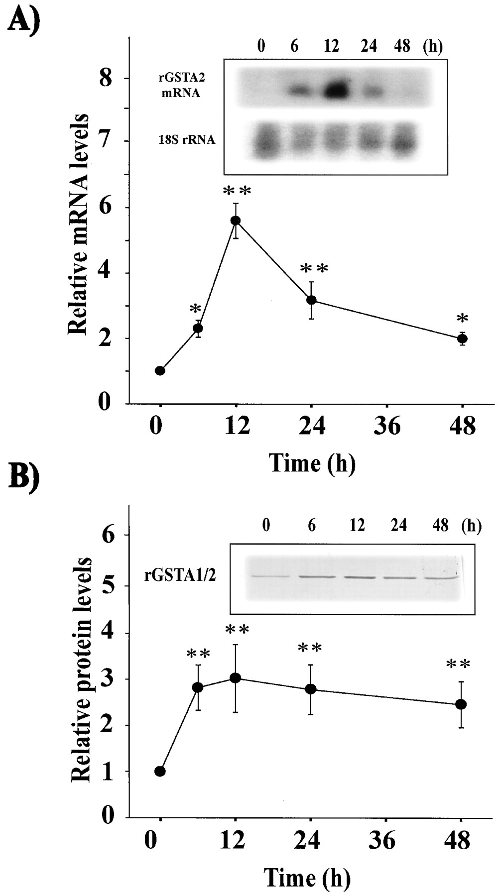
Induction of rGSTA2 by t-BHQ. A, the rGSTA2 mRNA levels. Northern blot analysis was performed with total RNA fraction (30 μg each) prepared from cells incubated with 30 μMt-BHQ for 6 to 48 h. The amount of RNA loaded in each lane was assessed by rehybridization of the stripped membrane with a 32P-labeled probe for 18 S rRNA. The relative rGSTA2 mRNA levels were assessed by scanning densitometry of Northern blots. B, expression of rGSTA1/2 subunit. Immunoblot analysis shows the level of rGSTA1/2 protein in H4IIE cells cultured in the presence of 30 μMt-BHQ for 6 to 48 h. Each lane was loaded with 10 μg of cytosolic proteins. The level of rGSTA1/2 was assessed by scanning densitometry of immunoblots. Data represent the mean ± S.D. with three separate experiments. One-way analysis of variance was used for comparisons of multiple group means followed by Newman-Keuls test (significant compared with control, *p < 0.05; **p < 0.01) (control level = 1).
Activation of PI3-Kinase and Akt.
The PI3-kinase activity was determined in H4IIE cells incubated with t-BHQ. The activity of PI3-kinase toward the substrate phosphatidylinositol was increased 10 min to 6 h after t-BHQ treatment (30 μM) and peaked at 30 min (Fig. 4A). These results demonstrated that oxidative stress produced from t-BHQ activated PI3-kinase.
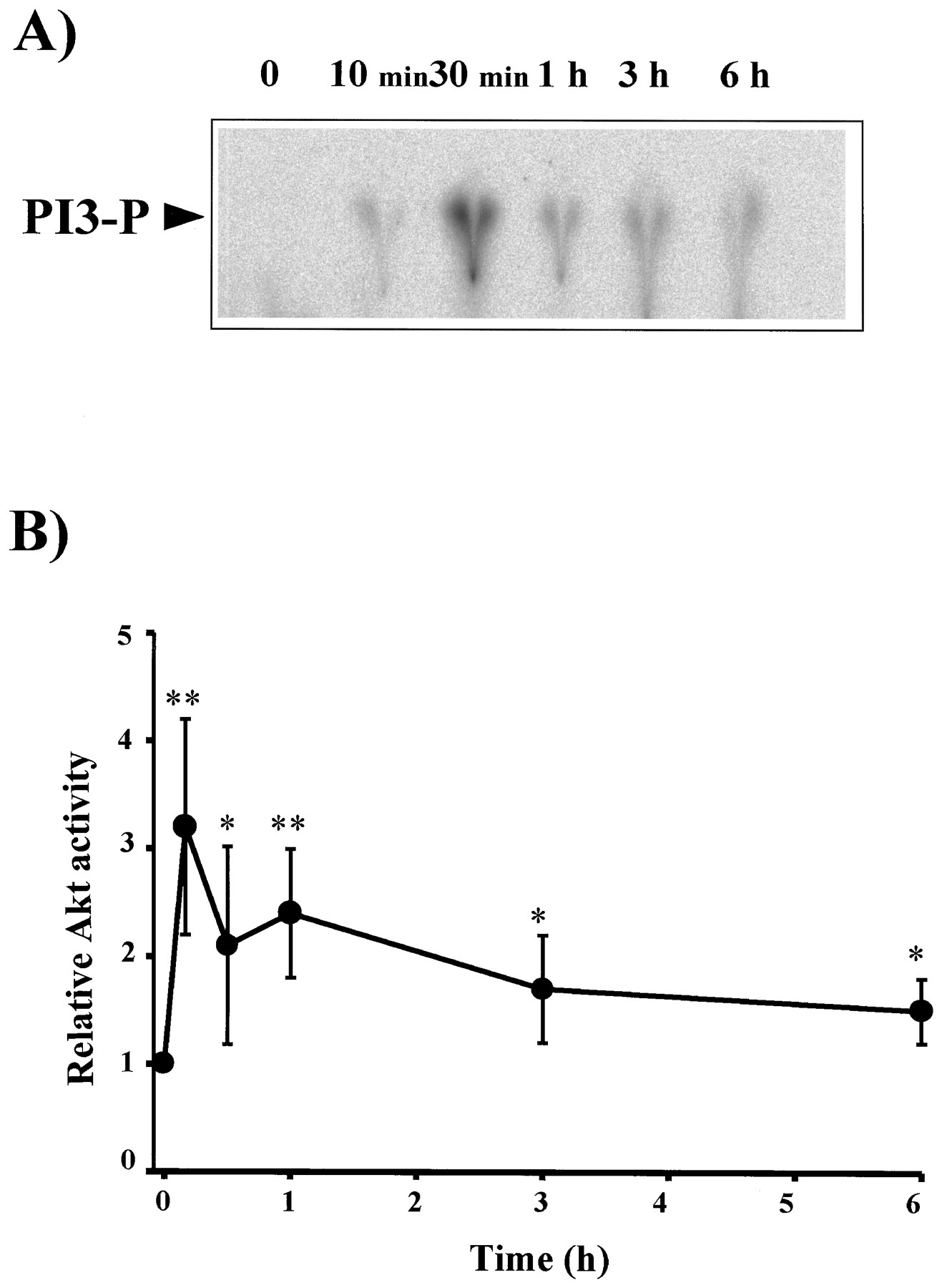
Activation of PI3-kinase and Akt byt-BHQ. A, PI3-kinase activity in anti-phosphotyrosine immunoprecipitates by TLC. The activity of PI3-kinase in cells treated with 30 μM t-BHQ. The PI3-kinase activity was measured using phosphatidylinositol as a substrate. Results were confirmed by repeated experiments. PI3-P, phosphorylated phosphatidylinositol. B, Akt activity after t-BHQ treatment. Activation of Akt was assessed by phosphorylation of a peptide substrate derived from the phosphorylation site of glycogen synthase kinase-3. Data represent the mean ± S.D. with 3 separate experiments. One-way analysis of variance was used for comparisons of multiple group means followed by Newman-Keuls test (significant compared with control, *p < 0.05, **p < 0.01). (Akt activity in control cells = 1).
The lipid products by PI3-kinase bind with high affinity and specificity to the Akt PH domain and phosphorylation of Akt serves to potently activate the enzyme (Kandel and Hay, 1999). The activity of Akt was assessed by the catalytic activity of immunoprecipitated Akt toward a synthetic phosphorylation site of glycogen synthase kinase-3 in cells treated with t-BHQ (Fig. 4B). t-BHQ activated Akt from 10 min through 6 h. The activation of Akt byt-BHQ was consistent with the increase in PI3-kinase activity.
Role of PI3-Kinase in ARE Activation and rGSTA2 Induction.
To determine whether the PI3-kinase cascade was involved in the activation of ARE-binding transcription factors, H4IIE cells were incubated witht-BHQ for 3 h in the presence of 500 nM wortmannin (Fig. 5A). Wortmannin inhibited the binding activity of ARE induced by t-BHQ, as evidenced by reduction of the DNA binding complex. The ARE binding activity was also suppressed by 50 μM LY294002 (Fig. 5A). Multiple experiments were carried out to determine the effects of PI3-kinase inhibitors on the ARE binding activity. A representative gel shift analysis showed that the band intensity was substantially reduced to that of control by the PI3-kinase inhibitors (Fig. 5A). These data demonstrated that the activity of PI3-kinase was essential for the regulatory pathway leading to ARE activation.

The effects of PI3-kinase inhibitors on ARE activation and rGSTA2 mRNA increase by t-BHQ. A, ARE gel shift analysis. H4IIE cells were stimulated with 30 μMt-BHQ for 3 h in the presence of wortmannin (WO, 500 nM) or LY294002 (LY, 50 μM). All lanes contained 10 μg of nuclear extracts and 5 ng of labeled rGSTA2 ARE DNA consensus sequence. The arrow indicates the ARE binding complex. B, Northern blot analysis and relative rGSTA2 mRNA levels. Cells were treated with 30 μMt-BHQ for 12 h in the presence of wortmannin or LY294002. Total RNA fraction (30 μg each) was subjected to Northern blot analysis. C, inhibition of PI3-kinase activation by wortmannin or LY294002. Cells were treated with 30 μM t-BHQ for 30 min in the presence or absence of wortmannin or LY294002 and the PI3-kinase activity was measured. Data represent the mean ± S.D. with three separate experiments. One-way analysis of variance was used for comparisons of multiple group means followed by Newman-Keuls test (significant compared with control, **p < 0.01; significant compared with t-BHQ, ††p < 0.01).
Whether suppression of ARE activation by PI3-kinase inhibition prevented the increase in the rGSTA2 mRNA was assessed (Fig. 5B). Either wortmannin or LY294002 significantly inhibited the increase in rGSTA2 mRNA by t-BHQ at 12 h. An additional experiment confirmed that the activity of PI3-kinase inducible by t-BHQ was completely inhibited by either wortmannin or LY294002 at the concentrations employed (Fig. 5C). Immunoblot analysis also showed that the PI3-kinase inhibitors prevented the induction of rGSTA1/2 (Fig.6, A and B). Hence, the change in the rGSTA2 mRNA level by inhibition of PI3-kinase activity paralleled with that in rGSTA2 protein.

The effects of PI3-kinase inhibitors ont-BHQ-inducible rGSTA1/2 expression. Cells were treated with 30 μM t-BHQ in the presence of a PI3-kinase inhibitor for 24 h. A, the level of rGSTA1/2 was assessed by immunoblottings. B, the relative rGSTA1/2 protein levels were plotted after scanning densitometry. Data represent the mean ± S.D. with three separate experiments. One-way analysis of variance was used for comparisons of multiple group means followed by Newman-Keuls test (significant compared with control, **p < 0.01; significant compared with t-BHQ alone, ††p < 0.01).
Activation of MAP Kinases.
t-BHQ activated ERK1/2 (Fig. 7). The level of active phosphorylated ERK1/2 was increased at 30 min after t-BHQ treatment and peaked at 1 h. To study whether other MAP kinase pathways were also stimulated by t-BHQ, we measured the activity of p38 kinase as a function of time. Activation of p38 kinase was measured by Western blot analysis. The level of phosphorylated p38 kinase was slightly enhanced in cells stimulated by t-BHQ for 5 min through 3 h. A peak of activation was observed at 1 h (Fig. 7). The activity of JNK was also assessed by phosphorylation of GST-c-Jun in cells stimulated with 30 μM t-BHQ. JNK activity was increased from 10 min, with the peak of activation observed at 3 h (Fig. 7). Additional studies showed that the extent of JNK activation was reduced 6 h after t-BHQ treatment, followed by a return to control values at 12 h (data not shown).

Activation of MAP kinases by t-BHQ in H4IIE cells. The extents of ERK or p38 MAP kinase activation were assessed by immunoblottings of the respective phosphorylated MAP kinase, whereas the JNK activity was determined using GST-c-Jun (1–79) as a JNK substrate. Results were confirmed by repeated experiments.
Effects of p38 MAP Kinase and ERK on ARE-Mediated rGSTA2 Induction.
We were interested in whether blockade of MAP kinase cascade led to a change in the ARE activation by t-BHQ. Cells incubated with PD98059 (50 μM) an ERK inhibitor failed to suppress ARE activation by t-BHQ (Fig.8A). SB203580, a specific p38 kinase inhibitor, at the concentration of 10 μM did not alter the ARE binding activity (Fig. 8A). To test whether blockade of MAP kinase cascade led to a change in the rGSTA2 expression stimulated byt-BHQ, cells were incubated with a specific MAP kinase inhibitor for 12 h. Neither PD98059 (50 μM) nor SB203580 (10 μM) inhibited the increase in rGSTA2 mRNA. PD98059 rather significantly increased the gene expression by t-BHQ (Fig.8B).

The effects of ERK and p38 MAP kinase inhibitors on the ARE activation and the increase in rGSTA2 mRNA byt-BHQ. A, gel shift analysis of ARE transcription complex. H4IIE cells were stimulated with 30 μM t-BHQ for 3 h in the presence of PD98059 (PD, 50 μM) or SB203580 (SB, 10 μM). All lanes contained 10 μg of nuclear extracts and 5 ng of labeled rGSTA2 ARE DNA consensus sequence. B, the effects of MAP kinase inhibitors on the rGSTA2 mRNA. Cells were incubated with 30 μMt-BHQ for 12 h in the presence of PD98059 or SB203580. Total RNA fractions (30 μg each) prepared from the cells were subjected to Northern blot analysis. Data represent the mean ± S.D. with three separate experiments. One-way analysis of variance was used for comparisons of multiple group means followed by Newman-Keuls test (significant compared with control, **p < 0.01; significant compared witht-BHQ alone, ††p < 0.01). C, inhibition of ERK1/2 and p38 kinase activities by the respective kinase inhibitors. H4IIE cells were incubated with t-BHQ (30 μM) in the presence of PD98059 (PD, 50 μM) or SB203580 (SB, 10 μM) for 1 h. The data shows that ERK1/2 and p38 kinase activities were inhibited by the respective inhibitors.
We previously demonstrated that PD98059 at the concentration of 50 μM significantly inhibited activation of ERK1/2 1 h after sulfur amino acid deprivation (M. H. Son, K. W. Kang, C. H. Lee, and S. G. Kim, unpublished observations). The activity of ERK1/2 toward PHAS-1 was prevented by PD98059 (M. H. Son, K. W. Kang, C. H. Lee, and S. G. Kim, unpublished observations). Whether PD98059 (50 μM) and SB203580 (10 μM) inhibited the activities of ERK1/2 and p38 kinase was assessed in H4IIE cells incubated with t-BHQ (30 μM) for 1 h. The inhibitors at the concentrations employed were active in inhibiting the respective kinase activities increased byt-BHQ (Fig. 8C).
Western blot analysis confirmed that neither PD98059 nor SB203580 prevented the induction of rGSTA1/2 (Fig.9, A and B). Although the rGSTA1/2 protein level was slightly greater than that caused by t-BHQ alone, the difference was not significant. The discrepancy between rGSTA1/2 protein and mRNA levels may result from the difference in their turnover rates as well as from the change in translational efficiency. Studies have shown that the extent of GST induction was much less than that of its mRNA increase (Kim et al., 1997; Cho and Kim, 2000; Cho et al., 2000).

The effects of MAP kinase inhibitors on rGSTA1/2 expression. A, the representative immunoblot shows the level of rGSTA1/2. Cells were treated with 30 μM t-BHQ for 24 h in the presence or absence of each MAP kinase inhibitor. B, relative rGSTA1/2 protein levels in H4IIE cells. Data represent the mean ± S.D. with three separate experiments. One-way analysis of variance was used for comparisons of multiple group means followed by Newman-Keuls test (significant compared with control, **p < 0.01).
Negative Gene Regulation by ERK.
The possible negative regulation of ERK for rGSTA2 expression was studied further. Northern blot analysis revealed that treatment of H4IIE cells with 50 μM PD98059 for 12 h markedly elevated the rGSTA2 mRNA level (i.e., 7.5-fold) compared with control (Fig.10A and 10B). The level of rGSTA2 mRNA was 2.1- to 5.7-fold increased in cells incubated with t-BHQ at the concentrations of 10 to 30 μM. PD98059 (50 μM) in combination with t-BHQ (10 μM) further increased rGSTA2 mRNA (i.e., a 10-fold increase relative to control), raising the possibility that the mechanism of rGSTA2 induction by t-BHQ differs from that by PD98059 (Fig. 10A and 10B). These data strongly supported the notion that ERK might negatively regulate the expression of rGSTA2.

The rGSTA2 mRNA level in H4IIE cells aftert-BHQ treatment with or without PD98059. A, representative Northern blot analysis. Cells were treated with 10–100 μM t-BHQ for 12 h in the presence or absence of 50 μM PD98059 (PD). B, relative rGSTA2 mRNA levels. Data represent the mean ± S.D. with three separate experiments. One-way analysis of variance was used for comparisons of multiple group means followed by Newman-Keuls test (significant compared with control, **p < 0.01; significant compared with the respective t-BHQ treatment, †p < 0.05, ††p < 0.01).
JNK1 Has No Effect on rGSTA2 Expression.
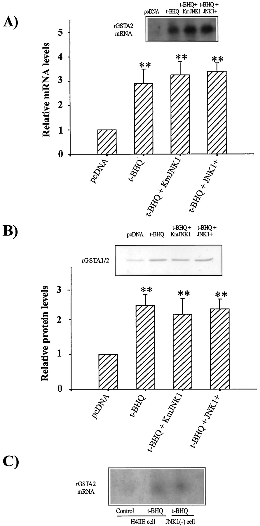
To establish the role of JNK on the rGSTA2 induction by t-BHQ, cells were transfected with an expression vector of JNK1-dominant negative mutant or of JNK1. Transfection of H4IIE cells with a JNK1-dominant negative mutant vector (10 μg per 2 × 10⁶ cells) failed to inhibit increases in rGSTA2 mRNA and protein levels by 30 μM t-BHQ at 12 h and 24 h, respectively (Fig.11, A and B). Transfection of H4IIE cells with a JNK1 overexpression plasmid did not altert-BHQ-inducible mRNA and protein levels (Fig. 11, A and B). Transfection of JNK1 dominant negative mutant in H4IIE cells completely blocked the expression of JNK1, as assessed by Western blot analysis. Diminished JNK1 expression was also confirmed in cells treated with 400 mM sorbitol for 5 min. Overexpression of JNK1 in cells transfected with a hemagglutinin-tagged JNK1+ plasmid was also verified by Western blotting using a monoclonal antibody recognizing hemagglutinin (data not shown). An experiment was also carried out in JNK1(−) stably transfected cells to further determine the role of JNK1. The extent of rGSTA2 induction by t-BHQ was not decreased in H4IIE cells stably transfected with a dominant negative mutant of JNK1 (Fig. 11C). These results indicated that JNK activation was not responsible for the induction of rGSTA2 by t-BHQ.

The effects of transfection with a JNK1 dominant negative mutant or JNK1 plasmid on rGSTA1/2 induction byt-BHQ. A, rGSTA2 mRNA levels. The mRNA levels were assessed 12 h after t-BHQ treatment (30 μM) following transfection of cells with each plasmid. B, immunoblot for rGSTA1/2 subunit. The level of rGSTA1/2 protein was determined 24 h after incubation of transfected H4IIE cells witht-BHQ, and the relative protein levels were plotted after scanning densitometry. Blocking of JNK1 expression or overexpression of JNK1 in transiently transfected H4IIE cells was confirmed by Western blot analyses. Data represent the mean ± S.D. with three separate experiments. One-way analysis of variance was used for comparisons of multiple group means followed by Newman-Keuls test (significant compared with control, **p < 0.01). C, Expression of rGSTA2 mRNA in JNK1(−) stably transfected H4IIE cells. The rGSTA2 mRNA levels were assessed 12 h aftert-BHQ treatment (30 μM) in H4IIE cells or in cells stably transfected with JNK1 dominant negative mutant [JNK1(−) cells].
Discussion
Cancer chemopreventive actions of phenolic antioxidants are considered to exert their beneficial effects through induction of phase II drug-metabolizing enzymes such as GST. A number of transcription factors are phosphorylated by members of kinase family triggered in response to a variety of stimuli including oxidative stress (Gupta et al., 1995; Tan et al., 1996). Expression of GST is primarily controlled by transcription factors, including Nrf family in response to oxidative stress (Wasserman and Fahl, 1997; Venugopal and Jaiswal, 1998). A previous study in this laboratory has shown that the transcription factor Nrf1/2 and small-Maf are involved in the induction of GST by decreased glutathione after sulfur amino acid deprivation (Kang et al., 2000). The role of Nrf as a transcription factor is further supported by the impairment of class α and μ GST induction byt-butyl-4-hydroxyanisole in Nrf2 knock-out mouse (Bolton et al., 2000) and the increase in ARE-mediated reporter gene activity by overexpression of Nrf2 in hepatoma cells (Hayes et al., 2000; Nguyen et al., 2000). The results of the current study showed that ARE binding to the ARE consensus sequence was also enhanced by t-BHQ treatment. Another study, however, showed that t-BHQ had no discernible effect on the intensity of ARE-binding protein band (Wasserman and Fahl, 1997). In their study, the ARE-binding was apparently saturated in HepG2 cells even without t-BHQ treatment. Discrepancy in the ARE activation by t-BHQ may result from different cell type or treatment regimen employed. The present study showed that the rGSTA2 mRNA was increased after ARE activation, which led to rGSTA2 induction at later times. Despite the identification of the transcriptional factors, which bind to ARE consensus sequence, the cellular signaling pathway for the induction of GST in response to oxidative stress has not yet been clearly defined.
PI3-kinase is an enzyme that functions in cell growth, survival, and transformation. The activity of Akt (PKB) is increased by oxidant insult, such as hydrogen peroxide (Wang et al., 2000), which is controlled by PI3-kinase. Akt/PKB activation seems to be regulated in a redox-sensitive manner (Ushio-Fukai et al., 1999). We were interested in whether t-BHQ as a representative prooxidant chemical could activate the PI3-kinase/Akt pathway and whether PI3-kinase regulated the ARE activation and subsequent transcriptional induction of rGSTA2 by t-BHQ. The present study clearly demonstrated that the activity of PI3-kinase was increased in cells treated witht-BHQ.
The activity of Akt was concomitantly increased by t-BHQ in parallel with PI3-kinase activation. Therefore, we hypothesized that activation of PI3-kinase and Akt might be responsible for the phosphorylation step required for the activation of ARE. Inhibition of PI3-kinase pathway by wortmannin or LY294002 suppressed the ARE binding activity and subsequent rGSTA2 induction by t-BHQ. The inhibition of ARE-mediated rGSTA2 induction by PI3-kinase inhibitors strongly supports the essential role of PI3-kinase for the regulation of phase II detoxifying enzyme expression in response to oxidative stress. We demonstrated for the first time that oxidative stress byt-BHQ activated PI3-kinase and Akt and that the activation of PI3-kinase was responsible for the activation of nuclear ARE-binding proteins consisting of Nrf and Maf and subsequent induction of rGSTA2. Activities of PI3-kinase and Akt preceded ARE activation. Whether Akt phosphorylates the components of ARE binding proteins or whether other kinases are involved between Akt activation and phosphorylation of ARE binding proteins remain to be established.
The beneficial effects of GST induction against toxic stimuli (e.g., carcinogens) have been extensively studied (Primiano et al., 1995). In addition to GSH conjugation of reactive intermediates, GST serves as a regulatory molecule for cellular signaling and may affect cell proliferation and cell cycle control. GST is a potent and selective inhibitor of JNK (Chie et al., 2000). For example, GST inhibited c-raf-induced oocyte maturation. GST-P blocked in vitro phosphorylation of Jun by JNK (Monaco et al., 1999). A specific region of GST-P is critical for the regulation of signal transduction. Activation of PI3-kinase and Akt by t-BHQ may represent the adaptive response, which includes the induction of a battery of phase II detoxifying enzymes. This is consistent with the notion that PI3-kinase/Akt may serve as the critical enzymes in protecting cells. Activation of PI3-kinase and Akt led to the induction of rGSTA2, which may represent an important adaptive mechanism in cells against oxidative stress.
It has been shown that t-BHQ stimulated ERK2 and weakly activated JNK1 in HeLa and Hep G2 cells (Yu et al., 1997). In the present study, t-BHQ induced activities of ERK, p38 MAP kinase, and JNK in H4IIE cells. In general, activation of ERK contributes to cell survival or proliferation. Extensive studies support the role of ERK activation as an antiapoptotic signal (Wilson et al., 1999). It has been reported that sulforaphane stimulated ERK activation. PD98059 blocked sulforaphane-induced ERK activation and ARE-mediated CAT activity in HepG2 cells transiently transfected with a plasmid construct containing an ARE enhancer linked to the minimal promoter-CAT gene (Yu et al., 1999). In that study, the inhibition of ERK diminished the activity of quinone reductase. In contrast to the previous report, we found that PD98059 failed to inhibit the ARE binding activity. Hence, it seems that a distinct signaling pathway contributes to the expression of each ARE-mediated phase II detoxifying enzyme. Alternatively, the difference may be caused by cell type-specificity or different assaying methods. Furthermore, treatment of cells with PD98059 markedly increased the gene expression in H4IIE cells. The expression of rGSTA2 was further increased byt-BHQ in combination with PD98059. An additive increase in rGSTA2 expression by t-BHQ and PD98059 raised the possibility that these agents do not share the induction mechanism. This is supported by the previous observation that the PI3-kinase pathway is not involved in the activation of MAP kinases (Danilkovitch et al., 2000; Kang et al., 2000). In fact, the increase in rGSTA2 mRNA by PD98059 failed to accompany the increase in the ARE binding activity (data not shown), which was in sharp contrast to the effect oft-BHQ. Hence, ERK seemed to be responsible for the negative regulation of rGSTA2 expression, and the induction of rGSTA2 by ERK inhibition may represent a significant mechanism for rGSTA2 induction. Discrepancy in the role of ERK activation between quinone reductase and rGSTA2 expression may result from the regulatory pathway distinct for each phase II enzyme expression. Wilkinson et al. (1998) reported that the phase II enzymes such as quinone reductase and GST were induced in the c-fos knock-out mouse. Considering the fact that c-Fos is the representative substrate phosphorylated by ERK, the negative regulation by ERK may result from the activation of c-Fos through phosphorylation by ERK.
Activation of p38 kinase or JNK is an early response of cells upon exposure to a variety of stresses such as heat, UV irradiation, DNA-damaging agents, and osmotic shock (Brewster et al., 1993; Zanke et al., 1996; Wesselborg et al., 1997; Fritz and Kaina, 1999). Although a variety of stressful stimuli concomitantly activate p38 MAP kinase and JNK, the p38 MAP kinase represents the stress-activated pathway. Activation of p38 MAP kinase precedes the induction of apoptosis. SB203580, a p38 kinase inhibitor, failed to inhibit ARE binding activity and rGSTA2 expression. Hence, the activation of p38 MAP kinase by t-BHQ was not responsible for the ARE-mediated rGSTA2 induction. A previous study in this laboratory has shown that p38 MAP kinase was maximally activated 1 to 3 h after oxidative stress induced by sulfur amino acid deficiency, followed by sustained diminished activation up to 12 h. Inhibition of p38 MAP kinase by SB203580 prevented the ARE-mediated rGSTA2 induction (Kang et al., 2000). In contrast to the previous observation of p38 MAP kinase-mediated rGSTA2 induction by GSH-depleted oxidative stress, the p38 MAP kinase was not responsible for ARE activation and the increase in rGSTA2 mRNA by t-BHQ. Hence, the regulatory pathway for rGSTA2 induction by t-BHQ seemed to differ from that by GSH depletion.
JNK-induced phosphorylation of c-Jun activates AP-1 and increases the expression of AP-1-targeted genes (Wesselborg et al., 1997; Fritz and Kaina, 1999). No significant change in t-BHQ-inducible rGSTA2 expression by overexpression of JNK1 dominant negative mutant or of JNK1 supported the conclusion that the JNK signaling was not involved in the induction of rGSTA2. Therefore, activation of JNK and p38 MAP kinase by t-BHQ observed in the present study might not be the functional cause of ARE activation or rGSTA2 induction.
Results of the present study raised the hypothesis that all of the ARE-containing genes may not be regulated by the activation of ARE binding proteins such as Nrf and Maf. We found out that the induction of rGSTA2 by certain chemopreventive agents (e.g., dithiothiones) resulted from the inhibition of ERK1/2 (Kang KW and Kim SG, unpublished observations), which was not dependent on the ARE activation. PD98059 slightly decreased quinone reductase activity (Yu et al., 1999), whereas rGSTA2 gene expression was ∼7-fold increased by PD98059 in the current study. These results raised the notion that the mechanistic basis of rGSTA2 expression may differ from that of quinone reductase. Induction of phase II enzymes may not be regulated by a single mechanism. Preliminary studies in this laboratory revealed that enhanced expression of the ferritin light-chain gene by oxidative stress was not dependent on the activation of its ARE in the gene (Kim HJ and Kim SG, unpublished observation). Expression of the heme oxygenase-1 gene containing ARE(s) has been shown to be regulated by all three MAP kinases. The induction of heme oxygenase-1 was controlled by JNK in response to GSH depletion (Oguro et al., 1998). On the contrary, cadmium or nitric oxide induces heme oxygenase-1 via the pathways of p38 MAP kinase and ERK (Alam et al., 2000; Chen and Maines, 2000). The induction of microsomal epoxide was significantly attenuated in JNK1(−) stably transfected cells (Kang KW and Kim SG, unpublished observations). All of these data support the possibility that distinct MAP kinase signaling modules function for the expression of respective phase II enzymes.
The present study provided strong evidence that the activation of ARE and induction of rGSTA2 by t-BHQ resulted from the increased PI3-kinase and Akt activities, but not from the activation of MAP kinases including ERK, p38 kinase, and JNK. Hence, the pathway involving PI3-kinase integrate oxidative input signals and exert distinct biological function. The PI3-kinase activity in conjunction with activation of Akt may represent an essential pathway for the ARE activation and subsequent induction of rGSTA2 by oxidative stress, whereas ERK may negatively regulate rGSTA2 expression without ARE activation.
Acknowledgments
We gratefully acknowledge the technical assistance of SH Suh, MA Sung, and JW Park.
Footnotes
- Received September 9, 2000.
- Accepted February 1, 2001.
-
Send reprint requests to: Sang Geon Kim, Ph.D., College of Pharmacy, Seoul National University, Sillim-dong, Kwanak-gu, Seoul 151–742, South Korea. E-mail: sgk{at}snu.ac.kr
-
This work was supported by funds from Korea Research Foundation (KRF, 2000–041-F00138) and Brain Korea 21, Ministry of Education, Republic of Korea.
Abbreviations
- GST
- glutathione S-transferase
- ARE
- antioxidant response element
- JNK
- c-Jun N-terminal kinase
- t-BHQ
- tert-butylhydroquinone
- PI3
- phosphatidylinositol 3
- MAP
- mitogen-activated protein
- ERK
- extracellular signal-regulated kinase
- AP-1
- activator protein-1
- MTT
- 3-(4,5-dimethylthiazol-2-yl)-2,5-diphenyl-tetrazolium bromide
- PAGE
- polyacrylamide gel electrophoresis
- MEM
- minimal essential medium
- PKB
- protein kinase B
- The American Society for Pharmacology and Experimental Therapeutics


