


Abstract
The release of transmitters at sympathoeffector junctions is not constant, but subject to modulation by a plethora of different mechanisms. In this respect, presynaptic receptors located on the sympathetic axon terminals are of utmost importance, because they are activated by exogenous agonists and by endogenous neurotransmitters. In the latter case, the transmitters that activate the presynaptic receptors of a nerve terminal may be released either from the very same nerve ending or from a different axon terminal, and the receptors involved are auto- and heteroreceptors, respectively. In terms of their structural and functional features, receptors of sympathetic axon terminals can be categorized as either ionotropic (transmitter-gated ion channels) or metabotropic (most commonly G protein-coupled) receptors. This review summarizes results on more than 30 different metabotropic and four different ionotropic receptors that have been found to control the amount of transmitter being released from sympathetic neurons. Each of these receptors may not only stimulate, facilitate, and reduce sympathetic transmitter release, respectively, but also interact with the functions of other receptors present on the same axonal varicosity. This provides a multitude of mechanisms that regulate the amount of sympathetic transmitter output. Accordingly, a sophisticated cross-talk within and between extra- and intracellular signals is integrated at axon terminals to adapt the strength of sympathoeffector transmission to a given situation. This will not only determine the function of the sympathetic nervous system in health and disease, but also therapeutic and untoward effects of drugs that bind to the presynaptic receptors in sympathetically innervated tissues.
I. Introduction
The release of a transmitter at a given synapse, whether in the central or peripheral nervous system, is far from being constant and subject to modulation by a variety of mechanisms. In contrast to the original view of one-way neurotransmission, where a chemical substance was suggested to be released from a neuronal site to act at the postsynaptic cell (e.g.,Loewi, 1921), we have learned during the last four decades that neurotransmitters may act back onto the nerve endings from which they have been released. In addition, other transmitters, drugs, and hormones may also change the amount of neurotransmitter being released from neurons by acting directly onto the sites of transmitter release. Because transmitters are most commonly released from nerve endings, also called presynapses, this phenomenon is known as presynaptic modulation. An early example of presynaptic modulation of neurotransmission was provided by Riker and coworkers (1957), who reported stimulatory effects of quarternary ammonium compounds at the neuromuscular junction. These authors also appear to be the first to use the term “presynaptic receptor” for the site of action of these stimulatory agents. In the same year, a first example of inhibitory presynaptic modulation of neurotransmission was obtained in the spinal cord where γ-aminobutyric acid (GABA1) was reported to diminish excitatory transmit-ter release from primary afferent nerve endings (Frank and Fuortes, 1957). Also at the same time, Brown and Gillespie (1957) found that the α-adrenoceptor antagonist phenoxybenzamine raised sympathetic transmitter release. However, at that time, it was not recognized that this effect may be due to the blockade of the autoinhibitory feedback modulation via presynaptic α-adrenoceptors, and the authors speculated that an inhibitory action on uptake mechanisms may underlie the effects observed. Since these early days of the investigation of presynaptic modulation, effects of either neurotransmitters or receptor agonists and antagonists on the amount of transmitter being released have been described for virtually each synapse investigated, and a number of excellent reviews have summarized these phenomena (e.g., Langer, 1977,1997; Westfall, 1977; Starke, 1981; Starke et al., 1989; Vizi et al., 1991; Wu and Saggau, 1997; Miller, 1998). A large part of our current knowledge about the function of presynaptic receptors has been obtained with monoaminergic neurons. As a consequence, a considerable number of reviews have previously dealt with the receptor-dependent modulation of the neuronal release of catecholamines in general (e.g., Langer, 1974,1981), and more specifically, with the presynaptic regulation of noradrenaline release (e.g., Starke, 1977, 1987; Fuder and Muscholl, 1995). In addition, there are excellent reviews on the pre- and postsynaptic modulation of adrenergic neurotransmission (Westfall, 1977; Vanhoutte et al., 1981) and neurotransmitter release from sympathetic varicosities (Stjärne, 1989). However, sympathetic neurons do release more than just noradrenaline (see below); and the presynaptic modulation of, for instance, ATP release has also been reviewed recently (Starke et al., 1996; von Kügelgen, 1996).
In light of the plethora of reviews dealing with either presynaptic receptors or neuromodulation in a more general sense, it appears somewhat surprising that we were unable to identify a single review that selectively describes the functions of presynaptic receptors in the sympathetic nervous system. Nevertheless, most types of receptors mediating presynaptic modulation in sympathetic neurons have been covered by the expert reviews mentioned above. Therefore, this paper does not aim at listing each and every report that described some kind of presynaptic receptor on sympathetic neurons. We will rather try to highlight recent developments in the field of presynaptic modulation in sympathetic neurons as a model to study general mechanisms of presynaptic modulation. For a detailed summary of reports on presynaptic receptors of peripheral noradrenergic neurons, the reader is referred to excellent previous reviews by Westfall (1977), Vizi (1979), Langer (1981), Starke (1977 and 1987), and Fuder and Muscholl (1995).
Presynaptic receptors may be activated by either exogenously applied receptor agonists or by endogenously released neurotransmitters. In the latter case, transmitters activating the presynaptic receptors of a given nerve terminal may be released from either the very same nerve ending or from a different axon terminal. In the first situation, the receptor involved is a presynaptic autoreceptor, whereas in the second situation, the receptor is called a presynaptic heteroreceptor. Although the functions of presynaptic heteroreceptors were elucidated first, the detection of presynaptic autoreceptors turned out to be decisively important, in particular for the description of receptor subtypes: α2-adrenoceptors and H3 histamine receptors were discovered as presynaptic receptors involved in the autoinhibition of transmitter release (Langer, 1974; Arrang et al., 1983). Several reviews have covered exclusively presynaptic auto- (e.g., Starke, 1987, Starke et al., 1989) and heteroreceptors (Fuder and Muscholl, 1995), respectively. Here, we will consider both types of presynaptic receptors.
By definition, presynaptic receptors are those located at the presynapse, in other words, at the neuronal structures from which transmitters are being released and that, therefore, contain large clusters of neurotransmitter containing vesicles. Transmitter release occurs at specialized regions of the presynapse called “active zones,” but the entire nerve ending is much larger than these restricted sites of transmitter release (Matthews, 1996). Is, thus, a presynaptic receptor only a binding site located at or close to the active zone? Is a receptor located somewhere else at the nerve terminal also a presynaptic one? And what about receptors located at the axon in close proximity to the nerve terminal (see below)? Finally, is a receptor detected within a synapse indeed a presynaptic one, or could it also be a postsynaptic one? Taken together, it appears somewhat difficult to define by just morphological means what may be a real presynaptic receptor. Therefore, various procedures have been used to identify by functional means whether a receptor is a presynaptic one. These experimental strategies include the following techniques: (i) the use of synaptosomes, which are isolated nerve terminals (Gray and Whittaker, 1962; Whittaker, 1993); (ii) the use of dissociated neurons in cell cultures where appropriate postsynaptic cells are lacking (seeBoehm and Huck, 1997a, for a review on sympathetic neurons); (iii) the application of tetrodotoxin in various in vitro preparations, which blocks the propagation of action potentials along the axons down to nerve terminals (see Methodological Considerations); and (iv) the destruction of nerve terminals by lesioning of the corresponding neuron and the subsequent demonstration of a loss of function or loss of ligand binding.
Dealing with presynaptic receptors, one also has to take into consideration the term “preterminal,” or presynaptic, receptors. As specified by the latter expression, this would designate a receptor that is located at regions of the axons that are close to the terminals but not at the terminals themselves. In analogy to what has been said above, a distinction between presynaptic and preterminal receptors can hardly be made by morphological means and must therefore rely again on functional criteria. Among the techniques mentioned above, the use of synaptosomes and the blockade of Na+-dependent action potentials, for instance, by tetrodotoxin should avoid the contribution of preterminal receptors to presynaptic modulatory effects.
On a physiological level, a presynaptic receptor is one that may, upon activation, modulate some function of the presynapse. Vesicle exocytosis and resulting transmitter release is only one task presynaptic nerve terminals have to deal with. Other functions of nerve terminals include, for instance, endocytosis, reuptake, and neurotransmitter synthesis and metabolism. Hence, any receptor that interferes with one of these presynaptic mechanisms may be called a presynaptic receptor. Nevertheless, in this text, we will only discuss receptors that somehow influence the amount of transmitter being released from sympathetic neurons. In this context, it should also be mentioned that transmitter release at nerve terminals may occur via at least two different mechanisms: vesicle exocytosis and reverse transport through the appropriate plasmalemmal transporter, which otherwise mediates neurotransmitter reuptake (Attwell et al., 1993). Here, we will mainly deal with receptors that regulate vesicular transmitter release, although in some cases a role of transporter-mediated release cannot be excluded.
When stating that the presynaptic modulation of transmitter release is the focus of attention of this review, one also has to consider the meaning of “release.” In principle, release denotes any passage of transmitter across the membrane into the extracellular space. As stated above, release of transmitters may be vesicular or transporter-mediated. Irrespective of the underlying mechanisms, release may occur spontaneously or in a stimulation-dependent manner. Stimulation paradigms most commonly used to experimentally induce transmitter release include the application of electrical fields, of depolarizing K+ concentrations, or of drugs triggering depolarization, for instance, through an activation of Na+ channels or via a blockade of K+ channels. In the presence of extracellular Ca2+, these types of stimulation will cause exocytotic transmitter release. In addition, a multitude of drugs used in experiments to assess sympathetic transmitter release do “stimulate” release in that they raise the amount of transmitter remaining in the extracellular fluid. Nevertheless, such a phenomenon does not necessarily reflect the type of stimulation-dependent release mentioned above, because, in many instances, it remains unclear whether the increase in the liberation of transmitter is related to an increase in neuronal activity. Therefore, such observations are usually described as increases in spontaneous transmitter release, and the underlying mechanisms remain mostly unknown. Unless indicated otherwise, such changes in spontaneous transmitter release will be neglected here.
At the sympathoeffector junction, the axons of postganglionic sympathetic neurons do not form conventional synapses as can be found, for instance, in the central nervous system. The sympathetic axons rather possess specialized regions that contain vesicles and are known as varicosities or boutons. Although these boutons may have close contact with effector cells such as smooth muscle cells, typical active zones are most commonly lacking, and opposed postsynaptic densities have not been identified at all (Hirst et al., 1992). Because of the fact that sympathetic axons do not form “real” synapses, the term presynaptic receptor may appear inappropriate. For this reason, several authors prefer to call the receptors located at sympathetic axonal varicosities “prejunctional” receptors (Fuder and Muscholl, 1995). In this paper, however, we prefer to use the term presynaptic receptor, because we believe that pharmacological characteristics and signal transduction mechanisms of the receptors located at sympathetic varicosities are the same as those of receptors located at other presynapses.
II. Anatomical and Functional Organization of the Sympathetic Nervous System
The autonomic nervous system regulates the function of all innervated effector tissues in vertebrate organisms with one exception, the skeletal musculature. It, thus, forms the major efferent component of the peripheral nervous system. Three major components contribute to the autonomic nervous system: (i) the sympathetic division originating in the thoracolumbar region of the spinal cord, (ii) the parasympathetic division, which arises in craniosacral regions of the spinal cord, and (iii) the enteric nervous system, an independent nervous system entirely residing within the intestine. Whereas the sympathetic and parasympathetic pathways originate in the central nervous system with neuronal somata located in cranial nerve nuclei and in the intermediate zone of the spinal cord, the enteric system is controlled by connections from the sympathetic and parasympathetic divisions. The most diverse of the autonomic systems is the sympathetic division, which sends axons to all parts of the organism, whereas the parasympathetic nervous system supplies only a limited number of target organs. Each of the autonomic pathways is interrupted by ganglia that receive a cholinergic input via preganglionic neurons and contain the somata of postganglionic neurons, which give rise to axons. These postganglionic axons run in bundles into the effector organs, where single axons give rise to a considerable number of branches.
In sympathetic ganglia, preganglionic axons form synapses with the somatodendritic region of postganglionic neurons. One preganglionic axon may innervate a number of postganglionic neurons, and one postganglionic neuron may receive input from a number of preganglionic axons. Thus, at the level of sympathetic ganglia, one can find neuronal convergence as well as divergence, and it appears, therefore, difficult to define specific pathways leading from the spinal cord directly to the effector organs. Nevertheless, postganglionic sympathetic neurons can be characterized by functional means and categorized into at least three groups: phasic, tonic, and long-after-hyperpolarizing neurons. The electrophysiological characteristics of these divergent types of neurons are caused by the differential expression of various voltage-dependent K+ channels. Phasic neurons are characterized by the presence of M-type K+channels, tonic neurons by small Ca2+-activated K+ channels, and long-after-hyperpolarizing neurons by the expression of small and long-lasting Ca2+-activated K+ channels. Phasic and long-after-hyperpolarizing neurons usually receive a strong preganglionic input and appear to function primarily as relay neurons. In contrast, tonic neurons receive weak efferent as well as afferent inputs and may, thus, integrate signals to mediate sympathetic reflexes. Phasic neurons predominate in paravertebral ganglia and exert in many cases vasoconstrictor function, whereas tonic neurons prevail in prevertebral ganglia and regulate primarily the contraction of visceral smooth muscle cells (Jänig and McLachlan, 1992).
The transmitter released from the preganglionic axon terminals is primarily acetylcholine (Feldberg and Gaddum, 1934), which depolarizes postsynaptic neurons most commonly in two phases: an early one in the millisecond range, mediated by an activation of nicotinic receptors, and a later one in the range of seconds, mediated by an activation of muscarinic receptors (Brown, 1983). In addition to acetylcholine, ATP is released within the ganglia (Vizi et al., 1997), but it has remained a matter of debate, whether ATP is really involved in ganglionic transmission; positive results have been obtained in enteric ganglia (Galligan and Bertrand, 1994), and negative results have been obtained in sympathetic ganglia (Inokuchi and McLachlan, 1995). In addition, several neuropeptides are present in autonomic ganglia, and it is mainly enkephalin, neurotensin, somatostatin, substance P, VIP, and CGRP that have been detected in sympathetic ganglia. These peptides are not involved, per se, in ganglionic transmission (Furness et al., 1992), but may exert modulatory effects: for instance, enkephalins are believed to dampen ganglionic transmission, whereas substance P, neurotensin, and VIP rather mediate excitatory actions (Benarroch, 1994).
In sympathetically innervated effector organs, there are numerous bundles containing one to 100 postganglionic axons, which are most commonly surrounded by Schwann cells. The axonal branches become varicose when they approach the cells that they innervate. Intervaricose axons are 0.1 to 0.4 μm in diameter and contain microtubules, whereas the varicosities themselves are larger and characterized by the presence of vesicles and mitochondria. Originally, axonal varicosities were believed not to form specialized synaptic contacts with their effector cells, but to release their transmitters at various distances from the postsynaptic target quasi-like local hormones. However, more recent evidence indicates that varicosities of sympathetic axons are in close contact with postsynaptic cells with a cleft of less than 100 nm and with only one sheet of basal lamina between pre- and postsynaptic structures. Nevertheless, typical presynaptic active zones and postsynaptic densities are most commonly lacking at these sympathoeffector junctions (Hirst et al., 1996). There are, however, indications from functional studies on cardiac pacemaker cells, which showed that added and neurally released transmitter, despite causing similar effects, activate different second messenger pathways (Hirst et al., 1996). This is thought to reflect the selective activation of postsynaptically clustered receptors coupled to one signaling pathway, on one hand, and activation of extrajunctional receptors linked to another signaling cascade, on the other hand.
Most varicosities of sympathetic axons contain noradrenaline and ATP as predominant neurotransmitters. Nevertheless, up to 15% of the neurons in paravertebral and about 1% of the neurons in prevertebral ganglia give rise to axons that use acetylcholine instead of noradrenaline as transmitter. The expression of either neurotransmitter phenotype is a multiple-step process. Although the precise underlying mechanisms remain to be elucidated, it is obvious that sympathetic neurons express cholinergic markers early during development. At later stages, the innervated target organ determines which of the two neurotransmitters is produced. Sweat glands release a cholinergic differentiation factor that leads to the expression of choline acetyl transferase and consequently to the biosynthesis of acetylcholine in sympathetic neurons (Ernsberger and Rohrer, 1999).
In addition to ATP, noradrenaline or acetylcholine, several neuropeptides are also stored in and released from postganglionic sympathetic axons. It should be noted, however, that the peptides and the amine transmitters are not stored within the same compartments: whereas amines are found preferentially in small (<70 nm) dense-cored vesicles, peptides are contained exclusively in large (>70 nm) vesicles, most commonly together with amines (De Potter et al., 1997). The type of peptide to be expressed in sympathetic neurons is also determined by the target organs: neurons innervating sweat glands contain VIP, CGRP, and/or substance P as cotransmitters to acetylcholine, whereas neurons that innervate, for instance, the heart, blood vessels, or the vas deferens store and release NPY, galanin, and/or somatostatin as cotransmitters to noradrenaline (Elfvin et al., 1993; Benarroch, 1994). Among these peptides, it is only NPY for which an unequivocal neurotransmitter role in sympathoeffector transmission has been established in various tissues and different species (Donoso et al., 1997; Franco-Cereceda and Liska, 1998; Kotecha, 1998; Hoyo et al., 2000). The expression of a certain neuropeptide phenotype is governed by the same mechanisms as the expression of the amine transmitters (Ernsberger and Rohrer, 1999).
The probability of transmitter release at central synapses is believed to be restricted in that only about one third of the action potentials invading the nerve terminals are able to cause significant vesicle exocytosis (Goda and Südhof, 1997). At sympathoeffector junctions, the probability of transmitter release appears to be even lower: individual varicosities respond to only 1 to 3% of the invading action potentials by releasing an amount of transmitter that is able to elicit postsynaptic responses. These postsynaptic responses, whether they are stimulation-dependent or spontaneously occurring, are all of similar size, which supports the view that sympathetic transmitter release is monoquantal due to the exocytosis of single vesicles. Intermittence of autonomic transmitter release was earlier suggested to be due to the failure of action potential propagation to surmount the abrupt impedance increase at the transition from the narrow diameter nonvaricose axon to the wider diameter varicosity. However, focal extracellular recording showed that action potentials are capable of invading sympathetic nerve terminals faithfully with every stimulus (Cunnane and Searl, 1994). Adjacent varicosities on the same nerve terminal were found to have different probabilities for transmitter release, which may be due to the observed nonuniformity in the Ca2+ influx between the varicosities (Bennett, 1998). In the course of repetitive stimulation, recruitment of previously silent varicosities occurs, so that the rate of arrival of action potentials in the secretory terminals determines the number of active varicosities (Cunnane and Searl, 1994). At the same time, trains of stimuli may activate more release sites on the same varicosity. Both effects may contribute to the facilitation observed in the autonomic system (Cunnane and Searl, 1994).
III. Methodological Considerations
Exogenous application of drugs that potentially modulate transmitter release at the sympathoeffector junctions may exert their actions at several levels of an organism. First, via several sites of action within the central nervous system, receptor agonists and antagonists can modulate neuronal activity in the autonomic nervous system and, thus, alter sympathetic transmitter release (Van Zwieten, 1996). Second, all agents can potentially influence ganglionic transmission, either via a presynaptic or a postsynaptic mechanism, which will finally cause a change in the amount of transmitter being released in the sympathetically innervated tissues. Third, the drugs may act directly on the postganglionic sympathetic axons to increase or decrease neuronal transmitter release. Hence, in vivo experiments will hardly reveal whether a drug acts specifically onto presynaptic receptors of sympathetic neurons. Therefore, it is primarily in vitro experiments that have been used to investigate this issue.
Mechanisms of transmitter release within autonomic ganglia are most commonly studied in in vitro preparations of isolated ganglia. There, acetylcholine, ATP, and some neuropeptides are released from the terminals of the preganglionic neurons. The two nonpeptide transmitters cause rapid ionic postsynaptic responses that are readily detectable by conventional electrophysiological recording techniques (Jänig and McLachlan, 1992; Galligan and Bertrand, 1994). Alternatively, the release of peptide and nonpeptide transmitters can be determined directly by various radiochemical, biochemical, or immunological techniques. In addition to the release from preganglionic axons, some exocytotic transmitter release within ganglia may also occur at the somatodendritic region of the postganglionic neurons (Zaidi and Matthews, 1997). In sensory neurons in primary cell culture, vesicle exocytosis has also been found to occur at neuronal somata (Huang and Neher, 1996). However, the majority of experiments that addressed the question as to whether sympathetic neurons do release their transmitters from dendrites or somata revealed that at least noradrenaline is released exclusively from the axons (Przywara et al., 1993a; Koh and Hille, 1997; Boehm, 1999).
The release of transmitters from postganglionic neurons onto effector cells is most commonly investigated in tissue preparations containing sympathetic axon terminals, such as different arteries or the vas deferens. Among the transmitters present in sympathetic varicosities, only ATP is able to elicit instantaneous ionic responses in the target cells. Hence, the determination of either evoked or spontaneously occurring excitatory junction potentials or excitatory junction currents in smooth muscle cells of arteries or vasa deferentia can only reflect the release of ATP (Cunnane and Searl, 1994). Noradrenaline may also cause an excitatory postsynaptic response which, however, does not occur spontaneously, requires strong neuronal stimulation, and is typically slower than the depolarization elicited by ATP (Sneddon, 2000). The response of the smooth musculature to sympathetic transmitter release can also be registered by measuring stimulation-dependent contractions, which also reveal a rapid, primarily ATP-mediated component, and a slow primarily noradrenaline-dependent component (von Kügelgen and Starke, 1991;Sneddon et al., 1996). As the postjunctional responses to noradrenaline appear to be delayed, real-time measurement of the release of endogenous noradrenaline has to rely on an alternative technique, the amperometric detection of the catecholamine via oxidizing carbon-fiber microelectrodes (Stjärne et al., 1994; Stjärne, 2000).
In contrast to the methods mentioned above that detect sympathetic transmitter release quasi “online,” preparations of sympathetically innervated tissues are frequently incubated in or superfused with physiological buffer, which is collected thereafter and subsequently assayed for its transmitter content. Using this procedure, endogenous noradrenaline can be determined by high-performance liquid chromatography followed by electrochemical detection of the amine, and ATP can also be measured by high-performance liquid chromatography and fluorimetric detection (Sneddon, 2000). Alternatively, ATP can be quantified by the luciferin-luciferase assay (von Kügelgen et al., 1994a). However, the most widespread technique is the determination of the release of a radiotracer. Pieces of tissues containing the postganglionic sympathetic axons are incubated in radiolabeled noradrenaline which is taken up by the varicosities and, at least to some extent, incorporated into the vesicles present in these varicosities. After removal of radioactivity adherent to the tissue under investigation by some unspecific mechanism, the release of radioactivity is generally accepted to parallel the neuronal release of endogenous transmitter. However, the reliability of the release of a radiotracer as a marker for the release of endogenous noradrenaline has also been questioned (e.g., Bitran and Tapia, 1997).
When measuring transmitter release in tissue preparations containing sympathetic nerve terminals, several problems may be encountered. For instance, the released neurotransmitter may originate not only from the axonal endings but also from the surrounding tissue. This is only a minor problem if the release of noradrenaline is determined, because noradrenaline, in general, is not stored in target cells, such as, for instance, cardiomyocytes (Wakade et al., 1988). However, ATP may be released either from smooth muscle due to neurogenic contractions (Vizi and Sperlagh, 1999) or within the vasculature from endothelial cells (Shinozuka et al., 1994). Furthermore, electrically evoked noradrenaline release from tissue preparations containing sympathetic axon terminals is greatly enhanced in the presence of antagonists at presynaptic autoreceptors, α2-adrenoceptors, and P2Y purinoceptors by as much as 100% or even more. This phenomenon is generally believed to reflect the interruption of the autoinhibitory feedback mediated by endogenous noradrenaline and ATP, respectively (e.g., von Kügelgen et al., 1993). This interpretation is supported by results obtained in experiments that applied electrical field stimulation using a single pulse only. Before the one pulse stimulation, there is no preexisting biophase concentration of endogenous agonist to be expected that could activate the autoreceptors. With the single pulse stimulation, antagonists at inhibitory autoreceptors accordingly failed to alter evoked noradrenaline release. However, when 10 or 100 pulses were applied instead of one, autoinhibition did develop, and the antagonists did augment stimulation-evoked release (Marshall, 1983). Hence, the investigation of pharmacological characteristics of release-modulating presynaptic receptors always has to take into account that an unknown concentration of endogenous agonist may be acting at presynaptic autoreceptors.
The problems mentioned above may be overcome by the use of primary cell cultures of dissociated postganglionic sympathetic neurons. Because these cell cultures are lacking target cells such as smooth muscle cells, transmitters are not released but from the neurons. Accordingly, cultures of sympathetic neurons have been employed successfully to directly demonstrate the sympathetic corelease of noradrenaline and ATP (von Kügelgen et al., 1994a). Moreover, in the monolayer cultures of dissociated neurons, autoinhibitory feedback modulation of transmitter release is either lacking or negligible, even though the presence of functional presynaptic autoreceptors can be demonstrated by the use of agonists (Boehm et al., 1991; Boehm and Huck, 1995). The most likely explanation for this lack of autoinhibition is the immediate dilution of the released neurotransmitter by the surrounding buffer to yield inactive concentrations only. Hence, in monolayer cell cultures, results obtained with exogenous agonists or antagonists at autoreceptors can be assumed to be hardly affected by the presence of endogenous agonists (see Boehm and Huck, 1997a, for a review).
The accumulation of a biophase concentration of endogenous noradrenaline in tissue preparations also prevented the detection of a feature of presynaptic α2-autoreceptors that has finally been described in cultures of dissociated sympathetic neurons: the rapid, agonist-induced desensitization of release modulating α2-adrenoceptors. This phenomenon has been investigated not only in cell cultures, but also in brain slices where no signs of agonist-induced desensitization could be detected, and it was the concentration of endogenously released noradrenaline in brain slices that was inferred as major reason for the failure of detecting desensitization (Boehm et al., 1995b).
Taken together, dissociated neurons appear to represent an ideal model to investigate receptors that modulate stimulation-evoked transmitter release, because (i) non-neural sources of endogenous modulators are absent and (ii) autoinhibitory feedback is virtually lacking. However, one also has to consider problems when using primary cultures of sympathetic neurons, because there are not only the axons and axon terminals, but the entire neurons. Dissociated postganglionic sympathetic neurons in cell culture develop only a small number of short dendrites (Furshpan et al., 1986; Lein et al., 1995) and a network of branching axons. The neuronal somata tend to form aggregates that are interconnected by axons that frequently occur in bundles. Within the regions of these aggregates of neuronal somata, numerous quasi synaptic contacts can be revealed by punctate staining with antibodies directed against synaptic vesicle proteins (Mochida et al., 1994; Zhou and Misler, 1995) or against syntaxin, a membrane protein participating in sympathetic transmitter release (Mochida et al., 1994). However, the branching axons with their presynaptic specializations are the sole sites of transmitter release as detected by amperometric recordings (Zhou and Misler, 1995) and by biochemical techniques (Przywara et al., 1993a). The neuronal somata, by contrast, hardly store and do not release noradrenaline (Przywara et al., 1993a; Boehm, 1999). It has also been shown in explant cultures of rat superior cervical ganglia that sprouting axons, growth cones, or axon terminals are the predominant sites of noradrenaline uptake and release (Vogel et al., 1972). Even though the neuronal cell bodies, per se, do not release transmitters, stimulatory or inhibitory effects at these somata and/or dendrites may contribute to the modulation of transmitter release from cultured neurons by receptor agonists and antagonists. Hence, one has to establish whether modulatory effects arise at presynaptic sites. This can be achieved by performing experiments in the presence of Na+channel blockers, which prevent action potential propagation and, thereby, abolish signaling between neuronal somata and axonal varicosities (Allgaier and Meder, 1995; Kristufek et al., 1999a). One can alternatively generate cultures devoid of neuronal somata and dendrites by putting whole ganglia into culture dishes. When neurites have sprouted out of the ganglia and built up a network, the ganglia containing the cell bodies can be removed to leave only the neuronal processes behind. Immunocytochemical investigations have revealed that these neurites are only axons and not dendrites (Boehm, 1999). Drug effects on transmitter release observed in these “axonal” cultures cannot involve sites of action other than preterminal or presynaptic ones.
In vivo, postganglionic sympathetic neurons that innervate sweat glands utilize acetylcholine, and not noradrenaline, as the predominant neurotransmitter, and the development of a certain neurotransmitter phenotype is known to be target-dependent (see above). In vitro, a large number of environmental factors may cause dissociated sympathetic neurons to develop noradrenergic and/or cholinergic neurotransmitter traits (Landis, 1990). In vitro sympathetic neurons that are cholinergic form functional cholinergic synapses, either onto adjacent neurons (O'Lague et al., 1974) or onto themselves (so-called autapses;Furshpan et al., 1976). In addition, synaptic transmission between cultured sympathetic neurons may also occur via ATP (Evans et al., 1992). Because acetylcholine and ATP are known to activate sympathetic neurons via ligand-gated ion channels that mediate cationic currents, i.e., excitatory postsynaptic currents, with a resulting depolarization, i.e., an excitatory postsynaptic potential, release of these transmitters in cell culture can be determined by electrophysiological methods (Furshpan et al., 1976; Evans et al., 1992).
If sympathetic neurons are put in culture together with target cells, they form not only neuroneuronal synapses, but also neuro-effector contacts. One possible type of target cells are cardiomyocytes, which beat spontaneously in vitro. Stimulation of a neuron in synaptic contact with cardiomyocytes may then cause either acceleration of the spontaneous beating frequency of the cardiomyocyte, when the neuron is primarily adrenergic, and/or hyperpolarization and deceleration, when the neuron is primarily cholinergic (Furshpan et al., 1986). The cardiomyocytes mentioned above not only show spontaneous contractions, but also concomitant oscillations of the intracellular Ca2+ concentration. The frequency of these Ca2+ oscillations may also be regulated by transmitter release from cocultured sympathetic neurons, and stimulation-evoked release of acetylcholine and/or noradrenaline has been shown to alter this frequency (Toth et al., 1993).
The electrochemical, biochemical, and radiochemical methods that are frequently used to determine sympathetic transmitter release in innervated organs have also been applied to sympathetic neurons in cell culture. Oxidizing microelectrodes, for example, have been used successfully to investigate not only the secretion of catecholamines from chromaffin cells (Wightman et al., 1991), but also the release of noradrenaline from primary cultures of neurons from rat superior cervical ganglia. When carbon fiber microelectrodes were positioned in the clefts between neuronal somata, which harbor a large number of axonal varicosities, spikes that corresponded to the quantal packets of transmitter were observed either in the presence of depolarizing K+ concentrations or after exposure to α-latrotoxin (Zhou and Misler, 1995). In addition, the release of endogenous noradrenaline was analyzed by reverse-phase high-performance liquid chromatography and electrochemical detection (May and Braas, 1995). Finally, release of ATP stored in sympathetic neurons in primary cell culture was quantified by the luciferin-luciferase technique by using commercially available bioluminescence kits (von Kügelgen et al., 1994a).
The method that has been employed most frequently with primary cultures of sympathetic neurons relies on the labeling of the endogenous transmitter pool by tritiated noradrenaline. The radioactivity released from cultures previously incubated in [3H]noradrenaline was found to consist primarily of authentic noradrenaline (Wakade and Wakade, 1988; Schwartz and Malik, 1993) and is, therefore, believed to reflect the release of endogenous transmitter. After labeling with [3H]noradrenaline, sympathetic neurons plated onto cover slips have been superfused with physiological buffers, and the superfusate was collected continuously to determine the time course of [3H] outflow (Boehm et al., 1991; Schwartz and Malik, 1991). Alternatively, neurons in culture dishes have been submerged in small amounts of balanced salt solutions which were then removed by pipetting to determine the radioactivity in the supernatant (Wakade and Wakade, 1988).
IV. Ionotropic Receptors
Neurons within sympathetic ganglia posses several types of ligand-gated ion channels, as evidenced by various techniques including reverse transcription of RNA and subsequent polymerase chain reaction, immunocytochemistry, autoradiography, and functional investigations such as electrophysiological recordings. Among the known ligand-gated cation channels, nicotinic acetylcholine receptors (for a review, seeMcGehee and Role, 1995), P2X receptors for extracellular ATP (Collo et al., 1996; Evans and Surprenant, 1996), and serotonin 5-HT3 receptors (Yang et al., 1992; Rosenberg et al., 1997) have been detected on sympathetic neurons, and among the ligand-gated anion channels, GABAA receptors (Adams and Brown, 1975; Ballanyi and Grafe, 1985; Amenta et al., 1992) and glycine receptors (Boehm et al., 1997). However, the detection of these receptor subtypes within the sympathetic ganglia or within primary cultures of isolated sympathetic neurons does not necessarily mean that these receptors are also present at the axon terminals of these neurons.
Among the neurotransmitters by which these receptors are activated, ATP and acetylcholine may be present in sympathetic neurons, and the appropriate receptors may, thus, be classified as autoreceptors. However, to our knowledge, presynaptic nicotinic autoreceptors have not been described on cholinergic sympathetic axon terminals, although such receptors have been investigated extensively at the neuromuscular junction (Wessler, 1992). Therefore, we will classify only the presynaptic P2X receptors as autoreceptors, whereas the nicotinic acetylcholine receptors will be dealt with as presynaptic heteroreceptors, because their function has been investigated on noradrenergic sympathetic axon terminals, which do not release acetylcholine.
A. Ionotropic Autoreceptors
1. P2X Nucleotide Receptors.
Receptors for purines and pyrimidines comprise binding sites for either adenine nucleosides (P1 receptors) or adenine and uracil nucleotides (P2 receptors). Within the family of nucleotide receptors, there are ligand-gated ion channels named P2X and G protein-coupled receptors named P2Y. P2X receptors are activated by adenine nucleotides only, whereas some of the P2Y receptors are sensitive to uridine nucleotides. Currently, at least seven different P2X receptor subunits are known that may coassemble into functional heteromeric receptors (Ralevic and Burnstock, 1998).
The idea that sympathetic neurons release ATP and noradrenaline as cotransmitters is about 30 years old (Burnstock, 1976). However, it was only some 15 years ago that ATP was also suggested to act as a presynaptic modulator of sympathoeffector transmission (Stjärne and Astrand, 1985). Thereafter, ATP and related nucleotides were found to reduce stimulation-evoked transmitter release in the majority of sympathetically innervated tissues (see below, P2Y receptors). However, in some cases, adenine nucleotides were reported either to enhance stimulation-evoked or to stimulate otherwise unstimulated sympathetic transmitter release. In fact, early reports dealing with presynaptic effects of P2 receptor agonists revealed that ATP (Miyahara and Suzuki, 1987) and α,β-methylene-ATP (Sperlagh and Vizi, 1991) augmented neuroeffector transmission in the rabbit ear artery and [3H]noradrenaline release from guinea pig ileum, respectively. A few years later, it was found that ATP triggered the release of tritiated noradrenaline from rat superior cervical ganglion neurons in cell culture (Boehm, 1994). This ATP-evoked release was largely but not entirely reduced by Na+channel blockade with tetrodotoxin and, thus, was assumed to be mediated primarily by action potential propagation from neuronal somata down to the axon terminals. Subsequent studies confirmed the ATP induced noradrenaline release under conditions of Na+ channel blockade (Boehm, 1999; von Kügelgen et al., 1999b). Furthermore, the ATP-evoked noradrenaline release was not abolished by the blockade of Ca2+ channels with Cd2+, and in “axonal cultures” devoid of neuronal somata, ATP also induced transmitter release and transmembrane Ca2+ influx (Boehm, 1999). This proved that axon terminals of postganglionic sympathetic neurons possess P2X receptors which stimulate transmitter release. By pharmacological criteria, these presynaptic receptors resembled P2X2 receptors (Boehm, 1999), but the definite composition of these presynaptic P2X receptors remains to be established.
Lumbar paravertebral sympathetic neurons (C. Allgaier, personal communication), and superior cervical ganglion neurons of the rat (unpublished results) express all known subtypes of P2X receptor subunits, as determined by RT-PCR. In in situ hybridizations, P2X1, -2, -4, and -6 receptor subunits were detected in superior cervical ganglia (Collo et al., 1996). Because P2X receptors are known to form functional oligomers, most likely trimers (Nicke et al., 1998), a large number of possible subunit combinations must be expected. Although the identity of the presynaptic P2X receptors remained elusive, their pharmacological characteristics appeared to be different from those of the P2X receptors located at the somata of sympathetic neurons (Boehm, 1999). Similar observations have also been made for presynaptic nicotinic receptors (see below).
The detection of presynaptic P2X autoreceptors in sympathetic neurons raises the question of whether these receptors may mediate feedback modulation of transmitter release. Under conditions that minimized ectonucleotidase activity and, thus, ATP degradation, P2X receptor blockade by suramin reduced electrically evoked noradrenaline release from rat superior cervical ganglion neurons, but only when the signaling cascades of inhibitory presynaptic P2Y receptors had been inactivated (Boehm, 1999). This suggests that endogenously released ATP may modulate sympathetic transmitter release from rat superior cervical ganglion neurons in a dual manner: it may cause positive feedback via P2X, and negative feedback via P2Y receptors (see below for P2Y autoreceptors).
In guinea pig atria, adenine nucleotides also evoked [3H]noradrenaline release, and this release was not abolished by Na+ or Ca2+ channel blockade. The receptor involved appeared to be P2X3-like, because the effect was also observed with α,β-methylene-ATP (Sperlagh et al., 2000). Moreover, blockade of these receptors by PPADS reduced electrically induced noradrenaline release when adenosine and P2Y receptors were also blocked. Thus, in guinea pig atria, presynaptic P2X receptors mediate an ATP-dependent positive feedback modulation of sympathetic transmitter release. In rat atria, however, ATP and related nucleotides were found to only reduce, and not to stimulate, noradrenaline release, and this effect was believed to be mediated by P2Y receptors (von Kügelgen et al., 1995). The reasons for these apparent species differences remain to be established, and it should be noted, in this context, that in neurons from paravertebral sympathetic ganglia of mice, for instance, ATP entirely fails to cause any kind of alteration in noradrenaline release (Nörenberg et al., 2001).
B. Ionotropic Heteroreceptors
1. Nicotinic Acetylcholine Receptors.
Nicotinic acetylcholine receptors are acetylcholine-gated cation channels composed of five subunits each having four membrane spanning domains. Currently, at least 16 different nicotinic receptor subunits are known, which are named by Greek letters (α1–α9, β1–β4, γ, δ, and ε). In neurons, α-subunits either assemble to form functional homomers or coassemble with β-subunits to build heteromeric receptors (Lukas et al., 1999).
It has long been known that acetylcholine and nicotinic agonists applied to sympathetically innervated organs cause noradrenaline release (see e.g., Starke, 1977; Fuder and Muscholl, 1995). These results have been confirmed most recently in the human dental pulp (Parker et al., 1998b) and in two different preparations of rat tissues: primary cultures of dissociated superior cervical ganglia (Kristufek et al., 1999b) and the vascularly perfused stomach (Yokotani et al., 2000). In the primary cultures, in contrast to sympathetically innervated tissues, neuronal somata, axons, and axon terminals are present. Therefore, it needs to be established whether nicotinic agonists stimulate transmitter release through a direct action at axonal varicosities. This has been achieved by the application of tetrodotoxin to suppress action potential propagation and by the use of “axonal cultures” (see Methodological Considerations). Interestingly, agonist potencies for the induction of transmitter release in the presence of tetrodotoxin as a measure for the activation of presynaptic nicotinic receptors was different from the agonist potencies for the induction of receptor-mediated currents as a measure for the activation of receptors located at neuronal somata. Hence, presynaptic nicotinic receptors may differ from their somatodendritic counterparts. Nevertheless, all of the pharmacological characteristics observed resembled those of recombinant nicotinic acetylcholine receptors composed of α3- and β4-subunits (Kristufek et al., 1999b), and similar results have been obtained for the perfused rat stomach (Yokotani et al., 2000). This corroborates the idea that nicotinic receptors in the autonomic nervous system are predominantly composed of α3- and β4-subunits (Lukas et al., 1999).
In the recent past, presynaptic nicotinic acetylcholine receptors have been investigated in detail in the central nervous system (Wonnacott, 1997; MacDermott et al., 1999). There, activation of these receptors most commonly facilitates stimulation-dependent and spontaneous release of glutamate or γ-aminobutyric acid. In the sympathetic nervous system, however, diverging results have been obtained with respect to the presynaptic nicotinic modulation of stimulation-evoked transmitter release, because inhibitory as well as facilitatory effects were observed (Fuder and Muscholl, 1995; Parker et al., 1998b). Furthermore, under physiological conditions, excitation of the parasympathetic nervous system causes a reduction of sympathoeffector transmission via presynaptic muscarinic receptors. Only after inhibition of acetylcholineesterase, this reduction is converted into a stimulation of sympathetic neurotransmission mediated by nicotinic instead of muscarinic receptors (Beauregard and Smith, 1996). Thus, the physiological role of presynaptic nicotinic acetylcholine receptors on sympathetic axon terminals remained largely unclear, although these receptors are believed to mediate most of the nicotine-dependent cardiovascular pathology (Haass and Kübler, 1996).
2. γ-Aminobutyric AcidAReceptors.
GABAA receptors are anion channels gated by γ-aminobutyric acid. In analogy to what has been stated about nicotinic receptors, GABAA receptors are composed of five of a repertoire of at least 18 different subunits, which are named by Greek letters (α1–α6, β1–β3, γ1–γ3, ρ1–ρ3, δ, ε, and θ). Although functions and pharmacological characteristics of GABAA receptors in the central nervous system are well known, their counterparts in the periphery are less well characterized (Barnard et al., 1998).
γ-Aminobutyric acid, in general, is an inhibitory transmitter in the central nervous system. Presynaptic inhibition via GABAA receptors has been demonstrated 40 years ago at the crayfish neuromuscular junction (Dudel and Kuffler, 1961), and this has been corroborated in numerous subsequent experiments (MacDermott et al., 1999). However, inhibitory effects of GABA on sympathetic transmitter release have been observed only in a few cases, and, most commonly, these effects of GABA were suggested to be mediated by metabotropic GABA receptors, i.e., GABABreceptors (see below). In contrast to this, GABA was reported to reduce [3H]noradrenaline release in the guinea pig sinus node via GABAA receptors. However, a more detailed analysis of this effect revealed that the action of GABA was an indirect one, because it was found to involve a modulation of acetylcholine release (Matsuyama et al., 1993). Therefore, sympathetic axon terminals do not appear to be equipped with presynaptic inhibitory GABAA receptors.
In rat pineal glands, in contrast, the GABAAreceptor agonist muscimol was found to enhance noradrenaline release, and the respective antagonist bicuculline applied alone reduced stimulation-evoked noradrenaline release. These results appear to indicate that activation of presynaptic GABAAreceptors may cause a facilitation of sympathetic transmitter release, an effect that was suggested to also involve an inhibition of the noradrenaline transporter (Rosenstein et al., 1990). In support of a possibly stimulatory effect of GABA in sympathetic neurons, GABA was shown to cause not only hyperpolarization, but also depolarization of superior cervical ganglion neurons (Ballanyi and Grafe, 1985). In accordance with this excitatory effect, we found in primary cultures of rat superior cervical ganglia that GABA stimulates release of previously incorporated [3H]noradrenaline (Fig.1). This effect depended on the extracellular Cl− concentration. Thus, this stimulatory effect of GABA appears to involve anion channels, in other words GABAA receptors. However, the secretagogue action of GABA was abolished in the presence of tetrodotoxin, which was used to suppress action potential propagation. It must, thus, be assumed that these stimulatory GABAA receptors are not located directly at the axon terminals, but rather at the somatodendritic region of sympathetic neurons. Similar results have been obtained with glycine in cultures of chick sympathetic neurons (see below).
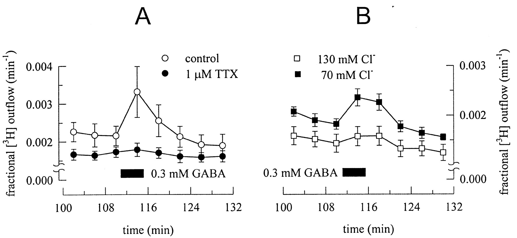
γ-Aminobutyric acid stimulates [3H]noradrenaline release from primary cultures of dissociated rat superior cervical ganglia. The cultures were loaded with 0.05 μmol l−1 [3H]noradrenaline at 36°C for 1 h. After labeling, the cultures were transferred to small chambers and superfused with a physiological buffer at 25°C at a superfusion rate of about 1.0 ml min−1. Collection of 4-min superfusate fractions was started after a 60-min washout period. GABA was present as indicated by the black bar. In A, the buffer contained either no or 1 μM tetrodotoxin (TTX), to suppress action potential propagation. In B, the Cl− concentration of the buffer was either 130 or 70 mM, and the omitted Cl− was replaced by gluconate. This procedure shifts the Cl−equilibrium by about 13 mV to more positive potentials and, thus, will facilitate Cl− efflux. Both graphs show the time course of fractional tritium outflow per minute, n = 6 in each case. For experimental details, see e.g., Vartian et al. (2001).
3. Glycine Receptors.
Glycine receptors are ligand-gated anion channels closely related to GABAAreceptors. They are composed of α- and β-subunits. Currently, at least four different α-subunits (α1–α4) are known, and alternative splicing of these may contribute to additional heterogeneity. α- and β-subunits form heterooligomers with a stoichiometry of 3α:2β, but α-subunits can also form homomeric receptors (Kuhse et al., 1995).
Glycine receptors are widespread within the central nervous system (Betz, 1991), but in the peripheral nervous system, they have been detected only in the neurons of ciliary (Zhang and Berg, 1995) and paravertebral sympathetic ganglia (Boehm et al., 1997) of chicken. In primary cultures of dissociated chick sympathetic ganglia, glycine caused not only anion currents, but also triggered transmitter release. Glycine-induced delivery of previously incorporated [3H]noradrenaline was abolished in the presence of tetrodotoxin. This indicated that the glycine receptors that stimulated sympathetic transmitter release were not located at the sites of exocytosis, i.e., not at the axon terminals themselves, but rather at a remote site, presumably the somatodendritic region of the neurons. There, a depolarizing effect of glycine was also evidenced by measuring glycine-evoked transmembrane Ca2+ entry (Boehm et al., 1997).
4. Serotonin 5-Hydroxytryptamine3Receptors.
Within the huge family of serotonin receptors, the 5-HT3 receptor is peculiar because it is a single ligand-gated cation channel among a large number of G protein-coupled receptors (Hoyer et al., 1994). Like nicotinic acetylcholine receptors, the 5-HT3 receptor is composed of five subunits, but there are only two subunit plus one alternatively spliced variant that have been characterized by molecular means (Fletcher and Barnes, 1998; Davies et al., 1999).
Despite an early description of stimulatory effects of serotonin on sympathetic axon terminals in rabbit hearts, which led to a huge increase in noradrenaline release, serotonin receptor agonists were generally found to reduce sympathetic transmitter release in cardiovascular preparations derived from, for instance, dogs, guinea pigs, and rats (for a review, see Fozard, 1984). Whether serotonin exerts excitatory or inhibitory cardiovascular effects is most frequently explained by species differences (Saxena, 1989). More recently, a considerable number of reports have confirmed inhibitory actions of serotonin that are mediated by G protein-coupled 5-HT receptors located at sympathetic axonal varicosities (Fuder and Muscholl, 1995).
In primary cultures of dissociated rat superior cervical ganglia, serotonin elicits rapidly activating cationic currents carried via 5-HT3 receptors (Yang et al., 1992). In analogy to the results obtained with P2X nucleotide receptors and nicotinic acetylcholine receptors (see above), one would expect that serotonin through its action on the 5-HT3 receptors may cause depolarization and ensuing transmembrane Ca2+ entry and transmitter release. However, we were unable to detect any significant secretagogue effect of serotonin in cultures of rat superior cervical ganglion neurons labeled with [3H]noradrenaline, which otherwise displayed prominent tritium overflow in the presence of ATP or acetylcholine (unpublished results). Similarly, serotonin did not affect noradrenaline release in cultures of mouse sympathetic neurons (Göbel et al., 2000). Nevertheless, in mesenteric blood vessels of the rat, a 5-HT3 receptor antagonist did significantly reduce neurogenic vasoconstriction, and it was therefore concluded that presynaptic 5-HT3 receptors may modulate sympathetic transmitter release (Potenza et al., 1998). Whether this phenomenon can also be observed in other preparations remains to be established.
V. Metabotropic Receptors
As mentioned above, postganglionic sympathetic axon terminals—with the exception of those innervating sweat glands—store noradrenaline, ATP, and several peptides, such as NPY, somatostatin, galanin, and endorphins (Elfvin et al., 1993; Benarroch, 1994). However, from a functional point of view only ATP, noradrenaline, and NPY have been consistently shown to contribute to sympathoeffector transmission (Franco-Cereceda and Liska, 1998). Therefore, receptors for these transmitters will be considered autoreceptors, whereas the receptors for the other peptides mentioned above will be assumed to rather represent presynaptic heteroreceptors. In this context, it appears necessary to reconsider the term “autoreceptor”: as stated above, this designates a receptor that is activated by a neurotransmitter, which is released from the very neuron on which the receptor is located. Hence, if ATP, noradrenaline, and NPY are released as cotransmitters from one neuron, each receptor on this neuron that is activated by one of these transmitters can be considered an autoreceptor. This also holds true, if the release of only one of the cotransmitters is being determined. In contrast to this interpretation, some authors categorize receptors for NPY regulating the release of noradrenaline from sympathetic neurons as heteroreceptors (Fuder and Muscholl, 1995). Because this review focuses on the presynaptic modulation of sympathetic transmitter release, all receptors activated by one of the sympathetic cotransmitters will be dealt with as autoreceptors.
As mentioned under Methodological Considerations(Section III.), in sympathetically innervated tissues, presynaptic autoreceptors mediate negative feedback modulation of transmitter release. This phenomenon is evidenced by a facilitatory effect of respective receptor antagonists either on stimulation-evoked transmitter release or on postsynaptic responses. Such results have been obtained with antagonists at α2 and P2Y receptors, but not with a recently developed antagonist at NPY Y2 receptor subtypes (Smith-White et al., 2001). Nevertheless, this apparent lack of NPY-dependent autoinhibitory feedback modulation of sympathoeffector transmission does not necessarily imply that presynaptic Y2 receptors cannot function as autoreceptors either under adequate experimental conditions or in vivo.
In analogy to what has been said above, facilitatory presynaptic autoreceptors may mediate positive feedback modulation of transmitter release. In addition to α-adrenoceptors, sympathetic axon terminals possess presynaptic β-adrenoceptors, the activation of which causes facilitation of stimulated transmitter release. Nevertheless, antagonists at these β-receptors do not alter stimulation-induced transmitter release or sympathoeffector transmission under conditions that otherwise permit noradrenaline-dependent autoinhibition (e.g.,Brock et al., 1997). Thus, released noradrenaline apparently fails to activate presynaptic β-receptors, and, therefore, these receptors are classified as heteroreceptors rather than autoreceptors.
Characteristics of presynaptic autoreceptors, although in a more general sense and not only in sympathetic neurons, have been reviewed in detail by Starke and coauthors (Starke, 1987; Starke et al., 1989), and those of presynaptic heteroreceptors by Fuder and Muscholl (1995). Therefore, we will summarize only results that have been obtained since the publication of these highly informative reviews, and the reader is referred to these papers for older references.
A. Metabotropic Autoreceptors
1. α2-Adrenoceptors.
For more than 50 years it has been known that adrenoceptors do not represent a homogenous group of binding sites but rather a family of receptor subtypes that were initially divided in α- and β-adrenoceptors (for a review, seeBylund et al., 1994). Some 20 years later, the description of presynaptic α-autoreceptors led to the subdivision of these adrenoceptors into α1- and α2-adrenoceptors (Langer, 1974; Starke, 1977), and nowadays at least four different subtypes of α2-adrenoceptors are known (see below).
Depending on the experimental conditions, sympathoeffector transmission (e.g., Todorov et al., 1999), as well as stimulation-evoked release of ATP (von Kügelgen, 1996) or noradrenaline (Starke, 1987) from sympathetic nerve terminals is greatly enhanced in the presence of antagonists at α-adrenoceptors. This phenomenon is generally accepted to reflect the interruption of the autoinhibitory feedback mediated by the released noradrenaline which activates the presynaptic adrenoceptors. Only in rare cases, the occurrence of α-adrenoceptor-mediated autoinhibition is questioned (Kalsner, 1990).
Inhibitory presynaptic adrenoceptors most commonly belong to the class of α2-receptors, but some presynaptic α-autoreceptors were believed to be α1- rather than α2-receptors. Most frequently, this conclusion was based on results obtained with either the α-adrenoceptor agonist methoxamine or the respective antagonist prazosine; these two agents display some selectivity for α1 receptors, but they were typically applied at high concentrations (Starke, 1987). Therefore, experiments have been performed to find out whether presynaptic noradrenergic autoreceptors may also comprise α1 receptors (Table1). In rabbit and rat kidney, α1 receptors were suggested to participate in the autoreceptor-mediated inhibition of noradrenaline release (Rump et al., 1992b; Bohmann et al., 1993), but no evidence was obtained for a contribution of α1-adrenoceptors to the autoinhibition in rat submaxillary glands and atria (Limberger et al., 1992). These apparent inconsistencies in pharmacological data may be caused either by effects of α1-adrenoceptor agonists on postjunctional receptors (Bohmann et al., 1993; Shinozuka et al., 1995) and noradrenaline reuptake (Schwartz and Malik, 1991) or by a heterogeneity of presynaptic α2-autoreceptors in different tissues and species.
Presynaptic adrenoceptors mediating inhibition of noradrenaline release from sympathetic neurons
Molecular cloning revealed that there is not only one α2-receptor, but rather a subfamily consisting of four different subtypes: these were designated α2A through α2D (Bylund et al., 1994; MacKinnon et al., 1994). In a given species, only three different subtypes are expressed, and the α2A- and α2D-subtypes, which show ≥89% sequence identity, are believed to represent species orthologs with α2A being expressed, for instance, in man and pig, and α2D being expressed, for example, in rats, mice, and cattle (O'Rourke et al., 1994). Prazosine differentiates between different α2-adrenoceptor subtypes and displays affinities for α2B- and α2C-receptors between 3 and 135 nM and affinities in the micromolar range for α2A- and α2D-receptors (MacKinnon et al., 1994). Thus, diverging results obtained with prazosine may reflect a heterogeneity of presynaptic α2-autoreceptors rather than a role of α1-receptors in the feedback modulation of noradrenaline release.
Experiments using a large number of α2-adrenoceptor agonists and antagonists were performed subsequently to compare pharmacological characteristics of native presynaptic α-autoreceptors with those of recombinant α2-adrenoceptor subtypes (Table 1). Most of the results obtained in these investigations indicated that presynaptic α2-autoreceptors most commonly belong to the α2A- or the α2D-subtype, depending on the species investigated (e.g., Funk et al., 1995; Limberger et al., 1995; Paiva et al., 1997). However, evidence was also obtained that α2-autoreceptors may correspond to the α2C-subtype (Trendelenburg et al., 1994; Rump et al., 1995a), and some authors suggested that the autoreceptors belong to the α2B-subgroup (Connaughton and Docherty, 1990; Smith et al., 1992; Blandizzi et al., 1993; Nakatsuka et al., 1995). Finally, in tissues obtained from mice that lacked either one or both of these two adrenoceptor subtypes, it was corroborated that, in fact, α2A- and α2C-receptors may contribute to autoinhibition of noradrenaline release, although the relative contribution of each of these two subtypes varies between different tissues. The α2A-receptor appears to be of greater importance in the brain as compared with the sympathetic nervous system. Moreover, the inhibitory effect of presynaptic α2C-receptor activation is more pronounced at low (<0.3 Hz) than at high (>0.3 Hz) stimulation frequencies, whereas the reverse holds true for the α2A-receptors (Hein et al., 1999).
Experiments aiming at the characterization of α2-autoreceptors were also performed in primary cultures of dissociated sympathetic ganglia (Table 1). Early experiments with neurons from lumbar paravertebral sympathetic ganglia of chicken embryos failed to detect release modulating adrenoceptors, although α2-adrenoceptors were clearly present as identified by receptor-mediated changes in cyclic AMP. These receptors were found to affect transmitter release only when neurons were kept in coculture together with cardiomyocytes (Wakade et al., 1988). More recently, sympathetic neurons in primary culture without any target cells, whether obtained from the frog (Lipscombe et al., 1989), from the chicken embryo (Boehm et al., 1991), from the rat (Hill et al., 1993; Schwartz and Malik, 1993; Boehm and Huck, 1995), or from the mouse (Trendelenburg et al., 1999b), were reported to possess α-adrenoceptors that inhibited either electrically or K+-evoked noradrenaline release. In chicken, rat, and mouse sympathetic neurons, these receptors were found to unequivocally belong to the α2 subfamily, as defined by actions of the selective agonists clonidine and UK 14,304 and by antagonistic effects of yohimbine or rauwolscine (Boehm et al., 1991, 1992; Hill et al., 1993; Allgaier et al., 1994b; Boehm and Huck, 1995). The α-adrenoceptor subtype of frog sympathetic neurons was also characterized as α2 by the antagonistic effect of yohimbine, even though this receptor was not activated by clonidine (Lipscombe et al., 1989). In primary cultures derived from mice, the noradrenergic autoreceptor subtype was characterized in further detail and was suggested to be, at least predominantly, an α2D subtype (Trendelenburg et al., 1999b). In recent experiments on cultured sympathetic neurons obtained from mice lacking α2D-receptors, an inhibition of [3H]noradrenaline by α2-adrenoceptor agonists was not detectable, and there was also no sign of any autoinhibitory feedback modulation of noradrenaline release (Trendelenburg et al., 2001). This corroborates that the genetic α2A/D-adrenoceptor subtype is the predominating receptor among the presynaptic α-autoreceptors in the sympathetic nervous system.
2. P2Y Nucleotide Receptors.
The superfamily of P receptors consists of receptors for nucleosides (P1) and nucleotides (P2). As mentioned above (see P2X nucleotide receptors), the family of P2 nucleotide receptors comprises ionotropic (P2X) and metabotropic (P2Y) G protein-coupled receptors. At least six different subtypes of P2Y receptors are known currently, which are designated P2Y1, -2,-4, -6,-11, and -12. Whereas P2Y1, -11, and-12 receptors are activated by adenine nucleotides only, P2Y2, -4, and -6 receptors are sensitive to uridine nucleotides (Ralevic and Burnstock, 1998; Hollopeter et al., 2001). Presynaptic actions of ATP (Cunha and Ribeiro, 2000) and of P2 receptors (Stone et al., 2000), in general, have been summarized recently, and a more specific review (von Kügelgen et al., 1999a) dealt with somatodendritic as well as presynaptic P2 receptors of postganglionic sympathetic neurons.
In spite of the well-established roles of ATP and noradrenaline as sympathetic cotransmitters, preliminary evidence for the occurrence of a purinergic, in parallel to the noradrenergic (see above), autoinhibitory feedback modulation of sympathetic transmitter release has been obtained only 15 years ago (Stjärne and Astrand, 1985;Fujioka and Cheung, 1987). More recently, this phenomenon was investigated in greater detail: the P2 receptor antagonists suramin and reactive blue 2 were found to enhance electrically evoked [3H]noradrenaline release from the mouse vas deferens, but only when trains of pulses were used, which allowed for an active biophase concentration of ATP or metabolites to accumulate. These antagonist effects were attenuated by pertussis toxin, which revealed a role of receptors coupled to inhibitory G proteins. Because antagonists at inhibitory adenosine receptors did not mimic the effects of suramin and reactive blue 2, it was obvious that the inhibitory feedback was mediated by P2 and not by P1 (i.e., adenosine) receptors (von Kügelgen et al., 1993). In the mouse vas deferens, inhibitory actions of adenine nucleotides on stimulated transmitter release were also reported, and again these effects were not altered by adenosine receptor antagonists, but were antagonized by P2 antagonists in the following order: reactive blue 2 > brilliant blue G > suramin ≫ PPADS. This provided additional direct evidence for the existence of inhibitory presynaptic P2 receptors (von Kügelgen et al., 1989, 1994b).
In other tissues, inhibitory and facilitatory effects of various adenine nucleosides and nucleotides on sympathetic transmitter release have been detected (Table 2). Initially, the presynaptic effects of adenine nucleotides were believed to be mediated by the degradation products, the nucleosides (see e.g.,Starke et al., 1989). However, in some cases, the effects of adenosine nucleotides and nucleosides were attenuated by both antagonists at adenosine (P1) and nucleotide (P2) receptors. This led Westfall and collaborators to suggest that these presynaptic effects were mediated by a third type of purinoceptor which was proposed to be named P3 (Shinozuka et al., 1988; Forsyth et al., 1991; Todorov et al., 1994). More commonly, the effects of adenine nucleotides on transmitter release are to some extent antagonized by P1 receptor antagonists, but those of the nucleosides are not affected by P2 receptor antagonists. Furthermore, in the presence of saturating concentrations of P1 receptor antagonists, P2 receptor antagonists still block the effects of nucleotides (e.g., Fuder and Muth, 1993; von Kügelgen et al., 1995; Koch et al., 1998). Hence, nucleosides may contribute to the inhibitory actions of adenine nucleotides, but it is evident that presynaptic P2 nucleotide receptors do mediate autoinhibitory feedback just as presynaptic α2-adrenoceptors do. Most commonly, these inhibitory presynaptic receptors are activated by adenine but not uridine nucleotides, and they are insensitive to prototypic P2X receptor ligands and can, thus, be assumed to be P2Y receptors (von Kügelgen et al., 1989; Saiag et al., 1998; see Table 2).
Presynaptic nucleotide receptors mediating modulation of noradrenaline release from sympathetic neurons
In primary cultures of postganglionic sympathetic neurons from chicken embryos, ATP was also reported to reduce electrically evoked [3H]noradrenaline release. However, this effect was only observed when ATP was used at concentrations of 1 to 6 mM and when the nucleotide was applied 12 min (but not 2 min) before electrical stimulation. When applied 2 min before the stimulus, ATP exerted facilitatory actions (Allgaier et al., 1995b). In cultures of sympathetic neurons derived from neonatal rats, ATP and related nucleotides per se stimulate rather than reduce stimulation-evoked release (Boehm, 1994, 1999; Boehm et al., 1995a; von Kügelgen et al., 1997). Only about one-half of this stimulatory action of ATP is mediated by presynaptic P2 receptors, and these are ionotropic P2X receptors, and not metabotropic P2Y receptors (see above). In cultured sympathetic neurons, uridine nucleotides were also found to stimulate [3H]noradrenaline release (Boehm et al., 1995a; von Kügelgen et al., 1997) through activation of P2Y6 receptors (Vartian et al., 2001). However, these effects are entirely tetrodotoxin-sensitive and, thus, are not mediated by presynaptic receptors. In sympathetic neurons from mice, the secretagogue actions of uridine nucleotides were also detected, but ATP failed to affect spontaneous as well as electrically evoked transmitter release. Once again, the uracil nucleotides did not act via a presynaptic, but through a somatodendritic receptor and, thus, failed to alter stimulation-evoked release (Nörenberg et al., 2001).
A synopsis of the data available on presynaptic P2 receptors (Table 2) reveals a considerable heterogeneity in pharmacological characteristics. The inhibitory presynaptic P2Y receptors appear to have in common that they are not activated by uridine nucleotides and that they are either insensitive to, or display an apparently low affinity, for suramin. Instead, these receptors are quite potently blocked by reactive blue 2 and related substances. An inhibitory P2Y receptor with similar pharmacological characteristics (i.e., sensitivity to adenine but not uridine nucleotides and to reactive blue 2; insensitivity to suramin and PPADS) has been detected recently in PC12 cells, which are ontogenetically related to sympathetic neurons (Vartian and Boehm, 2001). Among the P2Y receptors that are insensitive to uridine nucleotides, PC12 cells express only P2Y12 (unpublished observation), and it, thus, appears likely that the inhibitory P2Y autoreceptors in the sympathetic nervous system correspond to this recently cloned P2Y receptor subtype (Hollopeter et al., 2001).
3. Neuropeptide Y Y2 Receptors.
Neuropeptide Y (NPY) shares a family of common receptors with peptide YY and pancreatic polypeptide, which consists of at least five members. These are named Y1, Y2, and Y4 through Y6 receptors. These receptors are characterized by diverging rank orders of agonist potencies, and for Y1 receptors, selective antagonists have been available for some time (Michel et al., 1998). A specific Y2 receptor antagonist has been introduced recently (King et al., 2000; Smith-White et al., 2001).
NPY is costored together with ATP and noradrenaline in most sympathetic axon terminals, and its release from sympathetically innervated tissues (Modin et al., 1996) and from sympathetic neurons in cell culture has been demonstrated (Braas and May, 1999). In the vasculature and particularly in arteries, NPY has been found to mediate nonadrenergic and nonpurinergic neurogenic contractions, and this effect is most commonly mediated by NPY Y1 receptors located on smooth muscle cells. However, in some cases, postjunctional effects of NPY may also involve Y2 receptors (for a review, see Franco-Cereceda and Liska, 1998; Modin et al., 1996). Despite this unequivocal cotransmitter role of NPY, NPY-dependent autoinhibition of sympathetic transmitter release could not be demonstrated, because appropriate receptor antagonists have been lacking.
In numerous in vitro preparations containing axons of noradrenergic postganglionic sympathetic neurons, NPY was found to reduce action potential-dependent transmitter release or sympathoeffector transmission, and these presynaptic effects of NPY have been summarized by Fuder and Muscholl (1995). Inhibitory effects of NPY on either excitatory junction potentials (Smith-White et al., 2001), neurogenic arteriolar vasoconstriction (Kotecha, 1998), or on noradrenaline release (Maynard and Burnstock, 1994) are typically mimicked by preferential Y2 receptor agonists, such as NPY 13-36 or NPY 24-36. NPY 24-36 was also reported to reduce sympathetic acetylcholine release in the dog (Mahns et al., 1998), and a related NPY Y2 receptor agonist also reduced sympathetic vasoconstriction in the dog in vivo (Mahns et al., 1999).
NPY 13-36 reduced NPY release from the spleen of reserpinized pigs (Modin et al., 1996), i.e., from sympathetic axon terminals depleted of noradrenaline, which proves that presynaptic Y2receptors are indeed autoreceptors. Nevertheless, in guinea pig vasa deferentia, a selective Y2 receptor antagonist failed to alter excitatory junction potentials, which reveals a lack of NPY-dependent autoinhibitory feedback modulation of sympathoeffector transmission (Smith-White et al., 2001). Despite this lack of effect when applied alone, the antagonist attenuated the inhibitory action of a Y2 receptor agonist. Furthermore, in rat hypothalamic slices, this antagonist clearly enhanced K+-evoked NPY release to demonstrate autoinhibitory modulation of NPY release in the central nervous system (King et al., 2000). Thus, it remains to be established whether and under which conditions NPY may mediate an autoinhibition of sympathetic transmitter release.
Presynaptic NPY receptors have also been investigated in primary cell cultures of dissociated rat superior cervical ganglion neurons. In cocultures of such sympathetic neurons and cardiac myocytes, electrical stimulation of a “presynaptic” neuron reduced the frequency of spontaneous oscillations in intracellular Ca2+ in a “postsynaptic” myocyte. This neuroeffector transmission frequently involved more than one transmitter and was blocked by a combination of adrenergic and cholinergic antagonists. NPY was found to reduce the effect of neuronal stimulation upon the Ca2+ oscillations in the myocytes without exerting any effect on its own. Thus, these results reflect a NPY-dependent presynaptic inhibition of transmitter release (Toth et al., 1993). Equivalent results were obtained in experiments that investigated [3H]noradrenaline release from cultured rat superior cervical ganglion cells. NPY caused a concentration-dependent decline of electrically evoked tritium overflow (Oellerich et al., 1994). However, in neither study the subtype of NPY receptor mediating the presynaptic inhibition was characterized in further detail.
B. Metabotropic Heteroreceptors
1. β2-Adrenoceptors.
As mentioned above (α2-autoreceptors) adrenoceptors have originally been divided into the α- and β-subfamilies, and within the latter group one can discern between at least three different subtypes, β1 through β3(Bylund et al., 1994). It is widely accepted that axon terminals of postganglionic sympathetic neurons are equipped with facilitatory β-adrenoceptors. Although some presynaptic β1-adrenoceptors have been described on axon terminals of sympathetic neurons, these receptors most commonly belong to the β2-subtype (Fuder and Muscholl, 1995), and this pharmacological characterization has been confirmed in recent experiments determining noradrenaline release in the guinea pig trachea (de Haas et al., 1999) and human dental pulp (Parker et al., 1998a) and atria (Rump et al., 1994), as well as ATP release in guinea pig atria (Tokunaga et al., 1995). As a consequence, presynaptic β-adrenoceptors are relatively insensitive to noradrenaline and prefer adrenaline as endogenous agonist. Therefore, these receptors are thought to represent presynaptic heteroreceptors rather than autoreceptors, which become activated when adrenaline originating from the adrenal gland is stored in and released from sympathetic nerve terminals, a phenomenon that was believed to play a role in essential hypertension (see Starke et al., 1989; Fuder and Muscholl, 1995). Accordingly, blockade of presynaptic β-adrenoceptors does not alter sympathoeffector transmission (Brock et al., 1997) nor sympathetic transmitter release (Apparsundaram and Eikenburg, 1995; Parker et al., 1998a), even though in these reports facilitatory effects of the respective agonists were detected. In addition, the facilitatory effects of β-receptor agonists on sympathetic transmitter release appear to be variable and to change under several conditions: (i) β-adrenoceptors are subject to agonist-dependent desensitization. Thus, long-term exposure to β2-adrenoceptor agonists was found to abolish the facilitatory effect of presynaptic β-receptor activation (Encabo et al., 1996). Moreover, presynaptic β-adrenoceptors were shown to also desensitize more rapidly, within a time course of minutes (Lakhlani and Eikenburg, 1993). (ii) The facilitatory presynaptic action of β2-adrenoceptor agonists is frequency- and stimulation-dependent in that the effect decreases when either the frequency (Apparsundaram and Eikenburg, 1995) or the duration (Lakhlani and Eikenburg, 1993) of neuronal stimulation is increased. Furthermore, the facilitatory effect of the β-agonist isoprenaline was most pronounced with the first stimulus within a train of pulses (Brock et al., 1997). (iii) The action of presynaptic β-adrenoceptors also depends on the state of activation of α-adrenoceptors. Blockade of the latter receptors supports the facilitatory action of β-agonists (Costa and Majewski, 1988; Apparsundaram and Eikenburg, 1995), and this phenomenon may, of course, contribute to the aforementioned stimulation and frequency dependence. In summary, the physiological relevance of facilitatory presynaptic β2-adrenoceptors remained somewhat unclear (see also Fuder and Muscholl, 1995).
In contrast to the data summarized above, β-adrenoceptor agonists have also been reported to inhibit sympathoeffector transmission or sympathetic transmitter release. In the rat vas deferens, isoprenaline reduced neurogenic contractions but did not alter the postjunctional responses due to application of either ATP or noradrenaline (Huang et al., 1998). Similarly, in the guinea pig vas deferens, isoprenaline also reduced purinergic neurogenic contractions and neuronal ATP release, but at the same time, the β-agonist increased noradrenaline release and noradrenergic neurogenic contractions. Both the inhibitory and facilitatory effects were found to involve presynaptic β2-adrenoceptors (Driessen et al., 1996;Goncalves et al., 1996).
In primary cultures of chick sympathetic neurons, isoprenaline reduced electrically evoked noradrenaline release in a concentration-dependent manner, and this effect was antagonized by the β-adrenergic antagonist propranolol but not by the α2-adrenoceptor antagonist rauwolscine (Allgaier et al., 1994b). In general, results concerning a possible modulatory effect of β-adrenoceptor agonists on transmitter release appear to be more heterogeneous when obtained in cell cultures as compared with sympathetically innervated tissues. Thus, in explant cultures of rat superior cervical ganglia, isoprenaline increased the release of [3H]noradrenaline evoked by K+-depolarization. This action was antagonized by propranolol and by butoxamine, but not by sotalol or practolol, which indicated that the receptor involved was a β2-adrenoceptor (Weinstock et al., 1978). In contrast, in primary cultures of dissociated rat (Hill et al., 1993;Koh and Hille, 1997) and mouse (Göbel et al., 2000) sympathetic neurons, no facilitatory effects of β-adrenoceptor activation were detected. Hence, the precise functional roles of presynaptic β-adrenergic receptors in sympathetic neurons in cell culture remain to be confirmed.
2. Muscarinic Acetylcholine Receptors.
The family of muscarinic acetylcholine receptors comprises at least five different proteins showing the seven transmembrane domain structure typical of G protein-coupled receptors. They are named M1through M5 and can be discriminated from each other by using a series of, but not a single, subtype preferring antagonists (Caulfield and Birdsall, 1998). The fact that sympathetic axon terminals possess inhibitory muscarinic receptors is known about as long as the existence of presynaptic nicotinic receptors on these nerve terminals (see above). Therefore, they probably represent the best documented example of presynaptic heteroinhibition in the sympathetic nervous system. Data concerning the muscarinic modulation of noradrenaline release have been included in most of the previous reviews on presynaptic receptors (e.g., Starke, 1977; Westfall, 1977;Langer, 1981; Vizi et al., 1991), and reports on this topic that had been published between 1980 and 1994 have been critically summarized byFuder and Muscholl (1995). The emerging picture indicated that presynaptic muscarinic receptors may either facilitate or inhibit sympathoeffector transmission or stimulation-evoked sympathetic transmitter release. The facilitatory effect was most commonly reported to involve M1 muscarinic receptors, whereas the inhibitory effect was typically correlated with M2 receptors, and there were only a few exceptions to this rule. In a few cases, M1receptors were suggested to inhibit instead of facilitate sympathetic transmitter release, and sometimes M3 receptors were reported to also function as inhibitory presynaptic heteroreceptors on sympathetic axon terminals (see Fuder and Muscholl, 1995). In guinea pig carotid arteries, M1 and M2 receptors were confirmed to facilitate and inhibit [3H]noradrenaline release, respectively, and activation of endothelial M3receptors caused vasorelaxation (Casado et al., 1994). Thus, the investigation of presynaptic muscarinic receptors in sympathetically innervated tissues has to take several possible sites of action into account, and indirect effects may impinge on the apparent pharmacological characteristics of presynaptic receptors.
In recent reports (Table 3), several examples of facilitatory presynaptic muscarinic receptors have been described, and, when investigated, the receptors were found to belong to the M1 subtype (Somogyi et al., 1996, 1998). Inhibitory presynaptic receptors, in contrast, were mostly reported to be of the M2 subtype (Jumblatt and Hackmiller, 1994; Hey et al., 1994; Haunstetter et al., 1994; Zhang et al., 1997b;Lambrecht et al., 1999). However, Matko et al. (1994) suggested the inhibitory presynaptic muscarinic receptors on sympathetic axon terminals in the human papillary muscle to belong to the M3 subtype, and for guinea pig atria it was also M3 receptors that were suggested to reduce noradrenaline release (Nakatsuka et al., 1995). The presynaptic muscarinic receptor inhibiting neurogenic contractions in rabbit vasa deferentia has long been considered an M1subtype, but recent experiments suggested that this receptor rather is an M4 subtype (e.g., Budriesi et al., 2001). In many other cases, the subtypes of muscarinic receptors mediating an inhibition of sympathetic transmitter release were not determined (e.g., Kotecha, 1999; Lomholt and Nedergaard, 2000; Table 3).
Presynaptic muscarinic acetylcholine receptors mediating modulation of sympathetic transmitter release
With respect to muscarinic AChRs, divergent results have been obtained in primary cultures of postganglionic sympathetic neurons. In cultures derived from chick embryos, muscarinic agonists, including methacholine and arecoline, triggered [3H]noradrenaline release, and the stimulatory action of methacholine was antagonized by nanomolar concentrations of atropine and abolished in the presence of tetrodotoxin (Greene and Rein, 1978). As evidenced by the tetrodotoxin sensitivity, these effects were not mediated by presynaptic receptors.Bhave et al. (1988), in contrast, were unable to detect transmitter release from chick sympathetic neurons when challenged by activation of muscarinic receptors, even though this stimulation clearly increased the intracellular levels of inositolphosphates. In cultures of mouse sympathetic neurons, the muscarinic agonist oxotremorine M, at 100 μM and in the absence of any other secretagogue stimulus, triggered transmitter release. Whether this effect arose at a presynaptic site of action remained unclear (Nörenberg et al., 2001). Furthermore, it should be noted, that oxotremorine M quite potently activates nicotinic acetylcholine receptors (Reitstetter et al., 1994). Thus, the presence of release stimulating muscarinic receptors in primary cultures of postganglionic sympathetic neurons remains to be confirmed.
In rat, superior cervical ganglion neurons in cell culture (Boehm and Huck, 1995), atropine at nanomolar concentrations caused a marked increase (instead of a decrease, as above) in acetylcholine-evoked noradrenaline release. Such results had also been obtained in rabbit hearts (Lindmar et al., 1968) and are indicative of presynaptic, release-inhibiting muscarinic receptors. The presence of inhibitory presynaptic muscarinic receptors in superior cervical ganglion neurons was confirmed by Koh and Hille (1997) who showed that pirenzepine-insensitive (i.e., not M1) muscarinic receptors reduced noradrenaline release. Most recently, M2 receptors were found to reduce electrically evoked transmitter release in cultured paravertebral sympathetic neurons from neonatal mice (Göbel et al., 2000).
3. Adenosine A1 and A2Receptors.
Within the superfamily of purine receptors, one has to discriminate between receptors for purine nucleotides, P2 receptors, and those for nucleosides, P1 or adenosine receptors. The adenosine receptors can be further subdivided by pharmacological means into three groups designated A1, -2, and -3, and within the A2subgroup, one can differentiate between A2A and A2B receptors. All types of adenosine receptors are widely distributed in the central and peripheral nervous systems (Ralevic and Burnstock, 1998), and presynaptic adenosine receptors have been reported to modulate transmitter release in virtually every neurotransmitter system investigated (Fredholm and Dunwiddie, 1988).Fuder and Muscholl (1995) have provided a concise overview of presynaptic P1 receptors on peripheral noradrenergic nerves, which revealed that adenosine may either enhance or reduce stimulation-evoked transmitter release. In general, the facilitatory effect is mediated by A2 receptors, whereas the inhibitory action involves A1 receptors. Whereas the physiological relevance of presynaptic A2 receptors remained largely elusive, the inhibitory A1 receptors are believed to be of major importance either during increased neuronal activity or under hypoxic conditions (Fredholm and Dunwiddie, 1988;Fuder and Muscholl, 1995). Accordingly, activation of presynaptic A1 receptors is believed to underlie neuronal stunning after myocardial hypoxia (Burgdorf et al., 2001).
In more recent experiments (Table 4), the previous results have been largely confirmed. In various sympathetically innervated tissues from different species, presynaptic A1 adenosine receptors were found to reduce either sympathoeffector transmission (e.g., Hardy and Brock, 1999), or stimulation-evoked release of noradrenaline (e.g., Shinozuka et al., 2001) and ATP (e.g., Driessen et al., 1994), respectively. In addition, presynaptic A2A receptors were reported to enhance stimulated noradrenaline release (e.g., Mota et al., 2000), and in several tissues, such as the rat tail artery (Goncalves and Queiroz, 1996) and the rat vas deferens (Kurz et al., 1993), both types of receptors, inhibitory A1 and facilitatory A2A receptors, were detected within one preparation. However, in some organs, for instance, the rat heart (Snyder et al., 1998; von Kügelgen et al., 1995) and pancreas (Koch et al., 1998), no evidence could be obtained for the existence of facilitatory A2A receptors on sympathetic axon terminals. One report appears to contradict the general rule that presynaptic A1 receptors reduce transmitter release from sympathetic neurons: in the rabbit ear artery, an A1 adenosine receptor preferring agonist was found to enhance electrically evoked [3H]noradrenaline release, but the receptor subtype involved in this effect had not been confirmed by the use of selective antagonists (Maynard and Burnstock, 1994).
Presynaptic adenosine receptors mediating modulation of sympathetic transmitter release
Contradictory results have been obtained in primary cultures of postganglionic sympathetic neurons. In neurons from thoracolumbar paravertebral ganglia from either the chicken (Allgaier et al., 1995b) or the mouse (Trendelenburg et al., 1999b), adenosine receptor agonists failed to cause any alteration of noradrenaline release. Similarly, in cultures of dissociated rat superior cervical ganglia, adenosine receptor agonists were reported not to affect the outflow of previously incorporated [3H]noradrenaline (Boehm, 1994). However, adenosine did significantly diminish the release of endogenous catecholamines evoked by exposure to depolarizing K+ concentrations (Koh and Hille, 1997), but the receptor involved was not further characterized.
In several experiments, P1 receptor antagonists were found to alter stimulation-evoked sympathetic transmitter release in the absence of agonists. In the rabbit ear artery, for instance, an antagonist reduced noradrenaline overflow (Ishii et al., 1996), whereas in the dog pulmonary artery, another antagonist enhanced noradrenaline overflow noradrenaline overflow. In both cases, these effects were endothelium-dependent and were, thus, mediated by endothelial adenosine release. Furthermore, stimulation of α1-adrenoceptors in rat caudal arteries was shown to induce the release of purines, which in turn reduced evoked neuronal transmitter release (Shinozuka et al., 1995). Hence, under certain conditions, release of endogenous purines from effector organs may control sympathoeffector transmission. In addition, the inhibitory action of presynaptic A1 receptors increases, when sympathetic transmitter output is raised, either by α2-autoreceptor blockade or by high-frequency stimulation (Crosson and Gray, 1997). Taken together, these data suggest that a local purinergic system and presynaptic A1 receptors may serve to precisely regulate the strength of the neuronal input in sympathetically innervated tissues. Interestingly, in spontaneously hypertensive rats, the A1 receptor-mediated control of arterial noradrenaline release is lost (Shinozuka et al., 1995).
4. Angiotensin II AT1 Receptors.
The various forms of angiotensins (angiotensin I–IV and angiotensin 1–7) exert their actions via a family of membrane receptors with seven membrane spanning domains, which are commonly linked to heterotrimeric G proteins. This family comprises the receptor subtypes AT1 and AT2, which have both been characterized by molecular means and which are activated by angiotensins II and III. In addition, there is an AT4 receptor primarily activated by angiotensin IV, and a less well-characterized binding site, which was suggested to be named nonAT1-nonAT2receptor. In rodents but not in man, pig and dog, there are two highly homologous AT1 receptor types termed AT1A and AT1B. Most of the physiologic effects of active angiotensins are believed to be mediated by AT1 receptors. AT2receptors are thought to be important during fetal development, and AT4 receptors are found primarily within the central nervous system (De Gasparo et al., 2000).
It has long been recognized that angiotensins support the functions of the sympathetic nervous system, and this is mediated primarily by a presynaptic facilitation of neuroeffector transmission (Starke, 1977;Fuder and Muscholl, 1995). In fact, facilitation of noradrenaline release by angiotensin II has been reported for a variety of sympathetically innervated tissues obtained from a large number of different species. In several investigations, evidence was provided that the this stimulatory effect was mediated by AT1 receptors. However, angiotensins may also exert postsynaptic effects, and via indirect mechanisms, such as the release of prostaglandins, this may also contribute to the presynaptic modulation of sympathoeffector transmission. Therefore, the physiological and pathophysiological impact of presynaptic angiotensin receptor activation remained to be elucidated (for a critical overview, see Fuder and Muscholl, 1995).
In recent years, it has been confirmed in a variety of tissues that angiotensins facilitate noradrenaline and/or ATP release from sympathetic axon terminals and, thereby, augment neuroeffector transmission (Table 5). However, negative results have also been obtained. In primary cultures of mouse sympathetic neurons (Göbel et al., 2000) and in the human forearm (Chang et al., 1995), no presynaptic effects of angiotensin II could be detected. Furthermore, inhibitory presynaptic effects of angiotensins have also been reported, for instance, in the rabbit pulmonary artery (Sim and Soh, 1995) and in the guinea pig vas deferens (Ziogas and Cunnane, 1991). In this latter report, the inhibitory action of angiotensin II was mediated by prostaglandins, whereas in all the other investigations, the effects observed were suggested to be mediated by presynaptic angiotensin receptors. These presynaptic receptors were identified most frequently as AT1 receptors (Table 5), but there were also notable exceptions. For instance, in rat atria and caudal arteries, presynaptic angiotensin effects were antagonized by both AT1 and AT2 receptor antagonists (Gironacci et al., 1994;Cox et al., 1996a, 1996b), whereas in other reports, antagonist concentrations selective for either subtype failed to attenuate the facilitatory actions of angiotensin in rat hearts (Moura et al., 1997) and dog arteries (Guimaraes et al., 1998b). Hence, there still is some uncertainty concerning the subtypes of presynaptic angiotensin receptors in sympathetic axon terminals.
Presynaptic angiotensin receptors mediating modulation of sympathetic transmitter release
Under quasi physiological conditions, stimulation-evoked sympathetic transmitter release was reported not to be altered in the presence of AT1 receptor antagonists, even though activation of the respective receptors clearly enhanced transmitter output (e.g.,Munch et al., 1996; Shetty and DelGrande, 2000). However, during or subsequent to hypoxic conditions, noradrenaline release from human or guinea pig heart was markedly reduced by AT1receptor antagonists (Munch et al., 1996; Maruyama et al., 1999), and the facilitatory action of angiotensin was lost (Munch et al., 1996). This suggests that local angiotensin production during myocardial ischemia contributes to enhanced sympathetic transmitter release through an activation of presynaptic angiotensin AT1 receptors.
5. Bradykinin B2 Receptors.
Actions of bradykinin are mediated by at least two types of receptors, B1 and B2 bradykinin receptors, and the existence of a third member of the family, a B3 receptor, has also been suggested. B1 receptors are mainly restricted to the vasculature, whereas B2 receptors have been detected in most types of tissues including the central and peripheral nervous systems (Marceau et al., 1998).
More than 20 years ago, bradykinin was shown to reduce electrically induced noradrenaline release in rabbit pulmonary arteries and hearts (Starke, 1977). The nonapeptide was found more recently to increase depolarization-evoked transmitter release from sympathetic nerve terminals of rat vasa deferentia (Llona et al., 1991). In mouse vasa deferentia, in contrast, bradykinin was suggested to modulate sympathetic transmitter release in a bimodal way, causing inhibition via B1 and facilitation via B2 receptors (Maas et al., 1995). The facilitatory action through B2 receptors has been corroborated recently (Trendelenburg et al., 2000). Augmentation of stimulated noradrenaline release via B2 receptors has also been reported in the human kidney (Rump et al., 1995b).
The actions of bradykinin on sympathetic axon terminals have been investigated most frequently in various cardiac preparations derived from rats (Chulak et al., 1995; Vaz-da-Silva et al., 1996; Foucart et al., 1997; Kurz et al., 1997; Rump et al., 1997; Moura et al., 1999), guinea pigs (Seyedi et al., 1997, 1999), mice (Chulak et al., 1998; Cox et al., 2000; Trendelenburg et al., 2000), and man (Rump et al., 1997). There, bradykinin was always found to enhance stimulation-evoked noradrenaline release, at least under quasi physiological experimental conditions. In most of these reports, bradykinin was evidenced to act via B2 receptors (Chulak et al., 1995;Vaz-da-Silva et al., 1996; Rump et al., 1997; Kurz et al., 1997; Chulak et al., 1998; Moura et al., 1999; Seyedi et al., 1999). In a considerable number of the aforementioned reports, the investigation of underlying mechanisms revealed that the apparently presynaptic effects of bradykinin were indirect ones. In the rat heart, removal of the endocardium abolished the effect of the nonapeptide (Vaz-da-Silva et al., 1996; Moura et al., 1999), and its action was suggested to involve angiotensin II and AT1 receptors (Moura et al., 1999); in human and rat heart, inhibition of cyclooxygenases attenuated the release-enhancing action of bradykinin (Rump et al., 1997); and in guinea pig cardiac synaptosomes, the bradykinin effects involved calcitonin gene-related peptide and substance P (Seyedi et al., 1999).
Inhibition of kininases to prevent bradykinin metabolism was either found to augment the facilitatory action of bradykinin (e.g.,Vaz-da-Silva et al., 1996; Rump et al., 1997; Chulak et al., 1998) or to permit the detection of the facilitatory action of the peptide, which was otherwise not detectable (Rump et al., 1995b, 1997). In guinea pig cardiac synaptosomes, the angiotensin-converting enzyme inhibitor enalaprilat raised stimulation-evoked noradrenaline release when applied alone, and this effect was mediated by B2 receptors (Seyedi et al., 1997). Moreover, a transient hypoxia also raised noradrenaline release in guinea pig hearts, and this enhanced release of noradrenaline could not be further augmented by bradykinin but was reduced by blockade of B2 bradykinin receptors (Hatta et al., 1999). In rat hearts, bradykinin was suggested to raise noradrenaline release after a hypoxic period via B1 receptors (Foucart et al., 1997). Taken together, the facilitatory effect of bradykinin on noradrenaline release in the heart, whether through direct or indirect mechanisms, appears to be of major importance during cardiac ischemia.
A possibly modulatory effect of bradykinin on sympathetic transmitter release has also been investigated in primary cultures of postganglionic sympathetic neurons. When these were derived from neonatal mice, bradykinin failed to affect noradrenaline release (Göbel et al., 2000). However, in cultures of dissociated rat superior cervical ganglia, activation of B2bradykinin receptors was sufficient to stimulate transmitter release in the absence of any other secretagogue stimulus. However, this effect was abolished by Na+ channel blockade with tetrodotoxin and, thus, was mediated, not by presynaptic, but by somatodendritic B2 receptors (Boehm and Huck, 1997b).
6. Cannabinoid CB1 Receptors.
At least two types of cannabinoid receptors, CB1 and CB2, exist and, thus, may mediate the actions of endogenous and plant-derived agonists. Aside from their psychoactive and immunomodulatory effects, cannabinoids exert pronounced cardiovascular actions such as vasodilatation, tachycardia, and changes in blood pressure, all mediated most likely by CB1 receptors. Because central sites of action are not involved in these effects, cannabinoids are believed to elicit vasorelaxation through peripheral mechanisms, which may include inhibition of sympathoeffector transmission and direct actions on the vasculature (Hillard, 2000).
Inhibition of peripheral noradrenaline release by cannabinoids was reported for the first time more than 25 years ago (Graham et al., 1974), and these early results have been largely confirmed in the recent past. In rat kidneys (Deutsch et al., 1997) and vasa deferentia (Ishac et al., 1996) and in mouse vasa deferentia (Trendelenburg et al., 2000), cannabinoids reduced noradrenaline release via CB1 receptors. However, in the rat tail artery, this effect could not be detected, even though a CB1 receptor-mediated reduction of the generalized neurogenic vasopressor response was observed (Malinowska et al., 1997). In pithed rabbits, a CB1 receptor agonist also reduced blood pressure and noradrenaline spillover into the plasma as well as plasma noradrenaline concentrations (Niederhoffer and Szabo, 1999). In this report, a direct application of cannabinoids to the central nervous system of rabbits was shown to cause sympathoexcitation instead of inhibition as evidenced by increases in blood pressure and plasma noradrenaline concentrations.
In cardiac tissues, contrasting results have been obtained: in rat atria (Ishac et al., 1996) and human atrial appendages (Molderings et al., 1999), CB1 receptor activation caused an inhibition of [3H]noradrenaline release, whereas in mouse atria no such effect was detectable (Trendelenburg et al., 2000). Finally, in primary cultures of dissociated mouse postganglionic sympathetic neurons, cannabinoids were found to reduce sympathetic transmitter release via CB1 receptors (Göbel et al., 2000). In summary, the above results confirm that cannabinoids may cause sympathoinhibition through presynaptic CB1 receptors on sympathetic nerve terminals, the activation of which reduces transmitter release.
7. Dopamine Receptors.
To date, five different members have been identified within the family of dopamine receptors, which are designated D1 through D5 and categorized into two subfamilies, the D1- and the D2-like families, which harbor the D1 and -5 and D2, -3, and -4 subtypes, respectively (Levant, 1997). However, the endogenous agonist dopamine activates not only dopamine receptors, but is also an agonist at adrenergic receptors. Therefore, the well-documented inhibitory effect of dopamine in sympathetically innervated tissues may involve not only dopamine, but also α2-adrenoceptors. Nevertheless, dopamine receptors are quite widespread in the sympathetic nervous system (Willems et al., 1985), and presynaptic D2 receptors are generally believed to inhibit sympathoeffector transmission, as reviewed by Willems et al. (1985) and by Fuder and Muscholl (1995).
It has been confirmed recently in rat tail arteries that D2 receptor activation reduces not only noradrenaline release (Friedman and Duckles, 1994a, 1994b, 1995), but also neurogenic contractions (Friedman and Duckles, 1994b). The inhibitory function of presynaptic D2 receptors on sympathetic axon terminals has also been demonstrated in human tissues including the dental pulp (Parker et al., 1994), atria (Rump et al., 1995c; Abadie et al., 1996), and gastric as well as uterine arteries (Morgadinho et al., 1999). In contrast, in the guinea pig vas deferens, it was a presynaptic D1 receptor that was suggested to inhibit neurogenic contractions (Furukawa and Morishita, 1997).
One question related to the description of inhibitory presynaptic D2 receptors in the sympathetic nervous system is whether they may mediate an autoinhibitory feedback. In many instances, D2 receptor antagonists did not alter stimulation-evoked sympathetic transmitter release. However, in human uterine arteries, dopamine release has been demonstrated recently, and the D2 receptor antagonist sulpiride was reported to enhance the release of both dopamine and noradrenaline (Morgadinho et al., 1999). This suggested a physiological importance of endogenous dopamine, which was further corroborated by the fact that D2 receptor blockade was found to enhance the exercise-dependent increase in human plasma noradrenaline levels (Mannelli et al., 1999).
8. Endothelin Receptors.
Endothelin-1 was identified originally as an endothelium-derived vasoconstricting peptide, but the family of endothelins (endothelin-1, -2, and -3) has recently been suggested to act as neuropetides in the central and peripheral nervous systems. The actions of endothelins are mediated by either ETA or ETB endothelin receptors (Pollock et al., 1995). Postganglionic sympathetic neurons in cell culture have been shown to synthesize and release endothelin-1 and -3, and the peptides appear to be important for the development and survival of these neurons. Assuming that endothelins are released from axonal varicosities, presynaptic endothelin receptors should be considered autoreceptors. In support of this notion, neurogenic contractions of rat tail arteries in the absence of agonists were enhanced by an ETB receptor antagonist (Garcia-Villalon et al., 1999a), which is indicative of an autoinhibitory feedback. However, endogenous endothelins may stem, for instance, from the endothelium. Furthermore, evidence for a contribution of endothelins to sympathoeffector transmission has not been obtained, nor has the release from axon terminals been demonstrated (Damon, 1999). Therefore, presynaptic endothelin receptors will be dealt with as heteroreceptors.
Early reports on the modulation of neuroeffector transmission in sympathetically innervated tissues by endothelins have been reviewed byFuder and Muscholl (1995). These authors summarized data showing presynaptic facilitatory and inhibitory and postsynaptic effects of endothelins, and each of these effects was later corroborated. At the postjunctional level, endothelin-dependent augmentation of purinergic (Mutafova-Yambolieva and Radomirov, 1994) and noradrenergic (Zhang et al., 1996) responses has been observed in rabbit and dog arteries, respectively. At the presynaptic level, facilitation and inhibition by endothelins have been reported. In rat tail arteries, various endothelins were shown to augment the stimulation-induced release of noradrenaline and purine nucleotides (Mutafova-Yambolieva and Westfall, 1998a) and to potentiate neurogenic, but not noradrenaline-evoked, contractions via ETAreceptor activation (Garcia-Villalon et al., 2000b). In the guinea pig vas deferens, the stimulated release of ATP was enhanced in the presence of endothelins (Mutafova-Yambolieva et al., 1997), but the simultaneous release of noradrenaline remained unaltered (Mutafova-Yambolieva and Westfall, 1995). In both preparations, endothelins were also found to reduce stimulation-evoked sympathetic transmitter release when applied either in the presence of an ETA receptor antagonist (Mutafova-Yambolieva and Westfall, 1995) or at very low concentrations (Mutafova-Yambolieva and Westfall, 1998a). As a consequence, presynaptic ETB receptors were suggested to mediate the inhibition, whereas distinct presynaptic ET, but not ETA, receptors were proposed to mediate the facilitation of transmitter release. An inhibition of either noradrenaline release (Matsumura et al., 1996) or sympathetic neurotransmission (Garcia-Villalon et al., 1999a) via ETB receptors has been confirmed in dog kidneys and rat tail arteries, respectively. In contrast, in canine mesenteric arteries, the inhibitory effect of endothelin-1 on both neuroeffector transmission and noradrenaline release, was suggested to be mediated by presynaptic ETA receptors (Zhang et al., 1996). Thus, it appears obvious that endothelins may modulate sympathetic neurotransmission via facilitatory and inhibitory presynaptic receptors, but the receptor subtypes involved remain to be unequivocally identified.
9. γ-Aminobutyric AcidB Receptors.
In the central nervous system, GABA is well known to cause presynaptic inhibition of transmitter release via ionotropic GABAA receptors and metabotropic, G protein-coupled GABAB receptors (e.g., Miller, 1998). In the peripheral nervous system, much less evidence has been obtained in support of modulatory presynaptic effects of this amino acid transmitter. In an early report, Starke and Weitzell (1980)revealed that GABA reduced [3H]noradrenaline release in a bicucullin- and picrotoxin-insensitive manner and, thus, via a receptor different from the ionotropic GABAA receptor. The inhibitory effect of GABA on stimulation-evoked noradrenaline release from peripheral neurons was corroborated subsequently in bovine ovarian follicles (Kannisto et al., 1987), goat cerebral arteries (Miranda et al., 1989), rat pineal glands (Rosenstein et al., 1990), rat venae cavae (Schneider et al., 1991;Schlicker et al., 1993), pig retinae (Schlicker et al., 1993), and rat kidneys (Fujimura et al., 1999). In all of these cases, the receptor mediating the inhibitory action of GABA was demonstrated to be a GABAB receptor. Furthermore, presynaptic GABAB receptors were shown to mediate an inhibition of contractions in bovine ovarian follicles (Kannisto et al., 1986) and rat vasa deferentia (Kwan et al., 1996) and a reduction of neuronal vasoconstriction in goat cerebral arteries (Miranda et al., 1989) and rat renal arteries (Fujimura et al., 1999). In accordance with these latter results, GABA also attenuates the generalized neurogenic vasopressor response in pithed rats (Kohlenbach and Schlicker, 1990). Thus, sympathetic axon terminals appear to be equipped with inhibitory presynaptic GABABreceptors. However, the application of GABABreceptor antagonists does not affect stimulation-evoked noradrenaline release (Schneider et al., 1991), which suggests that there is no endogenous GABA being released in sympathetically innervated tissues upon electrical field stimulation. Accordingly, a possibly physiological relevance of the presynaptic GABABreceptors remains to be identified.
10. Histamine H3 Receptors.
Histamine exerts its effects via three different types of G protein-coupled receptors named H1 through H3 (Hill et al., 1997). Among these, the H3 receptor has been identified by its function as a presynaptic autoreceptor mediating feedback inhibition of histamine release in the central nervous system (Arrang et al., 1983). In the peripheral nervous system, histamine has long been recognized to act as a presynaptic modulator of noradrenaline release (Marshall, 1981), and more recent results concerning presynaptic histamine receptors on sympathetic axon terminals have been reviewed by Fuder and Muscholl (1995). The importance of presynaptic histamine H3 receptors in the cardiovascular system has been highlighted by Malinowska et al. (1998) and by Levi and Smith (2000).
Earlier reports on stimulatory and inhibitory presynaptic effects of histamine in the sympathetic nervous system favored a role of H1 and/or H2 receptors (Marshall, 1981). However, since the discovery of presynaptic H3 autoreceptors, it is this receptor subtype that is most commonly believed to mediate a histamine-dependent inhibition of sympathoeffector transmission (Fuder and Muscholl, 1995). Nevertheless, even in the recent past, there still appear to be notable exceptions to this general rule (Table6). For instance, in the rat vas deferens, Poli et al. (1994) reported that H1receptor agonists facilitated, whereas H2receptor activation inhibited, neurogenic contractions through presynaptic mechanisms. In the vas deferens of the guinea pig, however, neurogenic contractions were reduced by the activation of presynaptic H3 receptors (Luo et al., 1994). In the mouse uterine horn, histamine was also found to facilitate stimulation-evoked noradrenaline release via H1 receptors, even though this effect appeared to be progesterone-dependent (Montesino et al., 1995).
Presynaptic histamine receptors mediating modulation of sympathetic transmitter release
The vast majority of studies investigating presynaptic histamine receptors of sympathetic neurons used cardiovascular preparations and found inhibitory effects involving H3 receptors (Table 6). The only exception appears to be the chicken heart, for which H2 receptor activation was reported to cause positive chrono- and inotropic responses by eliciting noradrenaline release from sympathetic nerve terminals (Tanaka et al., 1995a, 1995b). The H3 receptor-mediated inhibitory action of histamine on sympathetic transmitter release in the heart appears to be of major pathophysiological importance, because cardiac release of histamine is greatly enhanced under ischemic conditions (Imamura et al., 1994). As a consequence, the postischemic rise in noradrenaline release (which is to a considerable extent nonexocytotic and mediated by monoamine transporters) is under tonic inhibitory control of histamine as revealed by an additional increase in noradrenaline release in the presence of H3receptor antagonists (Imamura et al., 1994; Hatta et al., 1997). Under quasi physiological conditions, however, these antagonists fail to alter noradrenaline release (Imamura et al., 1994). Thus, in hypoxic hearts of men and guinea pigs, a rise in endogenous histamine release is likely to mitigate noradrenergic overactivity through an activation of inhibitory presynaptic H3 receptors of sympathetic axon terminals (Levi and Smith, 2000). In rat hearts, however, such a protective effect of histamine could not be demonstrated (Mazenot et al., 1999a).
In addition to mediating a negative control of noradrenaline release, presynaptic H3 receptors were demonstrated to restrict sympathetic cholinergic transmission as evidenced by an inhibition of the neurogenic sudomotor response in the rat skin (Koss, 1994). In primary cultures of postganglionic sympathetic neurons derived from neonatal mice, however, histamine failed to regulate noradrenaline release (Göbel et al., 2000). Taken together, most of the experimental evidence available supports the view that histamine limits sympathoeffector transmission via presynaptic H3 receptors and suggests that this effect is of major importance under pathological conditions.
11. Imidazoline Receptors.
Imidazoline receptors are binding sites for drugs displaying imidazoline or guanidine structures and are categorized into two groups, I1 and I2 receptors. Whereas I1receptors appear to be restricted to the central nervous system, I2 receptors are found in various tissues and may correspond to binding sites located on monoaminoxidases (Parini et al., 1996). Drugs with imidazoline or guanidinium structures were not only found to enhance stimulated noradrenaline release from peripheral neurons through an interruption of the autoinhibitory feedback via presynaptic α2-adrenoceptors, but also to reduce transmitter release via an independent mechanism. The use of various agonists and antagonists indicated that sympathetic axon terminals of various rabbit arteries are equipped with inhibitory imidazoline receptors which can be separated by pharmacological means from α2-autoreceptors. However, these receptors also appeared to be different from the established I1 and I2 binding sites (for a review see Fuder and Muscholl, 1995; Molderings and Göthert, 1995b).
More recently, the existence of presynaptic imidazoline receptors has also been inferred from experiments performed in other rabbit tissues, such as iris-ciliary bodies (Ogidigben et al., 1994; Chu et al., 1996). Subsequently, inhibition of noradrenaline release by putative imidazoline receptor agonists has been reported for the kidney (Bohmann et al., 1994) and the aorta and vena cava of the rat (Molderings and Göthert, 1998). However, most, if not all, of the agonists commonly used may also activate α2-adrenoceptors, and in the rat vas deferens, the presynaptic inhibitory effects of imidazolines were suggested to involve adrenoceptors only (Avellar and Markus, 1996). Likewise, the inhibitory effects of imidazolines on stimulation-evoked noradrenaline release in pithed rats appeared to involve α2-adrenoceptors, but not imidazoline receptors (Hauser et al., 1995). Evidence for the existence of presynaptic imidazoline receptors on sympathetic axon terminals was also obtained in human atria, because the rank order of potency of various imidazolines and guanidines to inhibit noradrenaline release was the same as that previously reported for rabbit arteries. Furthermore, their effects were hardly attenuated under conditions that cause α2-adrenoceptor blockade, and the receptor involved was proposed to be a nonI1-nonI2 imidazoline receptor (Likungu et al., 1996; Molderings et al., 1997). In the recent past, the presynaptic imidazoline receptors of the human heart were found to be sensitive toward a blockade by CB1cannabinoid receptor antagonists, which appears to aggravate rather than facilitate the interpretation of the data available (Molderings et al., 1999; for a review, see Göthert et al., 1999).
Gaiser et al. (1999) have recently reinvestigated the question of presynaptic imidazoline receptors in rabbit pulmonary arteries in the absence of an α2-adrenergic autoinhibitory feedback. Under these conditions, which are believed ideal for the investigation of presynaptic receptors, the inhibitory effects of all agents that were used as agonists at imidazoline and/or α2-adrenergic receptors were antagonized equally well by α2-adrenoceptor antagonists. This led these authors to conclude that “there is no need to invoke presynaptic imidazoline receptors” to explain their results. Thus, the puzzle of presynaptic imidazoline receptors in the sympathetic nervous system still awaits unequivocal resolution.
12. Natriuretic Peptide Receptors.
The family of receptors for natriuretic peptides (i.e., atrial natriuretic peptide, brain natriuretic peptide, and C-type natriuretic peptide) comprises two members, the ANPA and the ANPB receptors. Both are membrane receptors with one transmembrane domain and intracellular guanylyl cyclase activity and, thus, belong to the superfamily of guanylyl cyclases (Lucas et al., 2000). The ANPA receptor is preferentially activated by atrial and brain natriuretic peptide, whereas C-type natriuretic peptide is the endogenous agonist for ANPB. Fuder and Muscholl (1995) summarized early evidence in favor of presynaptic effects of natriuretic peptides in the sympathetic nervous system, but they also reported on respective negative results. Since then, Trachte and Drewett (1994) demonstrated inhibitory presynaptic actions of both atrial natriuretic peptide and C-type natriuretic peptide, on the sympathoeffector transmission in the rabbit vas deferens. However, only the atrial natriuretic peptide caused increases in cyclic GMP, and this was taken as an indication to the absence of ANPB receptors in this tissue. In the guinea pig vas deferens, atrial and brain natriuretic peptides raised the stimulation-evoked release of ATP (Mutafova-Yambolieva et al., 1997), whereas in the rat tail artery, C-type natriuretic peptide reduced the overflow of purines (Mutafova-Yambolieva and Westfall, 1998b). In both tissues, the natriuretic peptides inhibited ectonucleotidase activity and, thereby, altered the composition of the purines released. Taken together, an unequivocal role of presynaptic receptors for natriuretic peptides on sympathetic axon terminals remains to be elucidated, and the natriuretic peptides may rather be assumed to control the sympathetic nervous system through central sites of action (Peng et al., 1996).
13. Opioid δ-, κ-, and μ-Receptors.
The family of receptors for opiates, opioids, and opioid peptides consists of three members, δ-, κ-, and μ-receptors, which were originally differentiated from each other by pharmacological means and which have, thereafter, been characterized by molecular cloning. A new nomenclature has been proposed that refers to these receptors as OP1, OP2, and OP3 receptors (Dhawan et al., 1996), respectively, but this terminology does not appear to be universally accepted and we will therefore rely on the old Greek letter system. An additional receptor has been cloned recently, which, like the opioid receptors, displays a seven-transmembrane domain structure and shows 50 to 60% sequence homology with the opioid receptors. This receptor is referred to as opioid receptor-like protein1, i.e., ORL1 (Quock et al., 1999).
Opioid receptor agonists have long been known to modulate transmitter release in the peripheral nervous system, and Illes (1989) critically reviewed the respective literature. Evidence has been obtained that each of the three known opioid receptor subtypes may be located on sympathetic axon terminals and upon activation reduce transmitter release. In particular, presynaptic δ-receptors inhibit sympathoeffector transmission in the cat spleen, guinea pig heart, hamster vas deferens, rat vas deferens, and various rabbit arteries and veins. κ-Receptors cause presynaptic inhibition in guinea pig hearts and in rabbit hearts, vasculature, and vas deferens. μ-Receptors diminish sympathetic transmitter release in the cat nictitating membrane and the rat vas deferens. In the mouse vas deferens, all three opioid receptors subtypes mediate an inhibition of transmitter release (Illes, 1989).
More recent experiments on presynaptic opioid receptors in the sympathetic nervous system have been summarized by Fuder and Muscholl (1995). Experiments that have been performed since then focused primarily on the possible existence of presynaptic ORL1 receptors on sympathetic axon terminals (Giuliani et al., 2000). The ORL1 receptor agonist nociceptin has been reported to reduce neurogenic contractions in vasa deferentia of mice (Berzetei-Gurske et al., 1996; Calo et al., 1996; Zhang et al., 1997a), rats, and rabbits (Nicholson et al., 1998), and in the rat anococcygeus muscle (Ho et al., 2000). In addition, nociceptin inhibited noradrenaline release in rat tail arteries (Bucher, 1998) and cardiac synaptosomes (Dumont and Lemaire, 2000), and in mouse vasa deferentia (Trendelenburg et al., 2000). A number of additional experiments verified that sympathetic axon terminals are equipped with various opioid receptor subtypes, which, in general, exert inhibitory effects (Table7). There is, however, one exceptional report demonstrating that μ-receptor agonists may facilitate noradrenaline release in the guinea pig colon (Cosentino et al., 1997).
Presynaptic opioid receptors mediating modulation of sympathetic transmitter release
Considering that opioid peptides have been found to be stored in sympathetic neurons (Elfvin et al., 1993; Benarroch, 1994), one has to pose the question as to whether endogenous opioids may modulate sympathoeffector transmission. In support of this possibility, antagonists at ORL1 receptors (Ho et al., 2000) and at δ- and μ-opioid receptors (Cosentino et al., 1995), when applied alone, were reported to enhance sympathetic transmitter release. However, most commonly, no antagonist effects have been observed in the absence of respective agonists (e.g., Illes, 1989;Bognar et al., 1995; Bucher, 1998). Thus, a major role of an endogenous opioid system in the regulation of sympathoeffector transmission appears unlikely, even though endorphins were reported to modulate, for instance, human catecholamine secretion during high intensity exercise (Angelopoulos et al., 1995).
14. Prostanoid Receptors.
Prostanoids comprise prostaglandins and thromboxanes, which are synthesized from arachidonic acid through the cyclooxygenase pathway in response to a variety of different stimuli. After being released from the cells where they have been produced, the prostanoids exert their actions via a family of receptors with seven membrane spanning domains linked to G proteins. Depending on their sensitivity toward the endogenous prostanoids prostaglandin D2, prostaglandin E2, prostaglandin F2α, prostaglandin I2, and thromboxane A2, these receptors are classified as DP, EP, FP, IP, and TP, respectively, and the EP receptors are further subdivided into EP1 through EP4(Narumiya et al., 1999).
Prostanoid-dependent presynaptic modulation of sympathetic transmitter release is well-documented and has been reviewed previously (Güllner, 1983; Fuder and Muscholl, 1995). In most cases, inhibitory actions of prostaglandins have been observed, and more recent data support the predominantly negative regulation of sympathoeffector transmission via prostanoid receptors. Thus, inhibition of noradrenaline release by prostaglandin E2 has been reported for the cardiovascular system in rabbits (e.g., Jensen and Nedergaard, 1999) and men (e.g.,Molderings et al., 1998), and the receptors involved were most commonly suggested to belong to the EP3 subtype (Table8). Presynaptic EP receptors were also described in the guinea pig vas deferens, where reductions in neurogenic contractions (Tam et al., 1997), excitatory junction potentials (Brock and Cunnane, 1996), or transmitter release (Driessen and Starke, 1994) have been observed. In addition, inhibitory presynaptic prostanoid receptors were found in erectile tissues of the rabbit (Italiano et al., 1995), in the rat stomach (Racke et al., 1995), and in human iris-ciliary bodies (Awe et al., 2000).
Presynaptic prostanoid receptors mediating modulation of sympathetic transmitter release
Facilitatory presynaptic effects of prostanoids were reported to occur in human saphenous veins (Molderings et al., 1994) and atrial appendages (Molderings et al., 1998) via DP receptors, in human iris-ciliary bodies via TP receptors (Awe et al., 2000), and in guinea pig vasa deferentia via IP receptors (Tam et al., 1997). Moreover, activation of EP1 receptors caused facilitation of stimulation-evoked noradrenaline release from primary cultures of chick sympathetic neurons (Allgaier and Meder, 1995). In contrast to these results, prostaglandin E2 was found to reduce transmitter release in primary cultures of postganglionic sympathetic neurons derived either from rats (Koh and Hille, 1997) or from mice (Göbel et al., 2000).
The presynaptic actions of prostanoids appear to be particularly important, because various pathologic conditions, such as hypoxia (Gomez-Nino et al., 1994; Nakhostine et al., 1995), may cause enhanced prostaglandin synthesis and release in sympathetically innervated tissues, and the endogenous prostaglandins, in turn, may control sympathetic transmitter release. Furthermore, prostaglandins may be involved in the effects of various other modulators of sympathoeffector transmission. Cytokines, for instance, reduce noradrenaline release in human (Abadie et al., 1997) and mouse (Foucart and Abadie, 1996) atria through the generation of prostaglandins. Likewise, the inhibitory effect of the antihypertensive agent cicletanine on sympathetic transmitter release was suggested to involve prostaglandins (Nasa et al., 1998). In contrast to its mainly facilitatory actions (see Table5), angiotensins may also inhibit neurogenic contractions in rabbit pulmonary arteries via the release of prostaglandins (Sim and Soh, 1995). Endogenous prostaglandins may also be involved in facilitatory actions. For instance, the enhancement of noradrenaline release in human and rat atria by bradykinin was proposed to be mediated by prostaglandins (Rump et al., 1997). Moreover, the stimulating actions of peroxides on sympathetic transmitter release in bovine iris-ciliary bodies rely on prostaglandin synthesis (Graham et al., 2000). Thus, it appears worthwhile to consider prostaglandin actions whenever modulation of sympathoeffector transmission is investigated.
15. Serotonin 5-Hydroxytryptamine1 Receptors.
The family of metabotropic serotonin receptors is huge and harbors at least six classes designated as 5-HT1 through 5-HT7, with the exception of 5-HT3, which is the serotonin-gated cation channel (see above). Among the 5-HT1 subfamily, one can discern at least five different subtypes (5-HT1A, 1B,1D, 1E,1F), among the 5-HT2receptors there are at least three different representatives (5-HT2A, 1B,2C), and finally two different 5-HT5 receptors (5-HT5A,5B) are known. Most, if not all of these receptors, are found in neurons, and it is primarily receptors of the 5-HT1 subfamily that are located on axon terminals and, thus, may mediate a modulation of transmitter release (Hoyer et al., 1994).
Early reports on the modulation of sympathetic transmitter release via presynaptic serotonin receptors have been reviewed by Fozard (1984) who differentiated between excitatory and inhibitory receptors. Although the excitatory actions appeared to involve one distinct receptor subtype, which is now known as 5-HT3, the inhibitory receptors were suggested to be mediated by a more heterogeneous group of receptors, which was classified as 5-HT1. Axon terminals of an ever growing number of sympathetically innervated tissues were reported more recently to be equipped with metabotropic 5-HT receptors (Fuder and Muscholl, 1995). These include predominantly blood vessels derived from rats, guinea pigs, rabbits, dogs, and pigs where presynaptic 5-HT1 or 5-HT1-like receptors were found to inhibit sympathoeffector transmission. Pharmacological and functional characteristics of inhibitory presynaptic 5-HT receptors in men have been reviewed separately (Göthert et al., 1996), and these receptors were suggested to belong to the 5-HT1B or 5-HT1D subtype, according to the recent subclassification (Hartig et al., 1996). In the recent past, the previous results have been largely confirmed by a few reports: In the pithed rat, serotonin was found to diminish the neurogenic vasopressor response (Terron et al., 1996), and in the human atrium, 5-HT1D receptors mediated an inhibition of noradrenaline release (Molderings et al., 1996). In the dog saphenous vein, 5-HT1B/1D receptors also reduced noradrenaline release and, to a lesser extent, neurogenic contractions (Medhurst et al., 1997). In rabbit saphenous veins, however, a 5-HT1A receptor mediated facilitation of neurogenic contractions, and 5-HT1B/1D receptors were not detectable by functional means (Cohen et al., 1999).
Describing presynaptic 5-HT receptors on sympathetic axon terminals, one has to consider a potentially physiological relevance of these. Serotonin may be released in considerable amounts from platelets, and platelet derived serotonin has been demonstrated to regulate the contractility of rat aortae (Yang and Mehta, 1994). Another potential source of serotonin are mast cells. In fact, mast cell degranulation in response to antigen stimulation leads to pronounced serotonin release in the rat heart which in turn inhibits noradrenaline release (Fuder et al., 1994). Finally, serotonin is taken up by, and may subsequently be released from, sympathetic axons (Gouliaev and Nedergaard, 1995). Thus, endogenous serotonin may stem from a variety of sources within sympathetically innervated tissues and may therefore contribute to the modulation of neuroeffector transmission. In accordance with this idea, antagonists at presynaptic 5-HT1 receptors were found to alter neurogenic contractions in the rabbit saphenous vein in the absence of respective agonists (Cohen et al., 1999).
16. Somatostatin Receptors.
The tetradecapeptide somatostatin is widely distributed within the central nervous system, and five different G protein-coupled somatostatin receptors (sst1–sst5) have been characterized by molecular cloning (Selmer et al., 2000). A certain proportion of noradrenergic neurons in sympathetic ganglia have been shown to contain somatostatin (Elfvin et al., 1993), but a corelease of these two transmitters has not been demonstrated.
Early evidence for a modulation of sympathetic transmitter release by somatostatin has been obtained in the rabbit ear artery (Maynard et al., 1991). There, somatostatin reduced neurogenic vasoconstriction, on one hand, and stimulation-evoked release of tritiated noradrenaline, on the other hand. However, the first effect may also involve a postsynaptic site of action as recently demonstrated in rabbit arteries (Ruiz et al., 2000). An inhibitory effect on transmitter release has been corroborated in rat mesenteric arteries (Calhau et al., 2000). In this study, an antagonist at somatostatin receptors has also been used but failed to alter stimulation-evoked release, thus, indicating that there was no feedback inhibition mediated by the peptide.
Inhibitory effects of somatostatin have also been detected in primary cultures of postganglionic sympathetic neurons, whether derived from chicken (Boehm and Huck, 1996a), rats (Koh and Hille, 1997), or mice (Göbel et al., 2000). In these reports, the neuropeptide reduced noradrenaline release as determined either after labeling of the transmitter pool with [3H]noradrenaline (Boehm and Huck, 1996a; Göbel et al., 2000) or by electrochemical detection (Koh and Hille, 1997). In two reports, the effects of somatostatin were found to be mimicked by the synthetic peptides seglitide (Boehm and Huck, 1996a) or octreotide (Calhau et al., 2000), and the receptors involved may, thus, correspond to the cloned receptor subtype sst2 or sst3. Nevertheless, a definite pharmacological characterization of inhibitory presynaptic somatostatin receptors on sympathetic axon terminals is still missing.
17. Receptors for Vasoactive Intestinal Peptide and Pituitary Adenylyl Cyclase-Activating Peptides.
Vasoactive intestinal peptide (VIP), pituitary adenylyl cyclase-activating peptides (PACAP), and additional peptides, such as secretin, share a family of common G protein-coupled receptors (Vaudry et al., 2000), which are termed PAC1, VPAC1, and VPAC2. Although VPAC receptors are equipotently activated by VIP and PACAPs, the PAC1 receptor is rather insensitive to VIP. Various forms of PACAPs and VIP are present in and released from sympathetic nerve terminals in, for instance, the canine heart (Anderson et al., 1993) and in cell cultures of sympathetic neurons (Brandenburg et al., 1997). Nevertheless, autoregulation of sympathetic transmitter release by VIP or PACAP has not been demonstrated. Therefore, the respective receptors are considered presynaptic heteroreceptors.
VIP is well known to cause vasodilatation and positive inotropic and chronotropic effects (Henning and Sawmiller, 2001). PACAP-27 has also been found to increase the heart rate, and this effect was suggested to involve a rise in noradrenaline release from cardiac sympathetic nerve terminals (Whalen et al., 1999). Likewise, the same peptide was reported to enhance sympathetic transmitter release from the dog pancreas in vivo (Yamaguchi and Fukushima, 1998). In contrast to these stimulatory actions, VIP has also been reported to reduce stimulation-evoked noradrenaline release and neurogenic contractions in rat portal veins (Bratveit and Helle, 1991).
Most of the results concerning the actions of VIP and PACAP on sympathetic transmitter release have been obtained in primary cultures of dissociated sympathetic ganglia. There, VIP was first found to enhance fast excitatory postsynaptic potentials at cholinergic synapses that form in between single rat sympathetic neurons (Takahashi et al., 1992). In chick sympathetic neurons, VIP enhanced [3H]noradrenaline release (Przywara et al., 1991). This facilitatory effect was confirmed by May and Braas (1995), who demonstrated that PACAP27 and -38 and VIP stimulated catecholamine and NPY secretion from rat superior cervical ganglion neurons, with the PACAPs being more potent than VIP. This indicated a role for a PAC1 receptor, which was corroborated in later experiments (Braas and May, 1999; Beaudet et al., 2000). In contrast to the results mentioned above, secretin was reported to reduce the depolarization-evoked release of noradrenaline from cultured rat sympathetic neurons, but the receptor subtype involved was not determined (Koh and Hille, 1997). Hence, presynaptic receptors for VIP and/or PACAP may either facilitate or inhibit sympathetic transmitter release, but the receptor subtypes mediating these opposite effects remain to be unequivocally identified.
18. Additional Receptors.
In addition to the plethora of presynaptic receptors that have been discussed systematically, there are single reports on several peptides that cause a presynaptic modulation of sympathetic transmitter release. In their review, Fuder and Muscholl (1995) mentioned presynaptic facilitation by ACTH and inhibition by neurotensin and insulin. The effect of insulin has been corroborated recently, because the hormone was found to reduce neurogenic vasoconstriction in rabbit ear arteries (Garcia-Villalon et al., 2000a). In addition, vasopressin has been reported to facilitate neurogenic vasoconstrictions in rabbit ear arteries (Garcia-Villalon et al., 1999b), on one hand, and in rat tail arteries (Garcia-Villalon et al., 2000b), on the other hand. In both cases, the effects were mediated by V1 vasopressin receptors. Finally, substance P released from sensory nerves was demonstrated to inhibit excitatory junction potentials in arterioles of the guinea pig small intestine (Coffa and Kotecha, 1999).
VI. Basic Mechanisms of Vesicular Sympathetic Transmitter Release
The remainder of this review is dedicated to the discussion of mechanisms that may underlie the presynaptic modulation of sympathoeffector transmission. To be able to discuss such mechanisms, one prerequisite is a detailed knowledge about the events that form the basis of sympathetic neurotransmitter release. Therefore, we shall first summarize ionotropic and metabotropic mechanisms underlying the delivery of transmitter into the neuroeffector junction to consider thereafter the major parameters possibly involved in the receptor-dependent stimulation, facilitation, or inhibition of the release process.
A. Ionotropic Mechanisms
Release of neurotransmitters from sympathetic axon terminals may either be vesicular or nonvesicular, the latter being most likely transporter mediated. This latter type will not be considered here, and the reader is referred to detailed reviews dealing with nonvesicular transmitter release (Attwell et al., 1993; Bonanno and Raiteri, 1994). Vesicular release may either occur spontaneously or as a result of a nerve impulse that propagates along an axon. In both cases, the vesicles that are exocytosed appear to be identical (Cunnane and Searl, 1994), and the mechanisms underlying vesicle exocytosis are thought to be the same and are discussed as metabotropic mechanisms below.
Activity-dependent release of neurotransmitters from presynaptic nerve terminals is initiated by an action potential that invades the varicosity and almost instantaneously, i.e., within less than 1 ms, triggers vesicle exocytosis. Three crucial steps are involved in excitation-secretion coupling in axon terminals: (i) the action potential transiently depolarizes the nerve terminal, (ii) this depolarization evokes transmembrane Ca2+ entry via voltage-activated Ca2+ channels, and (iii) the rise of intracellular Ca2+ up to submillimolar concentrations triggers exocytosis.
The stimulation paradigm most frequently used to investigate evoked transmitter release in tissue preparations containing sympathetic axon terminals and in cell cultures is the application of electrical fields. In cultured neurons, electrical field stimulation was shown to elicit action potentials with regular waveforms (Holz et al., 1988; Przywara et al., 1993b). Transmitter release triggered by electrical fields is abolished in the absence of extracellular Ca2+, which is consistent with Ca2+-dependent exocytosis, and in the presence of tetrodotoxin, which indicates the participation of Na+-dependent action potentials.
A second type of stimulation frequently used to experimentally induce sympathetic transmitter release is the application of depolarizing K+ concentrations. K+-evoked transmitter release is also absolutely Ca2+-dependent, but its tetrodotoxin sensitivity depends on the concentration of K+ used. At up to 30 mM K+, stimulation-dependent release of noradrenaline from sympathetic neurons is reduced by tetrodotoxin by up to 30%. At higher K+ concentrations, however, evoked transmitter release is not affected by this Na+ channel blocker (e.g., Allgaier and Meder, 1995; Kristufek et al., 1999). Thus, at moderate K+ concentrations, propagation of Na+-dependent action potentials may contribute to K+-evoked transmitter release. At higher K+ concentrations, however, the axon terminals are depolarized tonically and, as a consequence, Ca2+ enters the terminals via activated voltage-gated Ca2+ channels.
The free intracellular Ca2+ that is required for triggered neurotransmitter release is provided from the extracellular source via voltage-operated Ca2+ channels, which are clustered at the release zones. Upon opening, the voltage-gated Ca2+ channels allow high Ca2+ elevations in the proximity of the vesicle fusion machinery so that its Ca2+ sensor (putatively synaptotagmin, see below) is activated. However, Ca2+ elevations generated by individual action potentials may not be sufficient to saturate this sensor. This was concluded from the observation that sustained micromolar Ca2+ elevations trigger transmitter release more effectively than Ca2+ rises produced by presynaptic action potentials. This and the high cooperativity of Ca2+ ions in triggering vesicle fusion suggest that a partial inhibition of voltage-activated Ca2+ influx may have pronounced effects on transmitter release (Schneggenburger and Neher, 2000). Moreover, during an action potential with a typical duration of a few milliseconds, the synaptic membrane is only very briefly depolarized so that an only moderate inhibition of the kinetics of Ca2+current activation may profoundly affect the arising presynaptic Ca2+ elevation. Accordingly, blockers of voltage-gated Ca2+ channels that do not differentiate between various channel subtypes entirely abolish electrically induced transmitter release (e.g., Hirning et al., 1988;Smith and Cunnane, 1997). In contrast, spontaneous release of noradrenaline or ATP remains largely unaltered in either the presence of Na+ or Ca2+ channel blockers or in the absence of extracellular Ca2+.
Among subtype-specific Ca2+ channel blockers, the N-type-selective ω-conotoxin GVIA represents the most potent and efficient inhibitor of electrically as well as K+-evoked transmitter release from sympathetic nerve terminals (e.g., Hirning et al., 1988; Brock et al., 1989; Boehm and Huck, 1996b; Waterman, 1997; Serone and Angus, 1999; Molderings et al., 2000; Nedergaard, 2000). However, it is not only the N-type voltage-gated Ca2+ channel that may contribute to sympathetic transmitter release. When these channels are blocked, high-frequency stimulation (Smith and Cunnane, 1996; Waterman, 1997) is still able to elicit transmitter release, and this evoked release is reduced by ω-agatoxin IVA and ω-conotoxin MVIIC, blockers at P/Q-type Ca2+ channels (Smith and Cunnane, 1996;Waterman, 1997; Brock and Cunnane, 1999; Tanaka et al., 1999). Likewise, the application of activators of L-type Ca2+ channels to sympathetic neurons that had been treated with ω-conotoxin GVIA restores stimulated transmitter release (Boehm and Huck, 1996b). Hence, under certain conditions, voltage-gated Ca2+ channels other than N-type contribute to triggered exocytosis, but under quasi physiological conditions, the predominant or even exclusive role of the N-type channels in stimulation-evoked sympathetic transmitter release is undisputed.
Dihydropyridine blockers of L-type Ca2+ channels exert either no (e.g., Boehm and Huck, 1996b; Molderings et al., 2000) or small (Przywara et al., 1993a) inhibitory actions on electrically evoked sympathetic transmitter release. Nevertheless, significant levels of L-type Ca2+ channels are expressed in sympathetic neurons. However, they appear to be concentrated rather at the somatodendritic region than at the axon terminals where vesicle exocytosis occurs (Przywara et al., 1993a; Rittenhouse and Zigmond, 1999). Still, if transmitter release is triggered by K+ depolarization, the inhibitory actions of dihydropyridines and phenylalkylamines are markedly increased. This phenomenon is related to the voltage-dependent action of organic Ca2+ channel blockers (Holz et al., 1988; Boehm and Huck, 1993, 1996b), which is supported by the tonic K+ depolarization.
Depolarizations during action potentials are terminated by repolarizing K+ currents. Accordingly, the blockade of voltage-gated K+ channels by, for instance, tetraethylammonium causes a multifold prolongation of action potentials. This is likely to increase the probability of suprathreshold Ca2+ signals within the varicosities due to more long-lasting Ca2+channel-activating depolarizations. Thereby, K+channel blockade potentiates electrically evoked sympathetic transmitter release (Przywara et al., 1993b; Cunnane and Searl, 1994; Msghina et al., 1998). Typically, the evoked release of both ATP and noradrenaline is enhanced in the presence of K+ channel blockers (von Kügelgen et al., 1994a; Msghina et al., 1998). Investigations with various K+ channel blockers revealed that action potential repolarization in sympathetic neurons of the rat involves primarily delayed rectifier K+ currents that are sensitive to 4-aminopyridine and, to a variable extent, Ca2+-activated K+ currents sensitive toward a blockade by tetraethylammonium (Marsh and Brown, 1991). 4-Aminopyridine applied alone was found to be sufficient to induce sympathetic transmitter release (Huang, 1995), whereas tetraethylammonium fails to do so (Boehm and Huck, 1997). Another K+ channel blocker that triggers release from sympathetic neurons in cell culture is Ba2+(Boehm and Huck, 1997b). Ba2+ blocks G protein-gated inwardly rectifying K+ channels, on one hand, and M-type K+ channels, on the other hand. However, inwardly rectifying K+ channels are not expressed in sympathetic neurons (Ruiz-Velasco and Ikeda, 1998), and therefore the action of Ba2+ is rather selective for the inhibition of M-type K+channels. Another selective M-type K+ channel blocker, linopirdine, was also found to stimulate noradrenaline release from rat sympathetic neurons in primary cell culture (Kristufek et al., 1999a). However, the facilitating actions of Ba2+(Boehm and Huck, 1997b) and linopirdine (Kristufek et al., 1999a) on sympathetic transmitter release were shown to be entirely tetrodotoxin-sensitive. Thus, M-type K+ channels do not appear to be present at sympathetic axon terminals.
In contrast to the results obtained with blockers, openers of K+ channels were reported to reduce stimulation-evoked sympathetic transmitter release. Pinacidil, for instance, inhibited noradrenaline release from the rat vas deferens when stimulated by electrical fields, but not when evoked by K+ (Soares-da-Silva and Fernandes, 1990). Likewise, La3+, which not only activates K+ channels, but also blocks Ca2+ channels, reduced and finally abolished transmitter release from sympathetic neurons in cell culture (Przywara et al., 1992).
Neurotransmitter receptors that trigger or mimic one of the ionotropic steps involved in excitation-secretion coupling, i.e., action potentials and transmembrane Ca2+ entry, will induce transmitter release. Receptors that support or interfere with one of these steps will facilitate or reduce depolarization-evoked transmitter release, respectively. Therefore, presynaptic receptors may alter release by one ore more of the following mechanisms. (i) Interruption of the action potential invading the axon terminals. This can be achieved by the activation of some kind of noninactivating membrane conductance, which may then shunt the action potential and prevent further propagation. Suitable ion channels to provide such a conductance could be cation and anion channels. (ii) Shortening of action potentials, for instance, through the augmentation of repolarizing K+ currents. This will also reduce subsequent Ca2+ entry and, thereby, diminish Ca2+-dependent exocytosis. (iii) Prolongation of the action potential duration through mechanisms opposite to those mentioned before, which will in the end augment Ca2+ entry and ensuing release. (iv) Direct facilitation of transmembrane Ca2+ entry, which will raise the intracellular Ca2+ available to trigger exocytosis. (v) Direct inhibition of transmembrane Ca2+ entry, which will diminish the rise in intracellular and, thus, the amount of Ca2+-dependent exocytosis.
B. Metabotropic Mechanisms
A considerable part of our knowledge of the mechanisms involved in vesicle maturation and fusion has been obtained in experiments on the sympathicoblast-like PC12 cell line and its nonneoplastic counterpart, adrenal chromaffin cells. Due to the size of the secretory vesicles of these cells, a method based on the patch-clamp technique allows membrane-capacitance measurements to determine release triggered by quasi instantaneous Ca2+ elevations to micromolar levels evoked by flash-photolysis of caged Ca2+(Neher and Marty, 1982). Studies applying this method revealed the existence of at least three distinct pools of synaptic vesicles. First, a pool of vesicles that are immediately available for fusion upon Ca2+ elevation, termed the “readily releasable pool.” Second, a pool of vesicles that are localized in close proximity to the sites of exocytosis and, thus, are rapidly available for release. This pool was called the “proximal pool.” Third, a pool of vesicles that reside in some distance and only become available for fusion after a certain delay. This pool comprises a “reserve pool” of vesicles. The coordinated contribution of these vesicle pools to release triggered by experimental Ca2+elevations gives rise to three distinct kinetic components of the membrane capacitance response (e.g., Xu et al., 1999). These distinct kinetic components are not only observed when single Ca2+ flashes are applied, but can also be found during repetitive stimulation, suggesting that the three vesicle pools can be replenished. It is therefore thought that vesicles are recruited from the reserve pool to the proximal and subsequently to the readily releasable pool.
Within recent years, we have gained further insight into the mechanisms involved in this vesicle maturation process. Synaptic vesicles arriving at the presynapse are not immediately available for fusion with the plasma membrane. In this fusion-incompetent state, they are tethered to microfilaments by members of the synapsin protein family (Garner et al., 2000). The attachment to the cytoskeleton is regulated by Ca2+/calmodulin-dependent protein kinase II-mediated phosphorylation of synapsin, which leads to the release of the vesicular anchorage. Once vesicles have been released from the cytoskeleton, they have to undergo a series of reactions to enter the fusion-competent state. These reactions include binding to the release sites of the synaptic membrane, a process termed “docking.” A prerequisite for docking is the establishment of a close contact between synaptic vesicles and the presynaptic membrane. Proteins such as the small GTP-binding protein Rab3A together with its associate protein rabphilin3A have a putative role in the transport of the vesicles toward the release sites. In addition to the cytoskeletal anchorage, a cortical actin network prevents synaptic vesicles from diffusion into close contact with the plasma membrane. Depolarization and the concomitant Ca2+ rise are thought to cause disruption of this barrier by a mechanism involving Ca2+/calmodulin-mediated activation of the F-actin regulatory protein caldesmon and other F-actin-severing enzymes such as scinderin and gelsolin. Protein kinase C together with its substrates MARCKS and GAP-43 may also play a role in the regulation of the actin network (Vaughan et al., 1998).
Candidate proteins with a role in docking are the SNARE proteins (soluble N-ethylmaleimide-sensitive fusion [NSF] attachment protein receptors) syntaxin, synaptosomal associated protein of 25 kDa (SNAP)-25, and synaptobrevin/vesicle-associated membrane protein (VAMP). Syntaxin is an integral protein of the plasma membrane, and SNAP-25 is associated with the plasma membrane via palmitoylation. Synaptobrevin, on the other hand, is an integral protein of the vesicle membrane and, therefore, is also called VAMP. When coming into close contact, syntaxin, SNAP-25, and synaptobrevin bind to each other and form a four-helical bundle, which contains two α-helices provided by the SNAP-25 molecule and one α-helix each provided by syntaxin and synaptobrevin. Physiologically, the SNARE core complex is disassembled by the ATPase NSF, which requires cytosolic proteins called SNAPs (soluble NSF attachment proteins) for its association with the core complex. However, the proposed docking role of SNAREs is not compatible with results that showed that vesicles remain docked even when formation of SNARE complexes is experimentally impaired (Hunt et al., 1994; Coorssen et al., 1998). Alternative protein-protein interactions that may be involved in docking are the binding of Rab3-GTPases to the plasma membrane protein RIM (Rab interacting molecule) and the sec6/8 complex (Hsu et al., 1996).
In the subsequent “priming” reaction, the vesicle is prepared for the actual fusion, which will not take place until triggered by elevations of intracellular free Ca2+ to submillimolar levels (“triggering” reaction). Priming only occurs in the presence of Mg2+-ATP and is readily reversible after its omission (Hay and Martin, 1992). One candidate molecular reaction responsible for priming is the disassembly ofcis-SNARE complexes (i.e., complexes present in either the plasma or the vesicle membrane) by the ATPase NSF, which permits the subsequent formation of trans-complexes (formed between the SNARES of opposing membranes). In addition, phosphorylation of inositol polyphosphates by phosphatidylinositol kinases, such as phosphatidylinositol-4-phosphate-5-kinase (Hay et al., 1995), may be involved in vesicle priming, and phosphatidylinositol-4,5-biphosphate may have its role in recruitment of phosphoinositide-binding proteins, such as CAPS (Ca2+-dependent activator protein for secretion), to the site of exocytosis. Phosphatidylinositol 3-kinase and its phosphorylation product phosphatidylinositoltrisphosphate are believed to play a similar role by mediating the membrane binding of several interacting proteins (Klenchin and Martin, 2000).
As indicated earlier in this paper, voltage-gated Ca2+ channels are clustered at the sites of exocytosis in close contact with the fusion machinery. This arrangement appears to be maintained by the interaction of Ca2+ channels with SNARE proteins and is believed to be indispensable for sympathetic transmitter release (Martin-Moutot et al., 1996; Mochida et al., 1996). Ca2+ entry via these channels results in a pronounced focal increase of intracellular free Ca2+. Because Ca2+ is the trigger for the fusion reaction, the existence of a Ca2+ sensor that transmits the changes in free Ca2+ onto the fusion machinery was postulated. A candidate protein for this sensor function is the vesicle protein synaptotagmin. Indeed, a mutation in synaptotagmin I was recently shown to decrease the Ca2+ affinity of evoked transmitter release from hippocampal neurons (Fernandez-Chacon et al., 2001). Furthermore, in developing synapses, the appearance of the 4-fold cooperativity between Ca2+ and transmitter release (which is characteristic for mature synapses) also coincides with the expression of synaptotagmin I (Bennett, 1998). However, the mechanism by which synaptotagmin may trigger release is unknown, and it remained unclear whether its activity relies on the Ca2+-induced dissociation from or on the Ca2+-induced interaction with the SNARE fusion machinery (Chapman et al., 1995;Davis et al., 1999; Gerona et al., 2000; Leveque et al., 2000).
In addition to the evidence arguing against a docking role for the SNARE proteins, the purported indispensable role of the SNARE core complex for membrane fusion has also come under dispute. Several neurotoxins, such as tetanus toxin or the botulinum toxins, specifically cleave SNARE proteins and block neurotransmission (Humeau et al., 2000). However, it has emerged from work of the recent years that vesicle fusion is inducible even under conditions where core complex formation is blocked or impaired, but the fusion process then displays a significantly reduced Ca2+ sensitivity (Hayashi et al., 1994; Capogna et al., 1997; Gerona et al., 2000;Stewart et al., 2000). The most recent evidence for the implication of the SNARE core complex in the Ca2+ sensitivity of the fusion process comes from studies where levels of complexins were altered. Complexins are soluble proteins that bind to syntaxin especially when assembled in the core complex. Reim and coworkers (2001) demonstrated that in neurons with deletions in the complexin genes I and II, transmitter release is reduced, but can be restored to its normal extent by increasing the extracellular Ca2+. The importance of complexins for vesicle exocytosis was corroborated by Tokumaru et al. (2001), who reported that the presence of complexin results in the formation of high molecular weight SNARE complexes. This effect was blocked by a peptide derived from the syntaxin-binding motif of complexin, and the same peptide also abolished transmitter release.
A recent hypothesis suggests that SNARE proteins form complex oligomers, possibly in the form of a fusogenic ring (Humeau et al., 2000) where the four helix bundle of the core complex contains α-helices from two different SNAP-25 molecules so that individual core complexes are linked to each other and attached to the plasma membrane via the loop that connects the N- and C-terminal α-helix of SNAP-25 (Poirier et al., 1998). Tokumaru et al. (2001) proposed that complexins promote the formation of three-helical SNARE “precomplexes,” which have one α-helix of SNAP-25 available for interaction with other precomplexes. In such a scenario, various numbers of precomplexes are thought to form ring-like oligomers. Although there is evidence for the existence of such SNARE ring structures from electron microscopy, their mode of action in membrane fusion remained entirely unknown. It was speculated that SNARE rings may be analogous to the rings of hemagluttinin proteins that enable viral fusion by generating a microenvironment leading to lipid fusion (Tokumaru et al., 2001). Future studies will be needed to clarify the role of core complexes versus core complex oligomers in vesicle fusion, and it will be interesting to learn whether and how oligomer formation may affect transmitter release and how the activity of complexins in this process is regulated.
The assembly of the SNARE core complex appears to be under the control of several other regulatory proteins besides complexin. Syntaxin, for example, binds Munc18, and when so bound, it cannot enter the SNARE complex. Syntaphilin, which competes with SNAP-25 for binding to syntaxin, is believed to represent another SNARE clamp that inhibits core complex formation (Lao et al., 2000). Similarly, synaptobrevin is locked by binding to synaptophysin, and hrs-2 may have the same effect on SNAP-25. Such “locking”-interactions can be unlocked by other proteins or by Ca2+. The munc-18 clamp of syntaxin was proposed to be removed by rab/GTP (Zheng and Bobich, 1998), Munc13-1 (Brose et al., 2000) or by tomosyn (Fujita et al., 1998), whereas the interaction of hrs-2 with SNAP-25 is inhibited by Ca2+ (Bean et al., 1997).
Protein phosphorylation may be another important mechanism in the regulation of the core complex. SNAP-25, for example, represents a target for phosphorylation by protein kinase C. In contrast to syntaxin and synaptobrevin, SNAP-25 was shown to be phosphorylated by protein kinase C in vitro as well as by phorbol ester stimulation in PC12 cells, an effect that did not occur in the presence of the protein kinase C inhibitor, calphostin C. This phosphorylation reaction was found to modify the association of SNAP-25 with syntaxin and to increase noradrenaline release (Shimazaki et al., 1996). Furthermore, the protein kinase C α, a Ca2+-dependent isoform, was suggested to directly enhance Ca2+-dependent exocytosis (Chen et al., 1999), and additional Ca2+-dependent protein kinase C isoenzymes were reported to increase the size of the readily releasable pool of vesicles (Khvotchev et al., 2000), both in PC12 cells. This latter result was also corroborated in rat brain synaptosomes (Lonart and Südhof, 2000).
Phorbol esters are frequently applied to investigate the effects of protein kinase C activation on transmitter release. However, the effects of phorbol esters do not necessarily indicate an involvement of protein kinase C, but a possible role of other phorbol ester-sensitive proteins. Munc13-1 activation, for example, is also induced by phorbol esters. This protein may also act as a target of diacylglycerol and displaces the negative regulator Munc-18 from syntaxin. Munc-18 was suggested to lock syntaxin in a closed conformation, making it unavailable for SNARE partners. Accordingly, displacement of Munc18 by Munc13-1 may promote core complex formation (Brose et al., 2000). This mechanism may provide an alternative explanation for some of the stimulating effects of phorbol esters on transmitter release.
If protein kinase C acts to promote sympathetic transmitter release via a positive effect on the vesicle fusion machinery, one may also expect an enhancing effect of PKC on evoked release. However, studies on sympathetic neurons show that transmitter release induced by low-frequency stimulation does not involve protein kinase C, and only transmitter release during high-frequency stimulation is attenuated by protein kinase C inhibitors (Majewski et al., 1997). Therefore, rather than having a direct effect on the exocytotic machinery, protein kinase C phosphorylation may rather play a role in vesicle recruitment as corroborated in experiments on adrenal chromaffin cells (Smith et al., 1998; Smith, 1999).
Other kinases than protein kinase C may also regulate SNARE partners through phosphorylation. For instance, Ca2+/calmodulin-dependent protein kinase II was reported to phosphorylate syntaxin, VAMP, SNAP-25, α-SNAP, NSF, and synaptotagmin (Hirling and Scheller,1996), but the functional meaning of these phosphorylation reactions remained unknown. In this study, α-SNAP was also identified as a prominent substrate for phosphorylation by the cyclic AMP-dependent protein kinase A, whereas β-SNAP and SNAP-25 were less appropriate substrates. Protein kinase A-dependent phosphorylation of α-SNAP considerably reduced its binding to the SNARE core complex, and a loss of this SNARE complex adapter may be expected to inhibit NSF function, thereby, impeding the dissociation of the core complex.
Another target for phosphorylation by cyclic AMP-dependent protein kinase is snapin, which, when phosphorylated, shows increased binding to SNAP-25. Concomitantly, an enhanced association of synaptotagmin with the core complex can be observed (Chheda et al., 2001). Finally, in addition to the protein kinases discussed so far, there is also evidence for a role of cGMP-dependent protein kinase in the regulation of transmitter release. In a study by Yawo (1999), for example, this kinase was reported to mediate a noradrenaline-induced potentiation of transmitter release from the chick ciliary ganglion.
VII. Signaling Mechanisms of Presynaptic Ionotropic Receptors
The activation of presynaptic ligand-gated ion channels may impinge on the amount of transmitter being released by a multitude of mechanisms (Fig. 2). In principle, opening of ion channels located at or near the sites of transmitter release, i.e., at axonal varicosities, provides, on one hand, an increase in membrane conductance and may, on the other hand, lead to hyper- or depolarization. The increase in membrane conductance is well-suited to shunt action potentials and to prevent further propagation of the nerve impulse. This may provide an inhibitory effect on evoked transmitter release. Inhibition of transmitter release may also be caused by membrane hyperpolarization, because the difference between the actual membrane potential and the activation threshold for action potentials will be increased. However, membrane depolarization may also inhibit stimulation-evoked transmitter release, because voltage-gated Na+ and Ca2+channels may become inactivated, which will then hamper excitation-secretion coupling. In contrast to this inhibitory effect, membrane depolarization may also facilitate stimulated release, because the membrane potential will be closer to the threshold for action potential propagation and Ca2+ channel opening. Furthermore, depolarization to potentials where voltage-dependent Na+ and/or Ca2+ channels are activated will lead to transmembrane Ca2+entry and, thus, elicit Ca2+-dependent exocytosis. Finally, ligand-gated cation channels may be Ca2+-permeable themselves and, thus, may directly provide the transmembrane Ca2+ entry required to trigger release. Despite this variety of effects that presynaptic ionotropic receptors may elicit, all the reports on such receptors that have been mentioned above revealed stimulatory or facilitatory effects only.
Potential signaling mechanisms of ionotropic presynaptic receptors. (1) Activation of a ligand-gated ion channel located at or near axonal varicosities, provides a membrane conductance that is well suited to shunt action potential propagation. This effect is independent of the ion selectivity of the channel and will prevent action potential-evoked transmitter release. At this site, inhibition of transmitter release will also be achieved by membrane hyperpolarization, because the difference between the actual membrane potential and the activation threshold for action potentials will be increased, and the nerve impulse will not be propagated. Furthermore, membrane depolarization may also inhibit stimulation-evoked transmitter release, because voltage-gated Na+ channels may become inactivated. (2) Activation of a ligand-gated cation channel may depolarize axons or axon terminals to an extent that action potentials are initiated. The resulting transmitter release will then be tetrodotoxin-sensitive. (3) At axon terminals, activation of a ligand-gated cation channels may depolarize the membrane so that voltage-gated Ca2+ channels are opened, and the resulting transmembrane Ca2+ influx will trigger Ca2+-dependent exocytosis. This type of receptor-evoked transmitter release can be prevented by Ca2+ channel blockers. (4) Alternatively, ligand-gated cation channels may mediate transmembrane Ca2+ entry themselves to stimulate Ca2+-dependent exocytosis. In that case, blockade of voltage-gated Ca2+ channels will not affect stimulation-evoked transmitter release. According to the data available, this is the mechanism by which activation of presynaptic nicotinic acetylcholine receptors and of presynaptic ATP P2X receptors elicits sympathetic transmitter release. The intraterminal accumulation of Ca2+ will not only stimulate exocytosis per se, but may, in addition, lead to a facilitation of action potential-evoked transmitter release.
A. Signaling Mechanisms of Ligand-Gated Anion Channels
GABAA and glycine receptors were found either to stimulate noradrenaline release from cultured sympathetic neurons or to facilitate stimulation-evoked release in sympathetically innervated tissues (see Ionotropic Receptors). The first effect was abolished when Na+-dependent action potentials were blocked by tetrodotoxin and was, thus, mediated by receptors remote from the axon terminals. Nevertheless, the question as to how the opening of anion channels, whether at axon terminals or at neuronal somata, may stimulate or facilitate transmitter release remains to be answered. Whether the activation of an anion, and more specifically of a Cl−, conductance is accompanied by a hyper- or depolarization depends on the intracellular Cl− concentration and the resulting Cl− equilibrium potential. When intracellular Cl− is low, the Cl−equilibrium potential is negative to the resting membrane potential, and a Cl− conductance is hyperpolarizing; when intracellular Cl− is high, the Cl− equilibrium potential is positive to the resting membrane potential, and a Cl−conductance is depolarizing (e.g., Staley, 1992; Owens et al., 1996). If the Cl− equilibrium potential is even positive to the action potential threshold, activation of a Cl− conductance will lead to the generation of action potentials (Staley, 1992). The activation of ligand-gated anion channels, such as GABAA or glycine receptors, most commonly causes hyperpolarization of neurons, although there are notable exceptions to this rule: in developing neurons, for instance, GABA causes depolarization (e.g., Owens et al., 1996).
The transmitter release inducing action of glycin (Boehm et al., 1997) and GABA (Fig. 1) in sympathetic neurons can also be explained by high intracellular Cl−. Neurons can accumulate intracellular Cl− via Cl−cotransporters, which can be blocked, for instance, by furosemide (Ballanyi and Grafe, 1985; Kakazu et al., 1999). Accordingly, the secretagogue action of glycine in sympathetic neurons can be blocked by this drug (Boehm et al., 1997). Furthermore, a replacement of 60 mM NaCl by an iso-osmotic concentration of Na-gluconate, which can be expected to shift the Cl− equilibrium potential by about 13 mV to more positive values, enhanced the secretagogue action of GABA (Fig. 1). This corroborates a role of Cl− efflux, instead of influx, in transmitter release from sympathetic neurons triggered by the activation of GABAA or glycine receptors. A similar mechanism can be assumed to be involved in the facilitatory effect of GABAA receptor activation in rat pineal glands (Rosenstein et al., 1990).
B. Signaling Mechanisms of Ligand-Gated Cation Channels
Activation of nicotinic acetylcholine receptors and of ATP P2X receptors on sympathetic axon terminals triggers noradrenaline release. This effect is, as expected, entirely Ca2+-dependent, but occurs even in the presence of tetrodotoxin or of Ca2+ channel blockers (e.g., Boehm and Huck, 1995; Dolezal et al., 1996b; Boehm, 1999;Sperlagh et al., 2000). Furthermore, transmembrane Ca2+ entry into axonal varicosities elicited by the activation of these receptors is not prevented by Na+ or Ca2+ channel blockade (Dolezal et al., 1996b; Boehm, 1999). First, these results demonstrate that action potential propagation is not involved in the effects of receptor activation, and the receptors must therefore be localized at the axonal varicosities themselves. Second, the Ca2+ required for nicotine- or ATP-evoked transmitter release does not enter the neurons via voltage-gated Ca2+ channels, but directly via the transmitter gated ion channels, which are highly Ca2+permeable (Evans and Surprenant, 1996; Rogers et al., 1997). Finally, these results indicate that the presynaptic nicotinic acetylcholine receptors and P2X receptors must be located in close proximity to the sites of transmitter release. Vesicle exocytosis requires intracellular Ca2+ concentrations in the submillimolar range (Heidelberger et al., 1994), and due to limited diffusion of Ca2+ ions in neurons, such concentrations are restricted to the sites of transmembrane Ca2+entry (Augustine and Neher, 1992). In the case of action potential-dependent sympathetic transmitter release, the proximity between the site of Ca2+ entry and vesicle exocytosis is provided by a physical interaction between the SNARE protein syntaxin and N-type Ca2+ channels. If this interaction is disrupted, electrically evoked transmitter release is largely reduced (Mochida et al., 1996). In analogy, the presynaptic nicotinic acetylcholine and ATP P2X receptors must also be located directly at the sites of vesicle exocytosis, and it should be interesting to find out whether these ion channels may also interact with one of the SNARE partners.
In addition to triggering exocytosis themselves, activated ATP P2X receptors facilitated stimulation-evoked sympathetic transmitter release (Boehm, 1999; Sperlagh et al., 2000). At least two mechanisms may be responsible for this facilitatory effect. On one hand, P2X receptor activation will depolarize axon terminals, thus, shifting the membrane potential closer to the activation threshold for voltage-gated Ca2+ channels. Thus, smaller impulses invading the axon terminals will suffice to activate the voltage-gated Ca2+ channels. On the other hand, the intraterminal Ca2+ concentrations will be higher when the presynaptic ligand-gated cation channels are activated and this will support the Ca2+-dependent exocytosis. Such a mechanism may be considered a parallel to the well-known phenomenon of frequency-dependent facilitation of sympathetic transmitter release, and this effect is also supposed to involve accumulation of residual Ca2+ in the nerve terminals (e.g., Trendelenburg et al., 1999a).
VIII. Signaling Mechanisms of Presynaptic Metabotropic Receptors
With the exception of the receptors for natriuretic peptides, all presynaptic metabotropic receptors mentioned above belong to the superfamily of G protein-coupled receptors. Therefore, each of the signaling pathways that are known to be linked to G proteins may be involved in the receptor-dependent presynaptic modulation of sympathetic transmitter release. However, a detailed description of each of these pathways lies far beyond the scope of this review. We will, thus, concentrate on signaling mechanisms of only those presynaptic receptors that have consistently been shown to mediate either facilitation or inhibition of transmitter release. Furthermore, it does not appear helpful to us to include each signaling pathway that has been mentioned in conjunction with presynaptic modulation, and we will, thus, focus on the mechanisms that have been implicated frequently.
Signal-transducing G proteins are made up of three subunits (α, β, and γ), two of which (β and γ) form a heterodimer that dissociates only after denaturation and, thus, represents a functional monomer. Originally, it was the α-subunits that were believed to translate receptor activation into cellular responses, and, therefore, heterotrimeric G proteins were categorized into four subfamilies according to their α-subunits: Gs, Gi/o, Gq, and G12. Although we nowadays do know that βγ-subunits are important elements in signal transduction, this categorization is still in use (Neer, 1995). One can easily differentiate between the classes of G proteins involved in the cellular effects of receptor activation by using two bacterial toxins, cholera and pertussis toxin. Cholera toxin catalyzes an ADP-ribosylation of α-subunits of Gs, which first leads to a persistent activation and then to a loss of this subunit from the cells (Chang and Bourne, 1989). Pertussis toxin, in contrast, prevents the receptor-dependent activation of the family of Gi/o proteins (with the exception of Gz), an effect that is also mediated by ADP-ribosylation of the according α-subunits (Fields and Casey, 1997). The prototypic cellular effect of Gsactivation is stimulation of adenylyl cyclases with a resulting accumulation of cyclic AMP and subsequent activation of the cyclic AMP-dependent protein kinase. The typical consequence of Gi activation is the opposite of that of Gs activation, an inhibition of cyclic AMP accumulation (Neer, 1995). Toxin-insensitive G proteins, in contrast, are generally linked to a different signaling cascade, which involves activation of phosphatidylinositol-specific phospholipase C and subsequent generation of the second messengers inositoltrisphosphate and diacylglycerol, which then activates protein kinase C (Fields and Casey, 1997). In addition, most types of G proteins have been shown to be potentially involved in the receptor-dependent regulation of voltage-gated Ca2+ and K+channels.
In addition to the regulation of the signaling cascades mentioned above, G proteins may directly interfere with vesicle exocytosis, and several studies have demonstrated GTP-dependent regulation of Ca2+-independent release (Bittner et al., 1986;Ahnert-Hilger et al., 1992; Vu and Wagner, 1993; Glenn and Burgoyne, 1996). Carroll et al. (1990) analyzed the stimulatory effect of nonhydrolyzable GTP analogs on Ca2+-independent transmitter release and found that none of a variety of known second messenger systems typically linked to heterotrimeric G proteins (such as protein kinase C, cyclic AMP, cyclic GMP, and release of intracellular Ca2+) appeared to be responsible for this effect. In a cell free system (PC12 membranes), a nonhydrolyzable GTP analog was also found to induce exocytosis of docked vesicles in the absence of Ca2+ (Martin and Kowalchyk, 1997). Thus, G proteins may directly regulate vesicle exocytosis. However, as these effects were found to be insensitive to pertussis toxin and cholera toxin, they may involve small, instead of heterotrimeric, GTP-binding proteins (Bittner et al., 1986; Carroll et al., 1990).
A. Signaling Mechanisms of Facilitatory Metabotropic Receptors
Only a limited number of receptors have consistently been shown to mediate facilitation of sympathetic transmitter release, and the most prominent examples are β-adrenoceptors, angiotensin AT1 receptors (Table 5), bradykinin B2 receptors, muscarinic M1receptors (Table 3), and receptors for VIP and PACAP. In terms of their signaling cascades, these receptors can be divided into two major groups: β-adrenoceptors and VIP/PACAP receptors are most commonly linked to Gs proteins, and their activation leads to the accumulation of cyclic AMP, whereas AT1, B2, and M1 receptors typically cause activation of phospholipase C via proteins of the Gq family. Thus, either the stimulation of the cyclic AMP-dependent signaling cascade or inositoltrisphosphate- and diacylglycerol-dependent mechanisms may be involved in the receptor-mediated facilitation of depolarization-induced transmitter release (Fig. 3).
Signaling mechanisms of facilitatory metabotropic presynaptic receptors. Metabotropic presynaptic receptors that facilitate sympathetic transmitter release are most commonly linked to either Gs (e.g., β-adrenoceptors, A2Aadenosine receptors, and VIP/PACAP receptors) or Gq(angiotensin AT1, bradykinin B2, and muscarinic M1 receptors) proteins. Accordingly, their activation leads to the activation of adenylyl cyclases (1) and of phospholipases C (2), respectively. Thus, either the cyclic AMP-dependent signaling cascade or inositoltrisphosphate- and diacylglycerol-dependent mechanisms may be involved in the receptor-mediated facilitation of depolarization-induced transmitter release. (3) The elevation of cyclic AMP enhances spontaneous and depolarization-evoked transmitter release without altering Ca2+ influx via voltage-gated Ca2+ channels most likely via activation of cyclic-AMP-dependent protein kinase (e.g., Boehm et al., 1994). (4) Increases in inositoltrisphosphate and ensuing liberation of Ca2+ from intracellular stores do not facilitate sympathetic transmitter release. It is, rather, the activation of protein kinase C that mediates the facilitatory action of Gq-coupled receptors. Activation of protein kinase C augments Ca2+ influx via voltage-gated Ca2+channels to only a minor extent (e.g., Shapiro et al., 1996) and, thus, facilitates transmitter release more likely via a direct effect on the exocytotic machinery. Accordingly, protein kinase C activators have been found to raise sympathetic transmitter release in a Ca2+-independent manner (e.g., Vartian et al., 2001).
The intracellular accumulation of cyclic AMP is widely accepted to cause an enhancement of stimulation-evoked sympathetic transmitter release. In rat tail arteries (Bucher et al., 1990), kidneys (Rump et al., 1992a), urinary bladders (Somogyi et al., 1996), and vasa deferentia (Bitran et al., 1996), in guinea pig vasa deferentia (Hardy and Brock, 2000), in rabbit ear arteries (Abrahamsen and Nedergaard, 1995; Ishii-Nozawa et al., 1999), and in mouse (Costa and Majewski, 1990) and human atria (Rump et al., 1994), application of cell-permeable cyclic AMP analogs or activation of adenylyl cyclase was reported to enhance transmitter release or neuroeffector transmission, respectively. Likewise, in sympathetic neurons in cell culture, these manipulations also caused an increase in depolarization-evoked transmitter release (Schwartz and Malik, 1993; Boehm et al., 1994;Oellerich et al., 1994). Thus, β-adrenoceptors and VIP/PACAP receptors may be assumed to raise transmitter release via an increase in cyclic AMP. In accordance with this assumption, maximal stimulation of the cyclic AMP-dependent signaling cascade was found to occlude the facilitatory effect of β-adrenergic agonists on noradrenaline release in human atria (Rump et al., 1994) and rat kidney (Rump et al., 1992a). However, the release-enhancing action of PACAP in cultured rat sympathetic neurons was demonstrated to be mediated by an activation of phospholipase C, even though the peptide also elicited increases in cyclic AMP (Braas and May, 1999).
Evidence in support of a role of cyclic AMP in the receptor-dependent facilitation of sympathetic transmitter release has also been obtained with other receptors, such as prostanoid receptors. In general, activation of prostanoid EP1 receptors is accompanied by increases in intracellular Ca2+(Narumyia et al., 1999). In chick sympathetic neurons, however, EP1 receptors, which augmented stimulation-evoked noradrenaline release, also raised cyclic AMP (Allgaier and Meder, 1996).
Most commonly, actions of cyclic AMP are believed to be mediated by activation of cyclic AMP-dependent protein kinase. Accordingly, appropriate inhibitors were reported to antagonize the facilitatory effects of cyclic AMP accumulation on sympathetic transmitter release (e.g., Boehm et al., 1994). However, whether activation of β-adrenoceptors raises transmitter release via the cyclic AMP-dependent protein kinase has not been tested directly by using respective inhibitors. In addition, it remains to be established as to how the activation of the cyclic AMP-dependent protein kinase may lead to an increase in transmitter release. One crucial step in excitation-secretion coupling is transmembrane Ca2+ influx, and activation of cyclic AMP-dependent protein kinases may augment Ca2+currents, at least in cardiomyocytes (Osterrieder et al., 1982). However, neither forskolin nor cyclic AMP analogs augment Ca2+ currents in sympathetic neurons (Przywara et al., 1991; Boehm et al., 1994). It, thus, appears likely that the cyclic AMP-dependent protein kinase acts downstream of Ca2+ entry, most probably at the level of the exocytotic machinery. Indeed, cyclic AMP-dependent protein kinase is known to phosphorylate some of the SNARE proteins or interaction partners (see Metabotropic Mechanisms).
One phenomenon observed with the activation of presynaptic β-adrenoceptors needs to be considered separately. In the guinea pig vas deferens, presynaptic β2-receptors mediated an inhibition of neuronal ATP release and an enhancement of noradrenaline release (Driessen et al., 1996; Goncalves et al., 1996). Assuming that the facilitatory action was brought about by the mechanisms discussed above, one must ask how the inhibitory action arose? As will be explained in more detail below (see Signaling Mechanisms of Inhibitory Metabotropic Receptors), G protein activation may lead to an inhibition of voltage-gated Ca2+ channels via the liberation of G protein βγ-subunits. This effect can be observed with G protein heterotrimers containing stimulatory and inhibitory α-subunits (Zhu and Ikeda, 1994; Zamponi and Snutch, 1998). Thus, β-adrenoceptor activation may reduce transmitter release via the inhibition of voltage-gated Ca2+ channels and stimulate transmitter release through the generation of cyclic AMP. Why one of these opposite effects appears to predominate in conjunction with noradrenaline and ATP release, respectively, remains unknown.
Angiotensin AT1, bradykinin B2, and muscarinic M1receptors share common signaling mechanisms initiated by the G protein-dependent stimulation of phospholipase C. Accordingly, their activation has been shown to induce the formation of inositoltrisphosphate in sympathetic neurons (Stromberg et al., 1991;Wakade et al., 1991; Del Rio et al., 1999; Bofill-Cardona et al., 2000). In general, accumulation of inositoltrisphosphate leads to increases in intracellular Ca2+. However, in sympathetic neurons of chicken, inositoltrisphosphate was reported not to mobilize Ca2+ from intracellular stores (Wakade et al., 1990, 1991). Furthermore, increases in intracellular Ca2+ in sympathetic neurons, elicited, for instance, by caffeine are not associated with an increase in transmitter release (Wakade et al., 1990), and depletion of intracellular Ca2+ stores by thapsigargin also fails to alter electrically evoked noradrenaline release from sympathetic neurons (Boehm and Huck, 1997; Bofill-Cardona et al., 2000). Hence, the facilitatory actions of presynaptic AT1, B2, and M1 receptors are unlikely to be related to a receptor-mediated rise in intracellular Ca2+.
Another signaling mechanism shared by these receptors is the inhibition of M-type K+ (KM) channels. These K+ channels serve to regulate the excitability of sympathetic neurons, and an inhibition of these channels is known to mediate the excitatory action of M1 muscarinic receptor agonists at the somatodendritic region of the neurons (Brown, 1983). Linopirdine, a reportedly selective blocker of KM channels is known to enhance the release of various neurotransmitters including noradrenaline in the central nervous system (Aiken et al., 1996). In sympathetic neurons in cell culture, linopirdine also blocks KM channels and facilitates stimulation-evoked noradrenaline release (Kristufek et al., 1999b). However, the release-enhancing effect of linopirdine was entirely tetrodotoxin-sensitive, which indicates that it arose at the somatodendritic region and not at the axon terminals. Thus, KM channels are unlikely to contribute to the facilitatory actions of presynaptic AT1, B2, and M1 receptors.
Activation of phospholipase C causes not only the synthesis of inositoltrisphosphate, but also that of diacylglycerol, which then activates protein kinase C (Exton, 1996). Accordingly, phospholipase C-linked receptors could modulate transmitter release via protein kinase C. Direct activation of protein kinase C by phorbol esters has long been known to facilitate sympathetic transmitter release as revealed, for instance, in rat salivary glands (Wakade et al., 1985) and atria (Ishac and De Luca, 1988), in guinea pig hearts (Shuntoh and Tanaka, 1986), and in mouse atria (Musgrave and Majewski, 1989). The release-enhancing effects of phorbol esters decrease, when the stimulus used to trigger transmitter release is prolonged, and during long trains of high-frequency stimulation inhibitors of protein kinase C reduce evoked release (Wardell and Cunnane, 1994; Majewski et al., 1997). This suggests that, under quasi physiological conditions protein kinase C is not activated and not involved in sympathetic transmitter release. However, during repetitive and high-frequency stimulation, the kinase appears to become activated and to be involved in the frequency-dependent facilitation of transmitter release. By simple analogy, it appears straightforward to suggest that presynaptic AT1, B2, and M1 receptors may also facilitate transmitter release through an activation of protein kinase C. For M1 receptors (Costa et al., 1993; Somogyi et al., 1996), B2 receptors (Chulak et al., 1995), and AT1 receptors (Musgrave et al., 1991), this hypothesis has been corroborated by experimental results. However, B2 bradykinin receptors may also operate independently of protein kinase C, for instance, via phospholipase A2 (Kurz et al., 1997).
Considering that a presynaptic facilitation of transmitter output at sympathetic neuroeffector junctions may involve an activation of protein kinase C, one has to pose the question as to how this kinase may mediate such a facilitation. As mentioned above, possible targets for protein kinases are the voltage-activated Ca2+ channels involved in triggered transmitter release and proteins involved in vesicle exocytosis (Vaughan et al., 1998). Activation of protein kinase C by, for instance, phorbol esters fails to significantly augment the amplitudes of voltage-dependent Ca2+ currents in sympathetic neurons (Shapiro et al., 1996). Moreover, atypical protein kinase C isoforms have been shown to mediate an inhibition rather than an enhancement of voltage-gated Ca2+ currents (Boehm et al., 1996a). Therefore, activation of protein kinase C, whether by phorbol esters or through presynaptic receptors, is likely to augment transmitter release by a direct action on vesicle exocytosis. In accordance with this assumption, the release-stimulating action of phorbol esters on noradrenaline release from rat sympathetic neurons was found to be only partially Ca2+-dependent (Vartian et al., 2001).
Taken together, there is evidence to suggest that it is primarily two signaling mechanisms that appear to be involved in the facilitation of sympathetic transmitter release via presynaptic G protein-coupled receptors (Fig. 3): a Gs-dependent accumulation of cyclic AMP and a Gq-mediated activation of protein kinase C.
B. Signaling Mechanisms of Inhibitory Metabotropic Receptors
The majority of reports on presynaptic metabotropic receptors of sympathetic axon terminals demonstrate that their activation causes inhibition of transmitter release. Considering the potentially underlying signaling cascade, the first question to arise is related to the involvement of G proteins. As mentioned above, pretreatment with pertussis toxin is a convenient method to demonstrate that proteins of the Gi/o family are involved in a receptor-dependent action. Therefore, a considerable number of studies were performed to investigate whether this toxin may interfere with the functions of inhibitory presynaptic receptors (Table9). Unfortunately, the results obtained, even those concerning a single receptor subtype, were rather heterogeneous, and it, therefore, appears impossible to draw a clear-cut conclusion. Nevertheless, the remainder of this chapter disregards receptors for natriuretic peptides (which are guanylyl cyclases) and is written on the assumption that all the inhibitory effects are mediated by G proteins, whether they are pertussis toxin-sensitive or not.
Effect of pertussis toxin on the action of inhibitory presynaptic receptors on sympathetic axon terminals
Presynaptic inhibition via G proteins may occur primarily at two levels, at the level of excitation-secretion coupling and at the level of vesicle exocytosis (Fig. 4). Voltage-gated Ca2+ and K+channels are well known to be regulated by a variety of G proteins (Brown and Birnbaumer, 1990). An enhancement of repolarizing K+ currents may shorten the depolarizing phase of action potentials and, thus, diminish ensuing Ca2+ influx. This latter effect is also achieved by a direct inhibition of voltage-gated Ca2+channels. However, G proteins may also act downstream of Ca2+ entry by direct interference with protein-protein interactions involved in transmitter release, as suggested by studies on permeabilized cells. Nonhydrolyzable GTP analogs were found to alter catecholamine release from α-toxin-permeabilized chromaffin cells and PC12 cells in a pertussis toxin-sensitive manner (Ahnert-Hilger et al., 1987, 1992). The effect of one of these analogs, guanosine 5′[βγ-imido]triphosphate (GMPPNP), on the release induced by the addition of Ca2+ depends on the permeabilization method (Ahnert-Hilger et al., 1992). Whereas noradrenaline release in α-toxin permeabilized chromaffin cells is stimulated, GMPPNP displays an inhibitory effect if streptolysin O is used for permeabilization. As a possible explanation for this observation, the authors refer to the preferential loss of G protein α-subunits in streptolysin O-permeabilized cells, where larger pores than in α-toxin-treated cells are generated. In contrast, guanosine 5′[γ-thio] triphosphate (GTPγS) was found to inhibit Ca2+-induced noradrenaline release from permeabilized chromaffin cells, irrespective of the pore forming toxin used. In a recent study on motorneurons (Blackmer et al., 2001), the possibility is put forward that G protein-mediated inhibition of transmitter release may be due to an interaction of βγ-subunits with the SNARE fusion machinery. Hence, the GMPPNP- and/or GTPγS-induced inhibition of catecholamine release from PC12 or chromaffin cells may also rely on a similar action of G protein βγ-subunits.
Signaling mechanisms of inhibitory metabotropic presynaptic receptors. Metabotropic presynaptic receptors that inhibit sympathetic transmitter release do so via G proteins, which are in several, but not all, cases pertussis toxin-sensitive. Three crucial steps are involved in excitation-secretion coupling in axon terminals: (i) a transient depolarization by action potentials, (ii) transmembrane Ca2+ entry via voltage-activated Ca2+ channels, and (iii) the intraterminal accumulation of submillimolar Ca2+ concentrations, which trigger exocytosis. Activated G proteins may regulate each of these steps (Miller, 1998): they directly inhibit Ca2+ entry via voltage-gated Ca2+channels (1 and 2); they may enhance repolarizing K+currents to shorten the depolarizing phase of action potentials (3); and they may finally interfere with the process of exocytosis (4). Whereas no evidence has been obtained that the two latter mechanisms are involved in presynaptic inhibition in sympathetic neurons (3 and 4), the G protein-dependent closure of Ca2+ channels was found to be of major importance. This effect involves two signaling pathways: the first one (1) is membrane delimited and involves direct binding of G protein βγ-subunits to N-, P-, Q- and R-type Ca2+ channels, the second one (2) relies on a Ca2+-dependent diffusible second messenger system and affects all types of Ca2+ channels (Zamponi and Snutch, 1998; Shapiro et al., 1999). Of these two pathways that regulate the functions of voltage-gated Ca2+ channels, only the membrane delimited one (1) appears to be involved in the receptor-dependent presynaptic inhibition of sympathetic transmitter release (e.g., Koh and Hille, 1997).
In further support of a direct G protein-dependent inhibition of vesicle exocytosis, it was observed in central neurons that spontaneous release in either the presence of the Na+ channel blocker tetrodotoxin (and, thus, in the absence of action potentials) or in the presence of the Ca2+ channel blocker Cd2+ can also be reduced by presynaptic receptor activation and via pertussis toxin-sensitive G proteins (e.g., Boehm and Betz, 1997; Miller, 1998; Takahashi et al., 2001). However, spontaneous transmitter release from sympathetic neurons remains most commonly unaltered when inhibitory presynaptic receptors are activated (e.g., Brock and Cunnane, 1996; Trendelenburg et al., 2001a), and in some cases is even enhanced rather than reduced (Göbel et al., 2000). Thus, despite the availability of several purely metabotropic release inhibiting mechanisms, presynaptic inhibition in sympathetic axon terminals appears to rely predominantly, if not exclusively, on a modulation of ion channels.
A multitude of neurotransmitters and receptor agonists have been found to inhibit voltage-gated Ca2+ channels in vertebrate sympathetic neurons (Table10). Most of the receptors that mediate an inhibition of voltage-dependent Ca2+ currents were also reported to cause presynaptic inhibition of transmitter release with the following exceptions: receptors for VIP and PACAP and muscarinic M1 (Table 3) and angiotensin AT1 (Table 5) receptors were mostly found to mediate presynaptic facilitation and only in rare cases inhibition. In contrast, direct facilitation of voltage-dependent K+ currents in sympathetic neurons via receptors that mediate presynaptic inhibition has not been reported, and some of the receptors mentioned above rather cause inhibition of a certain K+ channel subtype, the KMchannel (see Signaling Mechanisms of Facilitatory Metabotropic Receptors; Hille, 1994). α2-Adrenoceptors as a prototypic example of inhibitory presynaptic receptors were directly shown not to affect depolarization-evoked K+ currents in sympathetic neurons (Bhave et al., 1990; Boehm and Huck, 1996a).
Receptors inhibiting voltage-gated Ca2+ channels in primary cultures of sympathetic neurons
Hence, it appears straightforward to suggest that the G protein-mediated inhibition of presynaptic voltage-gated Ca2+ channels is the major mechanism underlying the presynaptic inhibition of sympathetic transmitter release. In support of this idea, receptor-mediated reduction of depolarization-evoked noradrenaline release and inhibition of voltage-activated Ca2+ currents via the same receptor has been observed in primary cultures of postganglionic sympathetic neurons derived from different species (e.g., Lipscombe et al., 1989; Bley and Tsien, 1990; Boehm and Huck, 1995, 1996a, 1996b;Trendelenburg et al., 2001). However, a limitation of this approach is the fact that electrophysiological recordings measure Ca2+ currents at neuronal somata, but not those of nerve terminals where the Ca2+ channels are involved in triggered transmitter release. Hence, it appears reasonable to suggest that inhibitory presynaptic receptors may also operate independently of voltage-activated Ca2+ channels, as shown for several central neurons (Miller, 1998). Therefore, experiments have been performed that assessed the depolarization-induced rise in intracellular Ca2+, and these revealed that α2-adrenoceptors (Dolezal et al., 1994,1996a) and NPY receptors (Toth et al., 1993), for instance, reduce Ca2+ entry at both the somatodendritic and axonal compartments of sympathetic neurons. Other authors, however, found an inhibitory effect of α2-adrenoceptor activation on the triggered rise in intracellular Ca2+ at the axons only (Bhave et al., 1990) or failed to detect any akin effect (Schwartz, 1997) in cultured sympathetic neurons. In the mouse vas deferens, agonists at α2-adrenoceptors and P2Y receptors were also found to reduce the depolarization-evoked increase in intraaxonal Ca2+ (O'Connor et al., 1999).
To resolve the puzzle of the role of Ca2+ channel closure in the presynaptic inhibition of sympathetic transmitter release, a number of additional experiments have been performed in primary cultures of sympathetic neurons. The results obtained indicate that presynaptic α2-autoreceptors control transmitter release exclusively through an inhibition of voltage-gated Ca2+ channels. In rat superior cervical ganglion neurons, noradrenaline release triggered by the activation of presynaptic nicotinic acetylcholine receptors can still be observed when voltage-gated Ca2+ channels are entirely blocked by Cd2+ (Boehm and Huck, 1995; Kristufek et al., 1999b). Thus, this Cd2+-insensitive component of transmitter release cannot involve Ca2+ entry via voltage-activated Ca2+ channels and it is not affected by α2-adrenoceptor activation, although an α2-adrenergic agonist does reduce acetylcholine-evoked noradrenaline release in the absence of Cd2+ (Boehm and Huck, 1995). Moreover, when noradrenaline release is triggered by other means that also exclude an involvement of voltage-gated Ca2+ channels, such as the application of Ca2+ ionophores or α-latrotoxin, α2-adrenoceptor activation also fails to cause any inhibition (Boehm and Huck, 1996b). Thus, if the transmembrane Ca2+ entry occurs via routes other than voltage-gated Ca2+ channels, presynaptic autoinhibition cannot operate.
The G protein-dependent modulation affects mostly N- and P/Q-type Ca2+ channels, and only to a minor extent, if at all, L-type Ca2+ channels (Table 10; Zamponi and Snutch, 1998). In chicken sympathetic neurons, α2-adrenoceptor activation inhibits N-type Ca2+ channels only and reduces stimulation-evoked transmitter release. In these neurons, noradrenaline release can also be evoked under conditions where N-type Ca2+channels are inactivated. This is achieved by a pretreatment of the neurons with ω-conotoxin GVIA, which irreversibly blocks the N-type channels and by the subsequent application of the L-type Ca2+ channel agonist BayK 8644. In this case, α2-adrenoceptor activation fails to modulate noradrenaline release (Boehm and Huck, 1996b). Likewise, the presynaptic control of transmitter release from rat sympathetic neurons via α2-adrenoceptors (Koh and Hille, 1997), NPY receptors (Toth et al., 1993), and muscarinic M4receptors (Koh and Hille, 1997) is also lost after treatment of the neurons with the N-type Ca2+ channel blocker ω-conotoxin GVIA. Hence, when the Ca2+channels, to which the presynaptic receptors are linked, do not contribute to transmitter release, the receptor-mediated presynaptic inhibition is lost. This suggests that in sympathetic neurons of chicken and rat, α2-autoreceptors reduce transmitter release by no other mechanism than the inhibition of N-type Ca2+ channels (Boehm and Huck, 1996b).
In contrast, in sympathetic axon terminals of the guinea pig vas deferens, high-frequency stimulation elicits a considerable amount of transmitter release even when N-type Ca2+channels are blocked. This residual release appears to involve P-, Q-, and/or R-type Ca2+ channels and is inhibited by α2-adrenoceptor activation (Smith and Cunnane, 1998). This is in accordance with the idea, that N-, P-, Q-, and R-type Ca2+ channels are the primary targets of receptor-dependent G protein-mediated inhibition (Zamponi and Snutch, 1998).
Considering the closure of voltage-gated Ca2+channels as the decisive step in presynaptic inhibition in sympathetic neurons leads to the question as to how activated G proteins may cause Ca2+ channel closure. In rat sympathetic neurons, there appear to be two major pathways via which heterotrimeric G proteins may regulate Ca2+ channels: one is membrane-delimited and the other one involves the generation of a diffusible second messenger (Hille, 1994). The membrane delimited pathway is characterized by a voltage-dependence: the more the membrane is depolarized, the less the G proteins can inhibit Ca2+ channel opening. The underlying mechanism is a direct binding of G protein βγ-subunits to the α1-subunits of voltage-gated Ca2+ channels at a 1:1 stoichiometry; this renders the Ca2+ channels reluctant to open and shifts the current-voltage dependence to more positive values, but does not prevent full activation of the channels by larger depolarizations (Miller, 1998; Zamponi and Snutch, 1998). This voltage-dependence of the Gβγ-dependent inhibition of Ca2+ channels may also have pathophysiological consequences. At high action potential frequencies, the receptor-mediated closure of Ca2+ channels has been shown to be attenuated (Brody et al., 1997). The membrane-delimited pathway described above regulates only the gating of N- and P/Q-type Ca2+ channels of sympathetic neurons. The second messenger pathway controls, in addition, the opening of L-type Ca2+ channels and involves some Ca2+-dependent component, which remained to be identified (Hille, 1994; Shapiro et al., 1999). However, this latter pathway does not appear to be involved in the inhibition of sympathetic transmitter release via presynaptic receptors (Koh and Hille, 1997).
In addition to being controlled by the signaling pathways mentioned above, all ion channels may be regulated by protein phosphorylation and dephosphorylation (Levitan, 1994). In sympathetic neurons, the contribution of protein kinases and phosphatases to the G protein-mediated modulation of voltage-gated Ca2+channels appears to be species-dependent. α2-adrenoceptors, for instance, inhibit N-type Ca2+ channels in sympathetic neurons of the rat (Abrahams and Schofield, 1992) and the mouse (Shapiro et al., 1999) independently of protein kinase C, whereas in chicken sympathetic neurons, these receptors do so via activation of an atypical protein kinase C isoform (Boehm et al., 1996). Somatostatin receptors, in contrast, exert their inhibitory actions on Ca2+currents in sympathetic neurons of all three species independently of any protein kinase (Shapiro and Hille, 1993; Boehm and Huck, 1996;Shapiro et al., 1999), most likely via a direct interaction of G protein βγ-subunits with the channel protein, as described above.
Receptors linked to pertussis toxin-sensitive G proteins control not only the gating of voltage-dependent Ca2+channels, but also the synthesis of cyclic AMP by adenylyl cyclases in an inhibitory manner. As mentioned above, activation of adenylyl cyclases and cell-permeable cyclic AMP analogs are known to augment stimulation-evoked sympathetic transmitter release. It, therefore, appears reasonable to assume that an inhibition of adenylyl cyclases may also be involved in the presynaptic inhibition of transmitter release. In support of this idea, a reduction in cyclic AMP has been inferred as a mechanism of action of receptors that inhibited noradrenaline release in the central nervous system (Schoffelmeer et al., 1986). Likewise, in the rabbit iris-ciliary body, α2-adrenoceptors were suggested to reduce transmitter release through an inhibition of adenylyl cyclase (Jumblatt, 1994). However, most of the studies that have focused on the role of cyclic AMP in the presynaptic inhibition of noradrenaline release from sympathetic neurons found no evidence for a role of cyclic AMP in presynaptic inhibition (e.g., Bucher et al., 1990; Schwartz and Malik, 1993; Boehm et al., 1994; Oellerich et al., 1994). Nevertheless, an activation of the cyclic AMP-dependent signaling cascade may interfere with the function of inhibitory presynaptic receptors (seeMechanisms of Interaction).
IX. Interactions between Presynaptic Receptors
In light of the multitude of different receptors that may be present on sympathetic axon terminals, it appears logical to assume that activation of one of these receptors may influence the function of another receptor. As a consequence, the facilitatory or inhibitory action of one presynaptic receptor on sympathetic transmitter release may critically depend on the status of activation of another presynaptic receptor. Therefore, a number of studies have addressed possible interactions between various receptors on sympathetic axon terminals. Because in most experimental systems, sympathoeffector transmission is subject to a pronounced autoinhibitory modulation via presynaptic α2-adrenoceptors, this receptor subtype has been tested most frequently for eventual interactions with other presynaptic receptors.
A. Examples of Interaction
Considering the various types of presynaptic receptors described above, one may expect three principle ways of presynaptic receptor interactions: (i) interactions between two different ionotropic receptors, (ii) interactions between a metabotropic receptor and an ionotropic receptor, and (iii) interactions between two different metabotropic receptors. The majority of examples that will be mentioned below stem from the third type of interaction.
Activation of presynaptic nicotinic acetylcholine receptors and ATP P2X receptors triggers sympathetic transmitter release, and this effect does not involve the opening of voltage-gated Ca2+ channels. Thus, these two types of receptors must both be in close proximity to the neurotransmitter storing vesicles (see Signaling Mechanisms of Presynaptic Ionotropic Receptors). It has long been known that nicotinic acetylcholine receptors and ATP P2X receptors of sympathetic neurons may interact with each other in that activation of one of these receptors reduces current flow through the other one (Nakazawa, 1994). Moreover, noradrenaline release from primary cultures of chicken sympathetic neurons evoked by ATP was attenuated by antagonists at nicotinic receptors (Allgaier et al., 1995a). Recently, nicotinic acetylcholine receptors and ATP P2X receptors colocalized at postsynaptic sites were found to interact with each other (Khakh et al., 2000). Hence, it appears reasonable to assume that these two types of receptors may also regulate the function of each other within sympathetic axon terminals, although such an interaction has not been shown by direct means.
The functions of nicotinic receptors and P2X receptors may also be regulated via metabotropic, G protein-coupled receptors. In cultured sympathetic neurons of chicken embryos, prostaglandin E2 and substance P were found to reduce currents through nicotinic receptors and to inhibit transmitter release induced by activation of these receptors (Valenta et al., 1993; Tan et al., 1998). Similar results have been obtained by activation of NPY receptors in adrenal chromaffin cells (Nörenberg et al., 1995). In the PC12 cell line, adenosine was reported to augment transmitter release induced by the activation of P2X receptors via pertussis toxin-sensitive G proteins (Koizumi et al., 1994). However, it remained unknown whether these two latter types of interaction can also be observed at sympathetic axon terminals.
The possible types of interaction between presynaptic metabotropic receptors are multifarious, but one can discern between two principle forms that have been investigated: the interactions between inhibitory and facilitatory receptors and the interactions between two different inhibitory receptors. In both cases, the investigations have concentrated on the interactions between α2-autoreceptors and presynaptic heteroreceptors. Although a presynaptic facilitation of sympathetic transmitter release via β-adrenoceptors has been found in several tissues, its interaction with α2-autoreceptors has remained controversial. In mouse atria (Cox et al., 2000) and rat tail arteries (Mota et al., 2000), the enhancement via presynaptic β-adrenoceptors was not altered when α2-receptors were blocked. In contrast, in rabbit pulmonary arteries (Costa and Majewski, 1988) and in rat hearts (Apparsundaram and Eikenburg, 1995), a previously lacking facilitatory effect of β-adrenoceptor activation could be revealed by the application of α2-adrenoceptor antagonists. Thus, activation of inhibitory presynaptic receptors may in some cases interfere with the facilitation via presynaptic β-adrenoceptors.
Contradictory results have also been obtained with presynaptic AT1 receptors. In an early report, the facilitatory action of angiotensin on sympathetic transmitter release from rabbit hearts was lost when α2-adrenoceptors were blocked (Starke and Schümann, 1972). In rabbit pulmonary arteries, however, α2-receptor blockade was reported to enhance the facilitatory action of angiotensin (Costa and Majewski, 1988). More recently, it was corroborated that α2-autoreceptor blockade abolished the release-enhancing action mediated by AT1 receptor activation in guinea pig (Brasch et al., 1995) and mouse (Cox et al., 2000) hearts. Similarly, blockade of A1 adenosine receptors reduced the facilitatory effects of angiotensin in the rabbit vas deferens (Trachte and Heller, 1990). Furthermore, activation of inhibitory NPY or δ-opioid receptors supported the facilitatory effect of angiotensin in mouse atria (Cox et al., 2000). There, similar results have also been obtained with the presynaptic facilitation by bradykinin. Taken together, activation of inhibitory presynaptic receptors may be a prerequisite for the facilitation of sympathetic transmitter release via receptors that are linked to phospholipase C and/or protein kinase C.
Data on the interaction between α2-autoreceptors and other inhibitory receptors have been reviewed by Schlicker and Göthert (1998). A majority of reports concern the interaction between the autoreceptors and H3 histamine receptors. Most commonly, interruption of the noradrenergic autoinhibitory feedback was found to be a prerequisite for the presynaptic inhibition via H3 receptors to occur, as determined in pig retinae (Schlicker et al., 1990), rat tail arteries (Godlewski et al., 1997), and guinea pig ileal preparations (Blandizzi et al., 2000). Only in the human saphenous vein, such an interaction could not be detected (Molderings et al., 1992). Presynaptic inhibitory heteroreceptors other than H3 were also reported to interact with α2-receptors. In the guinea pig vas deferens, for instance, endogenous adenosine acting at A1receptors inhibited excitatory junction potentials only when α2-autoreceptors were blocked (Hardy and Brock, 1999). Along the same line, activation of α2-receptors was found to reduce the inhibitory effect of adenosine receptor agonists on noradrenaline release in rat tail arteries, and the same result was obtained with agonists at μ-opioid receptors (Bucher et al., 1992). Likewise, the inhibitory action of κ-opioid receptor activation on noradrenaline release from rabbit jejunal arteries was detected only when α2-adrenoceptors were blocked, even though δ-opioid receptors mediated presynaptic inhibition also in the absence of α2-adrenergic antagonists (Ramme et al., 1986). Such antagonists also enhanced the presynaptic muscarinic inhibition of noradrenaline release from either rabbit or rat blood vessels (Loiacono et al., 1985; Lomholt and Nedergaard, 2000). Finally, activation of either presynaptic serotonin or α2-adrenergic receptors reduced in each case the inhibitory action of the other receptor (Molderings and Göthert, 1990). Hence, activation of one inhibitory presynaptic receptor generally appears to attenuate or prevent an additional inhibitory action of another presynaptic receptor.
B. Mechanisms of Interaction
The mutual inhibitory interaction between the two presynaptic ligand-gated cation channels, nicotinic acetylcholine receptors and ATP P2X receptors, must be assumed to be mediated by direct protein-protein interactions. First, the interaction occurs instantaneously (Nakazawa, 1994), thus, being too fast to involve second messenger cascades or phosphorylation/dephosphorylation reactions. Second, the interaction can also be observed in cell-free membrane patches, which verifies that intracellular signaling components are not required (Nakazawa and Inoue, 1993). Third, the interaction between nicotinic receptors and P2X receptors occurs also when the receptors are heterologously expressed in Xenopus oocytes (Khakh et al., 2000). In contrast to this presumed protein-protein interaction, the regulation of the functions of ionotropic receptors via metabotropic receptors involves second messenger cascades. The best evidence to support this conclusion comes from electrophysiological experiments in which, for instance, prostaglandin E2 was applied to sympathetic neurons in a manner, that it could not reach the membrane area where the activity of nicotinic acetylcholine receptors was determined. Nevertheless, the eicosanoid did inhibit the frequency of channel opening (Tan et al., 1998). Furthermore, the inhibition of nicotinic receptors via NPY receptors was shown to involve cyclic AMP and cyclic AMP-dependent protein kinase (Nörenberg et al., 1995). Finally, the modulation of P2X receptors by adenosine was abolished by pertussis toxin implicating the involvement of inhibitory G proteins (Koizumi et al., 1994). Thus, the cross-talk from metabotropic onto ionotropic receptors appears to involve typical G protein-dependent signaling cascades depending on the type of metabotropic receptor being activated.
Although ionotropic receptors do not directly affect the functions of presynaptic metabotropic receptors, they may interfere with the inhibition of transmitter release via G proteins. As mentioned above (see Signaling Mechanisms of Inhibitory Metabotropic Receptors), receptor-dependent presynaptic inhibition is mediated in most cases by a closure of voltage-gated Ca2+channels via G proteins. Activation of presynaptic transmitter-gated cation channels, such as nicotinic acetylcholine receptors and ATP P2X receptors, triggers sympathetic transmitter release independent of voltage-gated Ca2+ channels. Hence, when sympathetic transmitter release is elicited by activation of these receptors, presynaptic G protein-coupled receptors can no longer reduce transmitter output through a blockade of Ca2+channels (Fig. 5A).
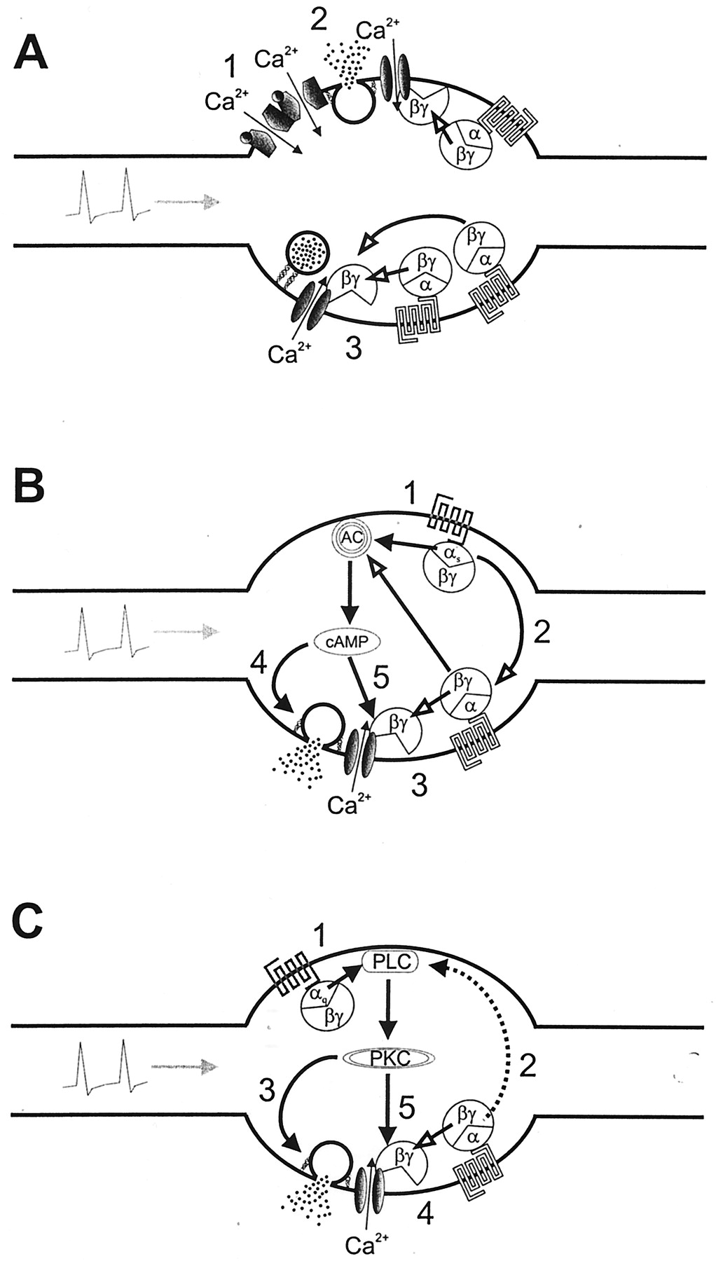
Mechanisms of interactions between presynaptic receptors. A, interactions between ionotropic and inhibitory metabotropic receptors, on one hand, and between two different inhibitory metabotropic receptors, on the other hand. (1) Nicotinic acetylcholine receptors and ATP P2X receptors interact with each other in a mutually inhibitory manner, so that transmitter release triggered by the activation of one of these two receptors will not be additive to the secretagogue action of the other receptor. This interaction is most likely mediated by direct protein-protein interactions (Khakh et al., 2000). (2) When transmitter release is evoked by activation of either nicotinic acetylcholine receptors or ATP P2X receptors, activation of some inhibitory metabotropic presynaptic receptor will not cause a reduction of stimulation-evoked release (e.g., Boehm et al., 1995). The reason for this is the fact that the Ca2+ required for exocytosis enters the axon terminal via the ligand-gated ion channels and not via voltage-gated Ca2+ channels (see Fig. 2). Therefore, the G protein-mediated closure of voltage-gated Ca2+ channels will not affect transmitter release. (3) Activation of two or even more inhibitory metabotropic presynaptic receptors does not cause more inhibition of transmitter release than the activation of only one of these receptors. The predominant mechanism in receptor-dependent presynaptic inhibition of sympathetic transmitter release is the blockade of voltage-gated Ca2+channels by G protein βγ-subunits (see also Fig. 4). However, this protein-protein interaction occurs at a 1:1 stoichiometry (Zamponi and Snutch, 1998). As a consequence, G protein activation will be ineffective once all of the Ca2+ channels are occupied by βγ-subunits. B, interactions between facilitatory Gs-coupled receptors and inhibitory metabotropic receptors. (1) The activation of metabotropic receptors linked to Gstype G proteins typically leads to the activation of adenylyl cyclase and the subsequent accumulation of cyclic AMP (see also Fig. 3). (2) In addition, G protein αs-subunits may exert a tonic inhibitory action on the signaling pathway of inhibitory receptors, as exemplified by α2-adrenoceptors. This effect is independent of changes in cyclic AMP (Boehm et al., 1996b). (3) The activation of inhibitory metabotropic receptors causes a blockade of voltage-gated Ca2+ channels and, thereby, diminishes transmitter release (see also Fig. 4). (4) The elevation in cyclic AMP, in contrast, will cause an increase in transmitter release through an action on the exocytotic apparatus (see also Fig. 3). (5) In addition, increases in cyclic AMP may interfere with the G protein-mediated inhibition of voltage-gated Ca2+ channels and, thereby, attenuate the reduction of transmitter release via inhibitory presynaptic receptors (Boehm et al., 1994). C, interactions between facilitatory Gq-coupled receptors and inhibitory metabotropic receptors. (1) The activation of metabotropic receptors linked to Gq type G proteins typically leads to the activation of phospholipase C via αq-subunits. (2) This enzyme can also be activated via G protein βγ-subunits derived, for instance, from inhibitory presynaptic receptors. By such a mechanism, activation of inhibitory receptors may support the facilitatory action of Gq-coupled receptors (e.g., Cox et al., 2000). (3) The activation of protein kinase C, for instance through diacylglycerol, will augment transmitter release by an action on the exocytotic machinery (see also Fig. 3). (4) The activation of inhibitory metabotropic receptors, in contrast, causes a blockade of voltage-gated Ca2+ channels by G protein βγ-subunits and, thereby, diminishes transmitter release (see also Fig. 4). (5) Activation of protein kinase C prevents the inhibitory effect of G protein βγ-subunits on Ca2+ channels (Zamponi and Snutch, 1998) and, thereby, may counteract the reduction of transmitter release via inhibitory presynaptic receptors (e.g., Cox et al., 2000).
The majority of interactions occur between two different types of metabotropic receptors, and these receptors may utilize a plethora of signaling cascades. Therefore, these interactions can involve diverse signaling mechanisms that finally converge to precisely control the amount of sympathetic transmitter being released (Fig. 5). As mentioned above, activation of presynaptic β-adrenoceptors may facilitate sympathetic transmitter release most likely via the synthesis of cyclic AMP. This action may be antagonized by the activation of receptors that inhibit adenylyl cyclases, such as α2-adrenoceptors, even though this signaling mechanism is not involved in the inhibition of transmitter release via these receptors (see Signaling Mechanisms of Inhibitory Metabotropic Receptors). In support of this competition between β- and α2-adrenoceptors at the level of adenylyl cyclases and cyclic AMP, it has been reported that increases in cyclic AMP raised noradrenaline release only when α2-adrenoceptors were blocked (Kazanietz and Enero, 1992).
Competition between the cyclic AMP-dependent signaling cascade and release inhibiting receptors may also occur at the level of voltage-gated Ca2+ channels. In chicken sympathetic neurons, an increase in cyclic AMP was found to counteract the inhibition of Ca2+ currents and of transmitter release, both via α2-adrenoceptors. This interaction did not arise at the level of adenylyl cyclases, because α2-adrenoceptor activation failed to inhibit adenylyl cyclase activity (Boehm et al., 1994). Hence, an activation of cyclic AMP-dependent signaling cascades, whether by receptor or direct adenylyl cyclase activation, may not only raise transmitter release by a mechanism downstream of Ca2+ entry, but may also attenuate the action of receptors that inhibit release. An additional competition between presynaptic receptors that stimulate transmitter release via increases in cyclic AMP and receptors that inhibit transmitter release has been detected at the level of G proteins. Whereas the first type of receptor operates through Gs proteins, the latter type of receptor typically involves pertussis toxin-sensitive G proteins. The removal of α-subunits of Gs-type G proteins from chicken sympathetic neurons in primary cell cultures (which can be achieved by a pretreatment with cholera toxin; see Chang and Bourne, 1989) caused sensitization of the α2-adrenoceptor-mediated inhibition of transmitter release. This effect did not involve cyclic AMP nor changes in any G protein subunit other than αs. Hence, the loss of stimulatory G protein α-subunits led to a sensitization of the inhibitory G protein-dependent signaling pathway (Boehm et al., 1996b). This indicates that G protein αs-subunits may mediate a tonic attenuation of the signaling cascade that is involved in the receptor-dependent presynaptic inhibition of sympathetic transmitter release (Fig. 5B).
M1 muscarinic receptors, B2bradykinin, and AT1 angiotensin receptors facilitate sympathetic transmitter release most likely through an activation of protein kinase C (see Signaling Mechanisms of Facilitatory Metabotropic Receptors). Frequently, this facilitation was lost when presynaptic α2-autoreceptors were blocked, i.e., when ongoing autoinhibition of transmitter release via these receptors was prevented. These results can be interpreted in at least two different ways. Either the activation of receptors linked to protein kinase C prevents the inhibitory action of α2-adrenoceptors, or α2-adrenoceptor activation is required to support the facilitatory effect of receptors linked to protein kinase C. The first type of interaction can be explained by results concerning the G protein-dependent modulation of voltage-gated Ca2+ channels. Activation of protein kinase C has been shown to prevent the inhibitory interaction of G protein βγ-subunits with N-type Ca2+ channels (Zamponi and Snutch, 1998) and, thus, abolishes the reduction of Ca2+ currents in sympathetic neurons via, for instance, α2-adrenoceptors and somatostatin receptors (Shapiro et al., 1996). Because closure of voltage-gated Ca2+ channels is believed to be the crucial step in presynaptic inhibition, this effect will impede the receptor-dependent inhibition and, thus, lead to an increase in transmitter release. The second type of interaction can be explained at the level of phospholipases C. These enzymes may be activated by G protein α-subunits of the Gq family and by βγ-subunits. These latter types of G protein subunits may derive from pertussis toxin-sensitive and -insensitive heterotrimers, which may be activated by a large variety of different metabotropic receptors (Exton, 1996). Hence, α2-adrenoceptor-dependent liberation of G protein βγ-subunits may synergize with a Gq-dependent activation of phospholipase C and, thereby, support the facilitatory effects mediated by receptors linked to this latter family of G proteins (Fig. 5C).
Interactions between two different inhibitory presynaptic receptors colocalized on sympathetic axon terminals typically occur in the way that activation of one of these receptors prevents the inhibitory action of the other one. This can again be interpreted in at least two ways. Either one of these inhibitory receptors interrupts the signaling cascade of the other one, or these two receptors share one common signaling cascade that becomes maximally stimulated by activation of only one of these two receptors. As stated above, there is good evidence to suggest that the G protein-mediated inhibition of voltage-gated Ca2+ channels is the predominant mechanism underlying receptor-dependent presynaptic inhibition. As indicated in Table 10, a considerable number of different receptors employ pertussis toxin-sensitive signaling cascades to block N-type Ca2+ channels (see also Hille, 1994), and N-type Ca2+ channels are the predominant channel subtype involved in excitation-secretion coupling in sympathetic axon terminals. Activation of two such receptors at the same time does not cause more inhibition of Ca2+ currents than the activation of one receptor only, as shown, for instance, for α2-adrenoceptors and somatostatin receptors in chicken sympathetic neurons (Golard and Siegelbaum, 1993). This appears even more remarkable as these two receptors use different signaling cascades to inhibit the Ca2+ channels: the inhibitory effect of α2-adrenoceptor activation involves an atypical, phorbol ester-insensitive protein kinase C, whereas somatostatin receptor activation does not (Boehm et al., 1996). At the level of noradrenaline release, there is again no additivity of the inhibitory actions mediated by these two receptors (Boehm and Huck, 1996a). Thus, the signaling cascades of different inhibitory presynaptic receptors appear to converge at the level of voltage-gated Ca2+ channels, which mediate the transmembrane Ca2+ entry required for triggered vesicle exocytosis. Therefore, activation of one of these inhibitory receptors will occlude an inhibitory action of another one (Fig. 5A).
X. Conclusion: Signal Integration in the Sympathetic Varicosity
At least 30 different metabotropic and four different ionotropic receptors have been found to control the amount of transmitter being released from a sympathetic axon terminal. The fact that one presynaptic receptor may not only stimulate, facilitate, or reduce transmitter release, but also interact with the functions of all other presynaptic receptors, provides a plethora of mechanisms that contribute to the regulation of sympathetic transmitter output. The possible simultaneous presence of various extracellular factors derived either from the sympathetic varicosity, from a nearby nonsympathetic nerve terminal, or from the target organ will activate one or more of the signaling cascades mentioned above at the same time and, thereby, add to the complexity of sympathoeffector transmission. Below, we will consider three examples to highlight how cross-talk within and between extra- and intracellular signals may be integrated at sympathetic axon terminals to adapt the strength of sympathoeffector transmission to a given situation.
1) ATP released from axon terminals may activate stimulatory P2X and inhibitory P2Y receptors and, thereby, cause positive as well as negative feedback modulation. Because the G protein-coupled P2Y receptors display a higher affinity for ATP than do P2X receptors (Ralevic and Burnstock, 1998), higher concentrations of the nucleotide will be required to enable the positive feedback. However, as soon as these high concentrations are reached and P2X receptors are activated, the negative feedback will be largely disabled because activated G proteins inhibit Ca2+ entry via voltage-gated Ca2+ channels but not that via P2X receptors (Fig. 5A). Together, these mechanisms will provide presynaptic amplification to guarantee efficient sympathoeffector transmission during neuronal activity. Nevertheless, this presynaptic amplification is self-limiting for two reasons. First, together with ATP, sympathetic axon terminals release nucleotidases, which rapidly hydrolyze the nucleotide (Todorov et al., 1997). Second, P2X receptors will desensitize in the presence of ATP, although to a varying degree depending on the receptor subtype (Ralevic and Burnstock, 1998). Thus, P2X receptors will become unresponsive, and ADP will be generated, which is an agonist at several P2Y, but not at P2X, receptors. Consequently, activation of the inhibitory P2Y receptors will lead to a reduction of transmitter release. Taken together, the mechanisms described above may serve to precisely shape neuromuscular transmission at sympathoeffector junctions and, thus, resemble the situation found within the skeletal musculature where presynaptic nicotinic acetylcholine receptors mediate a positive feedback to guarantee the efficiency of transmission and muscarinic receptors mediate a negative feedback to provide a temporal limitation of neuromuscular transmission (Wessler, 1989).
2) Several inhibitory presynaptic receptors are colocalized at axonal varicosities in sympathetically innervated tissues, but the activation of one of these receptors may occlude the action of the other ones (Fig. 5A). At first sight, such a redundancy does not appear to make sense. However, in various pathological conditions this presumed overendowment may provide a safety device to prevent overstimulation. In cardiac ischemia, for instance, α2-adrenoceptors loose their inhibitory action, and noradrenaline output increases, partly due to the contribution of transporter-mediated release. In that situation, presynaptic H3 histamine and A1adenosine receptors, for instance, are still capable of inhibiting sympathetic transmitter release (Imamura et al., 1996), and endogenous adenosine and histamine are believed to restrict the amount of noradrenaline being released (Hatta et al., 1997; Burgdorf et al., 2001). Under physiological conditions, in contrast, an additivity of the inhibitory actions of α2, H3, and A1 receptors may cause an exaggerated depression or even the abolishment of sympathetic transmitter release.
3) Other potentially pathogenetic factors in cardiac ischemia are bradykinin and angiotensin II, which both enhance noradrenaline release via B2 and AT1 receptors, respectively (Hatta et al., 1999; Maruyama et al., 1999). In this respect, the interaction of inhibitory presynaptic receptors with these facilitatory receptors may become cardioprotective. As indicated in Fig. 5C, the facilitatory receptors will loose their function as soon as hetero- or autoinhibitory modulation of sympathetic transmitter release is lacking. Hence, as soon as the inhibitory feedback is pathologically overridden, a further receptor-dependent facilitation of noradrenaline output appears impossible.
Independent of the importance of extra- and intracellular cross-talk at sympathetic axon terminals in physiology and pathology, the various types of interactions should also be borne in mind by experimental pharmacologists as well as by clinicians. For the first, it is important to know that ongoing autoinhibition via α2-adrenoceptors may largely or even completely conceal an eventual inhibitory action of some heteroreceptor (Fig. 5A). On the other hand, a certain extent of autoinhibition may be a prerequisite for the detection of the function of facilitatory heteroreceptors, such as AT1 receptors. In turn, under experimental conditions that minimize autoinhibition, such as the use of primary cell cultures, potentially facilitatory autoreceptors may be overlooked. In the clinical setting, diverse pharmacotherapeutic agents, such as antimigraine drugs like sumatriptan, which activate presynaptic 5-HT1 receptors, prostaglandin derivatives like sulprostone, which activate EP3receptors, or the somatostatin receptor agonist octreotide, which is used in the management of secretory neoplasms or gastrointestinal bleeding, will activate presynaptic heteroreceptors on sympathetic axon terminals. Hence, each of these drugs may interfere with the action of another one. Moreover, the actions of these drugs may be unexpectedly attenuated or reinforced, respectively, by agonists and antagonists at presynaptic autoreceptors, such as, for instance, clonidine and mianserin. Taken together, extra- and intracellular cross-talk at sympathetic axon terminals may not only determine the strength of sympathoeffector transmission in health and sickness, but also the therapeutic and untoward effects of drugs binding to either ionotropic or metabotropic presynaptic receptors.
Acknowledgments
Work in the authors' laboratory is supported by the Austrian Science Fund (Grants P12997, P13920, and P14951), by the Österreichische Nationalbank (Grant 8377), and by the Virologie Fonds of the Medical Faculty of the University of Vienna. We are indebted to Dr. Nina Vartian for help in collecting the literature. The perfect secretarial assistance of Maria Lachmann is gratefully acknowledged.
Footnotes
Address for correspondence: Stefan Boehm, Institute of Pharmacology, University of Vienna, Waehringerstrasse 13a, A-1090 Vienna, Austria. E-mail: Stefan.Boehm{at}univie.ac.at
Abbreviations
- GABA
- γ-aminobutyric acid
- PACAP
- pituitary adenylyl cyclase-activating peptides
- VIP
- vasoactive intestinal peptide
- NPY
- neuropeptide Y
- GMPPNP
- guanosine 5′[βγ-imido] triphosphate)
- GTPγS
- guanosine 5′[γ-thio]triphosphate
- The American Society for Pharmacology and Experimental Therapeutics
References
- ↵
- ↵
- ↵
- ↵
- ↵
- ↵
- ↵
- ↵
- ↵
- ↵
- ↵
- ↵
- ↵
- ↵
- ↵
- ↵
- ↵
- ↵
- ↵
- ↵
- ↵
- ↵
- ↵
- ↵
- ↵
- ↵
- ↵
- ↵
- ↵
- ↵
- ↵
- ↵
- ↵
- ↵
- ↵
- ↵
- ↵
- ↵
- ↵
- ↵
- ↵
- ↵
- ↵
- ↵
- ↵
- ↵
- ↵
- ↵
- ↵
- ↵
- ↵
- ↵
- ↵
- ↵
- ↵
- ↵
- ↵
- ↵
- ↵
- ↵
- ↵
- ↵
- ↵
- ↵
- ↵
- ↵
- ↵
- ↵
- ↵
- ↵
- ↵
- ↵
- ↵
- ↵
- ↵
- ↵
- ↵
- ↵
- ↵
- ↵
- ↵
- ↵
- ↵
- ↵
- ↵
- ↵
- ↵
- ↵
- ↵
- ↵
- ↵
- ↵
- ↵
- ↵
- ↵
- ↵
- ↵
- ↵
- ↵
- ↵
- ↵
- ↵
- ↵
- ↵
- ↵
- ↵
- ↵
- ↵
- ↵
- ↵
- ↵
- ↵
- ↵
- ↵
- ↵
- ↵
- ↵
- ↵
- ↵
- ↵
- ↵
- ↵
- ↵
- ↵
- ↵
- ↵
- ↵
- ↵
- ↵
- ↵
- ↵
- ↵
- ↵
- ↵
- ↵
- ↵
- ↵
- ↵
- ↵
- ↵
- ↵
- ↵
- ↵
- ↵
- ↵
- ↵
- ↵
- ↵
- ↵
- ↵
- ↵
- ↵
- ↵
- ↵
- ↵
- ↵
- ↵
- ↵
- ↵
- ↵
- ↵
- ↵
- ↵
- ↵
- ↵
- ↵
- ↵
- ↵
- ↵
- ↵
- ↵
- ↵
- ↵
- ↵
- ↵
- ↵
- ↵
- ↵
- ↵
- ↵
- ↵
- ↵
- ↵
- ↵
- ↵
- ↵
- ↵
- ↵
- ↵
- ↵
- ↵
- ↵
- ↵
- ↵
- ↵
- ↵
- ↵
- ↵
- ↵
- ↵
- ↵
- ↵
- ↵
- ↵
- ↵
- ↵
- ↵
- ↵
- ↵
- ↵
- ↵
- ↵
- ↵
- ↵
- ↵
- ↵
- ↵
- ↵
- ↵
- ↵
- ↵
- ↵
- ↵
- ↵
- ↵
- ↵
- ↵
- ↵
- ↵
- ↵
- ↵
- ↵
- ↵
- ↵
- ↵
- ↵
- ↵
- ↵
- ↵
- ↵
- ↵
- ↵
- ↵
- ↵
- ↵
- ↵
- ↵
- ↵
- ↵
- ↵
- ↵
- ↵
- ↵
- ↵
- ↵
- ↵
- ↵
- ↵
- ↵
- ↵
- ↵
- ↵
- ↵
- ↵
- ↵
- ↵
- ↵
- ↵
- ↵
- ↵
- ↵
- ↵
- ↵
- ↵
- ↵
- ↵
- ↵
- ↵
- ↵
- ↵
- ↵
- ↵
- ↵
- ↵
- ↵
- ↵
- ↵
- ↵
- ↵
- ↵
- ↵
- ↵
- ↵
- ↵
- ↵
- ↵
- ↵
- ↵
- ↵
- ↵
- ↵
- ↵
- ↵
- ↵
- ↵
- ↵
- ↵
- ↵
- ↵
- ↵
- ↵
- ↵
- ↵
- ↵
- ↵
- ↵
- ↵
- ↵
- ↵
- ↵
- ↵
- ↵
- ↵
- ↵
- ↵
- ↵
- ↵
- ↵
- ↵
- ↵
- ↵
- ↵
- ↵
- ↵
- ↵
- ↵
- ↵
- ↵
- ↵
- ↵
- ↵
- ↵
- ↵
- ↵
- ↵
- ↵
- ↵
- ↵
- ↵
- ↵
- ↵
- ↵
- ↵
- ↵
- ↵
- ↵
- ↵
- ↵
- ↵
- ↵
- ↵
- ↵
- ↵
- ↵
- ↵
- ↵
- ↵
- ↵
- ↵
- ↵
- ↵
- ↵
- ↵
- ↵
- ↵
- ↵
- ↵
- ↵
- ↵
- ↵
- ↵
- ↵
- ↵
- ↵
- ↵
- ↵
- ↵
- ↵
- ↵
- ↵
- ↵
- ↵
- ↵
- ↵
- ↵
- ↵
- ↵
- ↵
- ↵
- ↵
- ↵
- ↵
- ↵
- ↵
- ↵
- ↵
- ↵
- ↵
- ↵
- ↵
- ↵
- ↵
- ↵
- ↵
- ↵
- ↵
- ↵
- ↵
- ↵
- ↵
- ↵
- ↵
- ↵
- ↵
- ↵
- ↵
- ↵
- ↵
- ↵
- ↵
- ↵
- ↵
- ↵
- ↵
- ↵
- ↵
- ↵
- ↵
- ↵
- ↵
- ↵
- ↵
- ↵
- ↵
- ↵
- ↵
- ↵
- ↵
- ↵
- ↵
- ↵
- ↵
- ↵
- ↵
- ↵
- ↵
- ↵
- ↵
- ↵
- ↵
- ↵
- ↵
- ↵
- ↵
- ↵
- ↵
- ↵
- ↵
- ↵
- ↵
- ↵
- ↵
- ↵
- ↵
- ↵
- ↵
- ↵
- ↵
- ↵
- ↵
- ↵
- ↵
- ↵
- ↵
- ↵
- ↵
- ↵
- ↵
- ↵
- ↵
- ↵
- ↵
- ↵
- ↵
- ↵
- ↵
- ↵
- ↵
- ↵
- ↵
- ↵
- ↵
- ↵
- ↵
- ↵
- ↵
- ↵
- ↵
- ↵
- ↵
- ↵
- ↵
- ↵
- ↵
- ↵
- ↵
- ↵
- ↵
- ↵
- ↵
- ↵
- ↵
- ↵
- ↵
- ↵
- ↵
- ↵
- ↵
- ↵
In this issue


Jump to section
- Article
- Abstract
- I. Introduction
- II. Anatomical and Functional Organization of the Sympathetic Nervous System
- III. Methodological Considerations
- IV. Ionotropic Receptors
- V. Metabotropic Receptors
- VI. Basic Mechanisms of Vesicular Sympathetic Transmitter Release
- VII. Signaling Mechanisms of Presynaptic Ionotropic Receptors
- VIII. Signaling Mechanisms of Presynaptic Metabotropic Receptors
- IX. Interactions between Presynaptic Receptors
- X. Conclusion: Signal Integration in the Sympathetic Varicosity
- Acknowledgments
- Footnotes
- Abbreviations
- References
- Figures & Data
- Info & Metrics
- eLetters