Advertisement

Main menu

  • Home
  • Articles
    • Current Issue
    • Fast Forward
    • Latest Articles
    • Special Sections
    • Archive
  • Information
    • Instructions to Authors
    • Submit a Manuscript
    • FAQs
    • For Subscribers
    • Terms & Conditions of Use
    • Permissions
  • Editorial Board
  • Alerts
    • Alerts
    • RSS Feeds
  • Virtual Issues
  • Feedback
  • Submit
  • Other Publications
    • Drug Metabolism and Disposition
    • Journal of Pharmacology and Experimental Therapeutics
    • Molecular Pharmacology
    • Pharmacological Reviews
    • Pharmacology Research & Perspectives
    • ASPET

User menu

  • My alerts
  • Log in
  • Log out
  • My Cart

Search

  • Advanced search
Molecular Pharmacology
  • Other Publications
    • Drug Metabolism and Disposition
    • Journal of Pharmacology and Experimental Therapeutics
    • Molecular Pharmacology
    • Pharmacological Reviews
    • Pharmacology Research & Perspectives
    • ASPET
  • My alerts
  • Log in
  • Log out
  • My Cart
Molecular Pharmacology

Advanced Search

  • Home
  • Articles
    • Current Issue
    • Fast Forward
    • Latest Articles
    • Special Sections
    • Archive
  • Information
    • Instructions to Authors
    • Submit a Manuscript
    • FAQs
    • For Subscribers
    • Terms & Conditions of Use
    • Permissions
  • Editorial Board
  • Alerts
    • Alerts
    • RSS Feeds
  • Virtual Issues
  • Feedback
  • Submit
  • Visit molpharm on Facebook
  • Follow molpharm on Twitter
  • Follow molpharm on LinkedIn
Rapid CommunicationSpecial Issue Commemorating the 50th Anniversary of Molecular Pharmacology

G Protein–Coupled Receptor (GPCR) Expression in Native Cells: “Novel” endoGPCRs as Physiologic Regulators and Therapeutic Targets

Paul A. Insel, Andrea Wilderman, Alexander C. Zambon, Aaron N. Snead, Fiona Murray, Nakon Aroonsakool, Daniel S. McDonald, Shu Zhou, Thalia McCann, Lingzhi Zhang, Krishna Sriram, Amy M. Chinn, Alexander V. Michkov, Rebecca M. Lynch, Aaron C. Overland and Ross Corriden
Molecular Pharmacology July 2015, 88 (1) 181-187; DOI: https://doi.org/10.1124/mol.115.098129
Paul A. Insel
Departments of Pharmacology (P.A.I., A.W., A.C.Z., A.N.S., N.A., D.S.M., S.Z., T.M., L.Z., K.S., A.M.C., A.V.M., R.M.L., A.C.O., R.C.) and Medicine (P.A.I., F.M.), University of California, San Diego, La Jolla, California
  • Find this author on Google Scholar
  • Find this author on PubMed
  • Search for this author on this site
Andrea Wilderman
Departments of Pharmacology (P.A.I., A.W., A.C.Z., A.N.S., N.A., D.S.M., S.Z., T.M., L.Z., K.S., A.M.C., A.V.M., R.M.L., A.C.O., R.C.) and Medicine (P.A.I., F.M.), University of California, San Diego, La Jolla, California
  • Find this author on Google Scholar
  • Find this author on PubMed
  • Search for this author on this site
Alexander C. Zambon
Departments of Pharmacology (P.A.I., A.W., A.C.Z., A.N.S., N.A., D.S.M., S.Z., T.M., L.Z., K.S., A.M.C., A.V.M., R.M.L., A.C.O., R.C.) and Medicine (P.A.I., F.M.), University of California, San Diego, La Jolla, California
  • Find this author on Google Scholar
  • Find this author on PubMed
  • Search for this author on this site
Aaron N. Snead
Departments of Pharmacology (P.A.I., A.W., A.C.Z., A.N.S., N.A., D.S.M., S.Z., T.M., L.Z., K.S., A.M.C., A.V.M., R.M.L., A.C.O., R.C.) and Medicine (P.A.I., F.M.), University of California, San Diego, La Jolla, California
  • Find this author on Google Scholar
  • Find this author on PubMed
  • Search for this author on this site
Fiona Murray
Departments of Pharmacology (P.A.I., A.W., A.C.Z., A.N.S., N.A., D.S.M., S.Z., T.M., L.Z., K.S., A.M.C., A.V.M., R.M.L., A.C.O., R.C.) and Medicine (P.A.I., F.M.), University of California, San Diego, La Jolla, California
  • Find this author on Google Scholar
  • Find this author on PubMed
  • Search for this author on this site
Nakon Aroonsakool
Departments of Pharmacology (P.A.I., A.W., A.C.Z., A.N.S., N.A., D.S.M., S.Z., T.M., L.Z., K.S., A.M.C., A.V.M., R.M.L., A.C.O., R.C.) and Medicine (P.A.I., F.M.), University of California, San Diego, La Jolla, California
  • Find this author on Google Scholar
  • Find this author on PubMed
  • Search for this author on this site
Daniel S. McDonald
Departments of Pharmacology (P.A.I., A.W., A.C.Z., A.N.S., N.A., D.S.M., S.Z., T.M., L.Z., K.S., A.M.C., A.V.M., R.M.L., A.C.O., R.C.) and Medicine (P.A.I., F.M.), University of California, San Diego, La Jolla, California
  • Find this author on Google Scholar
  • Find this author on PubMed
  • Search for this author on this site
Shu Zhou
Departments of Pharmacology (P.A.I., A.W., A.C.Z., A.N.S., N.A., D.S.M., S.Z., T.M., L.Z., K.S., A.M.C., A.V.M., R.M.L., A.C.O., R.C.) and Medicine (P.A.I., F.M.), University of California, San Diego, La Jolla, California
  • Find this author on Google Scholar
  • Find this author on PubMed
  • Search for this author on this site
Thalia McCann
Departments of Pharmacology (P.A.I., A.W., A.C.Z., A.N.S., N.A., D.S.M., S.Z., T.M., L.Z., K.S., A.M.C., A.V.M., R.M.L., A.C.O., R.C.) and Medicine (P.A.I., F.M.), University of California, San Diego, La Jolla, California
  • Find this author on Google Scholar
  • Find this author on PubMed
  • Search for this author on this site
Lingzhi Zhang
Departments of Pharmacology (P.A.I., A.W., A.C.Z., A.N.S., N.A., D.S.M., S.Z., T.M., L.Z., K.S., A.M.C., A.V.M., R.M.L., A.C.O., R.C.) and Medicine (P.A.I., F.M.), University of California, San Diego, La Jolla, California
  • Find this author on Google Scholar
  • Find this author on PubMed
  • Search for this author on this site
Krishna Sriram
Departments of Pharmacology (P.A.I., A.W., A.C.Z., A.N.S., N.A., D.S.M., S.Z., T.M., L.Z., K.S., A.M.C., A.V.M., R.M.L., A.C.O., R.C.) and Medicine (P.A.I., F.M.), University of California, San Diego, La Jolla, California
  • Find this author on Google Scholar
  • Find this author on PubMed
  • Search for this author on this site
Amy M. Chinn
Departments of Pharmacology (P.A.I., A.W., A.C.Z., A.N.S., N.A., D.S.M., S.Z., T.M., L.Z., K.S., A.M.C., A.V.M., R.M.L., A.C.O., R.C.) and Medicine (P.A.I., F.M.), University of California, San Diego, La Jolla, California
  • Find this author on Google Scholar
  • Find this author on PubMed
  • Search for this author on this site
Alexander V. Michkov
Departments of Pharmacology (P.A.I., A.W., A.C.Z., A.N.S., N.A., D.S.M., S.Z., T.M., L.Z., K.S., A.M.C., A.V.M., R.M.L., A.C.O., R.C.) and Medicine (P.A.I., F.M.), University of California, San Diego, La Jolla, California
  • Find this author on Google Scholar
  • Find this author on PubMed
  • Search for this author on this site
Rebecca M. Lynch
Departments of Pharmacology (P.A.I., A.W., A.C.Z., A.N.S., N.A., D.S.M., S.Z., T.M., L.Z., K.S., A.M.C., A.V.M., R.M.L., A.C.O., R.C.) and Medicine (P.A.I., F.M.), University of California, San Diego, La Jolla, California
  • Find this author on Google Scholar
  • Find this author on PubMed
  • Search for this author on this site
Aaron C. Overland
Departments of Pharmacology (P.A.I., A.W., A.C.Z., A.N.S., N.A., D.S.M., S.Z., T.M., L.Z., K.S., A.M.C., A.V.M., R.M.L., A.C.O., R.C.) and Medicine (P.A.I., F.M.), University of California, San Diego, La Jolla, California
  • Find this author on Google Scholar
  • Find this author on PubMed
  • Search for this author on this site
Ross Corriden
Departments of Pharmacology (P.A.I., A.W., A.C.Z., A.N.S., N.A., D.S.M., S.Z., T.M., L.Z., K.S., A.M.C., A.V.M., R.M.L., A.C.O., R.C.) and Medicine (P.A.I., F.M.), University of California, San Diego, La Jolla, California
  • Find this author on Google Scholar
  • Find this author on PubMed
  • Search for this author on this site
  • Article
  • Figures & Data
  • Info & Metrics
  • eLetters
  • PDF
Loading

Visual Overview

Figure
  • Download figure
  • Open in new tab
  • Download powerpoint

Abstract

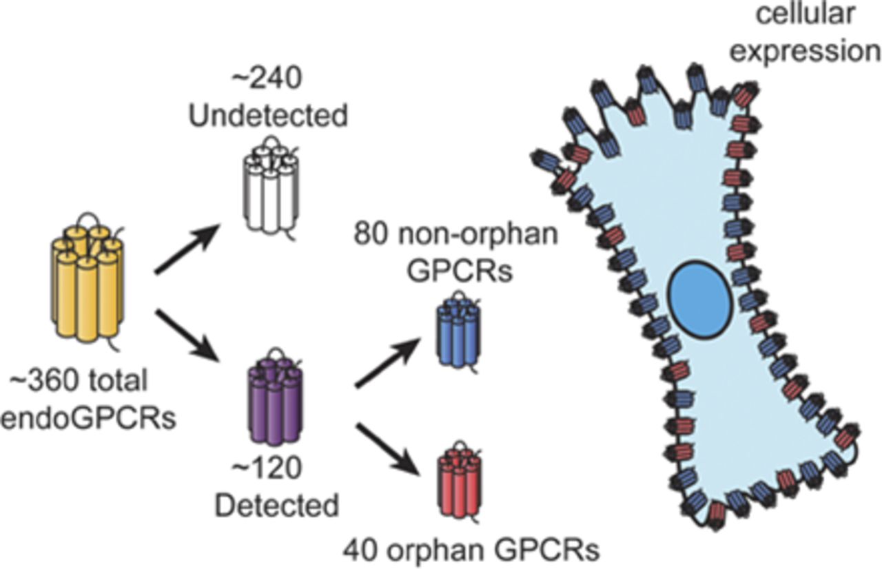
G protein–coupled receptors (GPCRs), the largest family of signaling receptors in the human genome, are also the largest class of targets of approved drugs. Are the optimal GPCRs (in terms of efficacy and safety) currently targeted therapeutically? Especially given the large number (∼120) of orphan GPCRs (which lack known physiologic agonists), it is likely that previously unrecognized GPCRs, especially orphan receptors, regulate cell function and can be therapeutic targets. Knowledge is limited regarding the diversity and identity of GPCRs that are activated by endogenous ligands and that native cells express. Here, we review approaches to define GPCR expression in tissues and cells and results from studies using these approaches. We identify problems with the available data and suggest future ways to identify and validate the physiologic and therapeutic roles of previously unrecognized GPCRs. We propose that a particularly useful approach to identify functionally important GPCRs with therapeutic potential will be to focus on receptors that show selective increases in expression in diseased cells from patients and experimental animals.

Introduction

G protein–coupled receptors (GPCRs, also termed 7-transmembrane or heptahelical receptors) have been of major interest for investigators in many disciplines, including molecular pharmacology. Early studies on GPCRs assessed the action in cells and plasma membrane preparations of neurotransmitters, hormones, and pharmacological agents in terms of their ability to regulate the generation of second messengers (e.g., cAMP, Ca2+) and, in turn, cellular events via enzymes (e.g., protein kinases) and ion channels. Results obtained by the Human Genome Project and for the genomes of other eukaryotes have revealed that GPCRs are the largest family of signaling receptors in humans and other species (Fredriksson et al., 2003; Vassilatis et al., 2003; Insel et al., 2012; Foster et al., 2014b). The receptors include those that interact with endogenous ligands (endoGPCRs); GPCRs regulated by exogenous factors, such as photons of light, odorants, and tastants (chemosensory receptors); and GPCRs that lack known physiologic ligands (termed orphan receptors). It is estimated that among the approximately 800 GPCRs in humans, ∼380 are endoGPCRs, of which about one-third are orphan receptors, even though there have been substantial efforts at deorphanization (Fredriksson et al., 2003; Kroeze et al., 2003; Ozawa et al., 2010; Amisten et al., 2013; Civelli et al., 2013).

In parallel with work that has involved the cloning, genomic characterization, heterologous expression, and studies of GPCR actions and regulation, other efforts have emphasized the utility of GPCRs as therapeutic targets. Indeed, GPCRs are the largest class (∼30%) of the targets of approved drugs (Overington et al., 2006; Lundstrom, 2009; Rask-Andersen et al., 2014). Reasons for the utility of GPCRs as therapeutic targets include the many different types of chemical entities with which they interact, the accessibility of GPCRs on the plasma membrane from the extracellular milieu, their ability to initiate signaling pathways that undergo amplification in target cells, and the selectivity in their expression by different types of cells. This latter property aids in facilitating tissue- and cell-selective actions of GPCR-targeted drugs.

Despite the widespread use of GPCRs as therapeutic targets, one can ask the following: Are the optimal GPCRs (in terms of efficacy and safety) targeted by current therapeutic approaches? This question derives in part from the current therapeutic targeting of only a fraction of the endoGPCRs. Moreover, orphan GPCRs have largely been ignored as therapeutic targets. It is thus necessary to identify the repertoire of GPCRs—in particular, endoGPCRs—expressed by individual tissues and, more importantly, native cells. Studies to assess this gap in knowledge test the hypothesis that certain GPCRs are enriched in native cells, regulate cellular (and tissue) function, and can be targeted therapeutically. In this article, we review the approaches and data that have begun to provide information to test this hypothesis. In addition, we discuss problems and limitations of available data and future directions that may help definitively answer the question posed earlier.

Methods and Approaches to Assess GPCR Expression

Analyses of functional responses, second messengers, or other signaling events represent hypothesis-testing approaches by asking if a particular receptor is biologically active and provide indirect ways to assess GPCR expression by tissues and cells. Radioligand binding assays facilitate the direct identification and quantification of GPCRs. However, functional, signaling, and radioligand binding assays are biased approaches: one chooses a GPCR of interest and then uses agonists, antagonists, and radioligands for the receptor being assessed. Thus, one can only study receptors for which appropriate reagents are available.

By contrast, hypothesis-generating approaches are not based on prior knowledge of a GPCR being present, but instead rely on unbiased analyses of the expression of receptor mRNA or protein. Such approaches can define the GPCR expression profile/repertoire and can quantify receptor expression. Table 1 lists several approaches used to assess GPCR expression.

View this table:
  • View inline
  • View popup
TABLE 1

Unbiased methods to assess expression of GPCRs

Numerous studies have used DNA microarrays (“DNA chips”) to define the transcriptomes of cells and tissues. Such microarrays contain probes (specific DNA sequences) that hybridize with the genes of humans, mice, or other species. Hybridization of the probes to target genes is quantified by chemiluminescence, fluorescence, or another method, facilitating quantitation of the abundance of individual mRNAs/cDNAs. Commercially available microarrays that assess most or all genes in a transcriptome are not optimized to detect GPCRs, but such arrays have been used to characterize GPCR expression.

Proprietary and commercial GPCR microarrays, to be discussed later, and real-time polymerase chain reaction (PCR) analyses for individual GPCRs offer an alternative approach to identify chemosensory and nonchemosensory GPCRs. For example, Regard et al. (2008) used Taqman quantitative real-time PCR to quantify the transcripts of 353 nonodorant GPCRs in 41 adult mouse tissues and predicted previously unanticipated roles for less well studied receptors—an idea consistent with our hypothesis that previously unrecognized GPCRs are enriched in native cells, contribute to physiology, and are potential therapeutic targets.

With improvement in the ability to perform sequencing and a rapid decrease in its cost, new techniques such as high-resolution RNA sequencing have begun to be used to identify and quantify expression of GPCRs and other members of the transcriptome. Such studies have recently included the profiling of GPCRs expressed in single cells (Manteniotis et al., 2013; Spaethling et al., 2014).

An alternative to assessing the expression of GPCR mRNA is the use of unbiased proteomic approaches, such as mass spectrometry. Although such technology has not, as yet, been used to define and quantify overall GPCR expression in cells and tissues, initial results suggest this may be a feasible approach (Eisen et al., 2013; Feve et al., 2014).

Microarrays for the Detection of GPCRs

Commercial microarrays have been created that are optimized to detect and quantify the mRNA of ∼350 nonchemosensory GPCRs of mice, rats, and humans. We and others have found these arrays to be quite useful to assess GPCR expression. Figure 1 shows a comparison of the detection of GPCRs by such a targeted array (a Taqman GeneSignature array; Life Technologies, Carlsbad, CA) with results obtained using an Affymetrix Mouse Genome 430A microarray (Affymetrix, Santa Clara, CA) (which detects ∼14,000 mouse genes). We found that highly expressed GPCRs on the latter microarray show a positive correlation (and an R2 of 0.37) with the GPCR array data. However, assessment of GPCRs expressed at lower levels reveals that the Affymetrix array has many false-positive and false-negative results (and R2 = 0.01). GPCR arrays thus seem to be more useful to detect and quantify GPCRs than nontargeted arrays. Data from others support this conclusion (Maurel et al., 2011).

Fig. 1.
  • Download figure
  • Open in new tab
  • Download powerpoint
Fig. 1.

Comparison of GPCR expression data obtained using a GPCR-specific array and a generic cDNA array. The data were collected in two experiments, with data obtained using cDNA prepared from murine wild-type S49 lymphoma cells and that could be compared for GPCRs present on an Affymetrix Mouse Genome 430A microarray and a Taqman GeneSignature array. The 32 GPCRs highly expressed (>log26) on the Affymetrix array show a positive correlation (R2 = 0.37) between the two arrays. GPCRs expressed at lower levels (<log26) show numerous false-positive and false-negative results in the detection of GPCRs by the Affymetrix array compared with the GPCR array (R2 = 0.01). ΔCT, cycle threshold relative to the mRNA used to normalize expression of each GPCR; GCRMA, guanine cytosine robust multiarray analysis.

What Do Available Data Reveal about GPCR Expression in Tissues and Native Cells?

GPCR expression has thus far been determined in a number of tissues (Table 2) and a smaller number of individual cell types (Table 3). Our laboratory has assessed the expression of nonchemosensory GPCRs by numerous individual cell types, including lymphoid cells, dendritic cells, neutrophils, vascular smooth muscle cells, several types of fibroblasts, adipocytes, renal epithelial cells, trigeminal neurons, and several types of cancer cells.

View this table:
  • View inline
  • View popup
TABLE 2

GPCR expression in tissues

View this table:
  • View inline
  • View popup
TABLE 3

GPCR expression in cells

The notion that GPCRs and GPCR signaling pathways are altered (and are potential therapeutic targets) in cancer in addition to various endocrine tumors (for which GPCR-targeted drugs are commonly used) has been largely ignored by investigators in oncology. Even so, several recent reviews have emphasized the importance of GPCRs and GPCR signaling in cancer (Lappano and Maggiolini, 2011; Feigin, 2013; O'Hayre et al., 2013, 2014). O’Hayre and colleagues (2013) noted that nearly 20% of human tumors have GPCR mutations, 4% of tumors have activating mutations of the Gαs gene, and activating mutations of Gαq family members occur in melanomas. GPCRs may contribute to cancer not only by effects on the growth, death, metabolism, and function of malignant cells, but also by actions on cells of the tumor microenvironment, including vascular cells, immune cells, and cancer-associated fibroblasts. Although not widely explored, such actions may have functional importance for the malignant phenotype (Hanahan and Weinberg, 2011) and thus have therapeutic potential.

Several general conclusions regarding GPCR expression in tissues and cells can be drawn from published findings and our unpublished results:

  1. Most tissues and individual cell types express at least 100 different GPCRs, including GPCRs that link to each of the major classes of heterotrimeric G proteins (Gs, Gi/o, Gq/11, and G12/13).

  2. Among the GPCRs with the highest expression are those not previously known to be expressed in the previously mentioned tissues and cells and, thus, not the subject of prior studies.

  3. Many of the highest expressed GPCRs are orphan receptors.

  4. Cells from animals or patients with particular diseases show prominent changes (increases and decreases) in expression of numerous GPCRs, thus revealing disease-specific changes in GPCR expression.

  5. Additional methods [quantitative real-time PCR, antibody-based and functional (including signal transduction and cellular response) assays]) confirm data obtained with GPCR arrays, including, for example, evidence that the expression of several Gs-coupled GPCRs correlates with their ability to maximally stimulate cellular cAMP production.

Such findings thus indicate that analysis of GPCR expression of tissues, and especially of individual cell types, appears to be a highly useful means to identify previously unrecognized GPCRs that may be functional, contribute to pathophysiology, and serve as novel drug targets.

What Are Some of the Problems and Limitations of Efforts to Define GPCR Expression in Tissues and Cells?

Despite the potential importance of the findings related to the discovery of “new GPCRs,” i.e., endoGPCRs not previously known to be expressed, in individual tissues and cell types, a number of issues must be considered in studies of GPCR expression:

  1. The source of material analyzed for GPCR expression: The results in Table 2 were primarily derived from studies of whole organs, tissues, and tissue biopsies, all of which are heterogeneous cellular preparations. The contribution of the different cell types in such preparations is thus not clear. Studies of individual cell types (Table 3) obviate this concern. Even in such studies, however, one analyzes a population of cells. The use of techniques such as high-resolution RNA sequencing (Manteniotis et al., 2013; Spaethling et al., 2014) and perhaps proteomic methods (Davies et al., 2007; Wu et al., 2012; Eisen et al., 2013) to assess GPCRs in individual cells will thus be an important future direction for defining the profile and variability in GPCR expression in particular cell types.

  2. Reproducibility and consistency of methods used to prepare mRNA, cDNA, or protein and to assess gene and protein expression: Standards have been developed to facilitate reproducibility in microarray (Brazma et al., 2001; Hansen et al., 2007; Rung and Brazma, 2013) and proteomic studies (Davies et al., 2007; Taylor et al., 2007), but results from different laboratories can differ. Such differences are generally attributed to variations in the methods used for the preparation and processing of samples. Primer design may not be ideal for the detection of all relevant GPCRs by arrays. For example, Taqman-based GPCR arrays may not detect certain, including functionally relevant, GPCRs. The use of different approaches for data analysis and statistics by different laboratories can contribute to discordant results (Pan, 2002; Motulsky, 2014). The dearth of well validated antibodies to detect native GPCRs, including for use in proteomic studies, is an important problem in GPCR research (Hutchings et al., 2010; Talmont et al., 2012; Eisen et al., 2013; Marchalant et al., 2014). Use of antibodies to study GPCRs is challenging because the receptors are typically expressed in cells at lower levels than many other cellular proteins. Criteria, including studies with cells or animals engineered to have a knockout of a particular GPCR, have been proposed to help validate GPCR-targeted antibodies (Michel et al., 2009). Moreover, since protein levels in cells are not necessarily predicted by mRNA abundance, differences in protein expression could derive from factors that include altered protein translation and/or degradation (de Sousa Abreu et al., 2009; Maier et al., 2009). Especially given the difficulties in obtaining well validated GPCR antibodies, a possible solution is to measure levels of actively translated mRNA using polysome profile analysis together with DNA high-density arrays or GPCR-specific quantitative PCR (Mašek et al., 2011; Gandin et al., 2014).

  3. Normalization of results for GPCR expression: Most assays used to determine mRNA and protein expression rely on normalization to “housekeeping” genes/proteins. An extensive literature has discussed problems related to normalization in such studies. One such problem is a change in expression of the gene/protein used for normalization—a particular concern in studies of development, differentiation, or disease (Khimani et al., 2005; Brattelid et al., 2007). Two issues in the use of arrays to assess GPCR expression are how to define the limit of detection of a receptor in terms of ∆cycle-threshold [∆C(t)] relative to the reference used for normalization, and the most appropriate statistical tests for data analysis (Khimani et al., 2005; Rubie et al., 2005).

  4. Approaches used to classify GPCRs in profiles of their expression in tissues and cells: One approach is to cluster results for GPCR expression on the basis of the coupling of receptors to heterotrimeric G proteins. Such information is available in articles and databases, such as the International Union of Basic and Clinical Pharmacology/British Pharmacological Society Guide to Pharmacology (Pawson et al., 2014). The International Union of Basic and Clinical Pharmacology/British Pharmacological Society systematically annotates each GPCR and provides reviews from expert subcommittees for each of the target families in the database. Results that supersede such data are published on an ongoing basis; therefore, one must be vigilant in updating conclusions regarding the coupling of GPCRs to G proteins. Problems associated with classifying results on this basis include the evidence that some GPCRs couple to multiple G proteins, the limited data regarding the G protein linkage of orphan GPCRs, and the ability of GPCRs to act via β-arrestin instead of (or in addition to) G proteins. Other ways to classify GPCRs include the Glutamate, Rhodopsin, Adhesion, Frizzled/taste2, and Secretin system, evolutionary relationships, ligand interactions, structural data, and susceptibility to post-translational modifications (Fredriksson et al., 2003; Davies et al., 2007; Secker et al., 2010; Lin et al., 2013; Venkatakrishnan et al., 2013).

  5. Which types of GPCRs should be studied? As shown by the studies in Tables 2 and 3, most efforts have focused on the use of array- or PCR-based methods and have emphasized the expression of nonchemosensory GPCRs. Recent studies, however, have identified chemosensory receptor expression in tissues not typically thought to be in involved in sensation (Reimann et al., 2012; Foster et al., 2014a,b; Pronin et al., 2014; Rajkumar et al., 2014; Malki et al., 2015). With the discovery of small-molecule metabolites [in some cases, products of microbiota (e.g., Natarajan and Pluznick, 2014)] that interact with GPCRs and that may have been considered odorants or tastants, the distinction between chemosensory and nonchemosensory GPCRs is blurring. Thus, future studies may need to incorporate analyses of both types of GPCRs.

  6. Prioritization of results regarding GPCR expression to guide subsequent studies that validate the expression and evaluate the functional role of individual receptors: The discovery that native cells typically express >100 GPCRs creates an embarrassment of riches, but also a challenge in terms of choosing individual GPCRs for subsequent studies. A key goal is to identify GPCRs that are important for cellular function and that may be therapeutic targets. The use of RNA interference and gene editing approaches (i.e., CRISPR/Cas9) to knock down receptor expression in cells of interest and then to assess the impact of receptor knock down on functional activity (signal transduction or cellular responses) provides a way to survey a population of GPCRs and identify physiologically (and potentially pharmacologically) important GPCRs (Willets and Nash, 2013). Additional features to help such prioritization include 1) the level of expression (more highly expressed GPCRs are predicted to contribute to a greater extent to cell function and may be easier to study); 2) knowledge of other cell types that express receptors of potential interest [to achieve greater selectivity in the site of action, including by comparing expression in cells that are related to one another, such as, for example, in different types of macrophages (Lattin et al., 2008; Groot-Kormelink et al., 2012; Hohenhaus et al., 2013)]; 3) choosing GPCRs whose expression is shared in a cell type found in humans and experimental animals (e.g., mice, rats); and 4) availability of reagents (e.g., agonists, antagonists, antibodies) to conduct signaling and functional studies. A useful feature, especially for studies of disease settings, is to focus efforts on GPCRs with prominent differences in expression between the normal cells and cells from diseased animals or patients. Differential GPCR expression can also be helpful in choosing GPCRs for studies of development and differentiation of tissues and cells.

  7. Determining the function of highly expressed orphan GPCRs: Some of the most exciting (and unexpected) data that we have obtained from our studies of GPCR expression in native cells is the high expression of a variety of orphan GPCRs. This is perhaps not surprising since orphan receptors represent about one-third of the endoGPCRs, and limited reagents have been available to assess function mediated by most orphan GPCRs. Since the endogenous ligands for such receptors are unknown, investigating their physiologic roles can be difficult (Ahmad et al., 2014). Molecular approaches, such as RNA interference or overexpression in heterologous cells, can help define signaling mechanisms, especially if a GPCR is constitutively active. Antibodies to orphan GPCRs might be used to block signaling and obviate the need for a ligand, although, as noted earlier, validation of GPCR antibodies can be difficult. Another challenge with orphan GPCRs has been the difficulty in setting up screens for ligands, as the G protein coupling of an orphan may not be known; chimeric G proteins may be a way to develop high-throughput screens (Yin et al., 2004).

Summary and Conclusions

Even though they have been widely studied and highly useful as therapeutic targets, GPCRs continue to be very important molecular entities. The recognition from genomic studies that there are many more GPCRs than were previously known or characterized, and that many of these are orphan receptors provides opportunities to discover physiologic and therapeutic roles for newly recognized GPCRs. Studies of GPCR expression in tissues, but especially in native cells, can reveal that previously unrecognized GPCRs contribute to cell function in health and disease. Numerous challenges exist in studies of GPCR expression, but if “new” GPCRs can be validated and shown to be functionally active, we anticipate that such GPCRs may prove to be as important—and perhaps even more so—than the GPCRs that have been the focus of efforts in physiology and pharmacology, and that are so valuable as therapeutic targets.

Acknowledgments

The authors thank their collaborators in recent efforts of their laboratory related to GPCR expression: Alessandra Franco, Randall French, Thomas Kipps, Jihyung Lee, Andrew Lowy, Victor Nizet, Hemal Patel, Laura Rassenti, Eyal Raz, Patricia Thistlethwaite (all from the University of California, San Diego), Christopher Walker, Debbie Hay (University of Auckland, Auckland, New Zealand), Michael Feigin (Cold Spring Harbor Laboratories, Cold Spring Harbor, NY), Ana Kilic, and Alexander Pfeifer (University of Bonn, Bonn, Germany).

Authorship Contributions

Conducted experiments: Wilderman, Zambon.

Performed data analysis: Wilderman, Zambon.

Wrote or contributed to the writing of the manuscript: Insel, Wilderman, Zambon, Snead, Murray, Aroonsakool, McDonald, Zhou, McCann, Zhang, Sriram, Chinn, Michkov, Lynch, Overland, Corriden.

Footnotes

    • Received January 23, 2015.
    • Accepted March 2, 2015.
  • Work in the authors’ laboratory on this topic has been supported by research and training grants from the National Institutes of Health [Grants CA189477, CA121938, GM68524, HL091061, HL066941, HL007444, and GM-68524], the Department of Defense [Grant W81XWH-14-1-0372], and with financial assistance from Roche, Pfizer-CovX, Bristol Myers Squibb, the American Heart Association, and an ASPET-Astellas Award in Translational Pharmacology.

  • dx.doi.org/10.1124/mol.115.098129.

Abbreviations

endoGPCR
GPCR activated by endogenous ligands
GPCR
G protein–coupled receptor
PCR
polymerase chain reaction
  • Copyright © 2015 by The American Society for Pharmacology and Experimental Therapeutics

References

  1. ↵
    1. Ahmad R,
    2. Wojciech S, and
    3. Jockers R
    (2014) Hunting for the function of orphan GPCRs - beyond the search for the endogenous ligand. Br J Pharmacol, in press.
    1. Amisten S,
    2. Neville M,
    3. Hawkes R,
    4. Persaud SJ,
    5. Karpe F, and
    6. Salehi A
    (2015) An atlas of G-protein coupled receptor expression and function in human subcutaneous adipose tissue. Pharmacol Ther 146:61–93.
    OpenUrlCrossRefPubMed
  2. ↵
    1. Amisten S,
    2. Salehi A,
    3. Rorsman P,
    4. Jones PM, and
    5. Persaud SJ
    (2013) An atlas and functional analysis of G-protein coupled receptors in human islets of Langerhans. Pharmacol Ther 139:359–391.
    OpenUrlCrossRefPubMed
    1. Assie G,
    2. Louiset E,
    3. Sturm N,
    4. René-Corail F,
    5. Groussin L,
    6. Bertherat J,
    7. Thomas M,
    8. Lefebvre H,
    9. Feige JJ,
    10. Clauser E,
    11. et al.
    (2010) Systematic analysis of G protein-coupled receptor gene expression in adrenocorticotropin-independent macronodular adrenocortical hyperplasia identifies novel targets for pharmacological control of adrenal Cushing’s syndrome. J Clin Endocrinol Metab 95:E253–E262.
    OpenUrlCrossRefPubMed
    1. Atwood BK,
    2. Lopez J,
    3. Wager-Miller J,
    4. Mackie K, and
    5. Straiker A
    (2011) Expression of G protein-coupled receptors and related proteins in HEK293, AtT20, BV2, and N18 cell lines as revealed by microarray analysis. BMC Genomics 12:14.
    OpenUrlCrossRefPubMed
  3. ↵
    1. Brattelid T,
    2. Tveit K,
    3. Birkeland JA,
    4. Sjaastad I,
    5. Qvigstad E,
    6. Krobert KA,
    7. Hussain RI,
    8. Skomedal T,
    9. Osnes JB, and
    10. Levy FO
    (2007) Expression of mRNA encoding G protein-coupled receptors involved in congestive heart failure—a quantitative RT-PCR study and the question of normalisation. Basic Res Cardiol 102:198–208.
    OpenUrlCrossRefPubMed
  4. ↵
    1. Brazma A,
    2. Hingamp P,
    3. Quackenbush J,
    4. Sherlock G,
    5. Spellman P,
    6. Stoeckert C,
    7. Aach J,
    8. Ansorge W,
    9. Ball CA,
    10. Causton HC,
    11. et al.
    (2001) Minimum information about a microarray experiment (MIAME)-toward standards for microarray data. Nat Genet 29:365–371.
    OpenUrlCrossRefPubMed
    1. Carr JC,
    2. Boese EA,
    3. Spanheimer PM,
    4. Dahdaleh FS,
    5. Martin M,
    6. Calva D,
    7. Schafer B,
    8. Thole DM,
    9. Braun T,
    10. O’Dorisio TM,
    11. et al.
    (2012) Differentiation of small bowel and pancreatic neuroendocrine tumors by gene-expression profiling. Surgery 152:998–1007.
    OpenUrlCrossRefPubMed
    1. Chen Y,
    2. Palczewska G,
    3. Mustafi D,
    4. Golczak M,
    5. Dong Z,
    6. Sawada O,
    7. Maeda T,
    8. Maeda A, and
    9. Palczewski K
    (2013) Systems pharmacology identifies drug targets for Stargardt disease-associated retinal degeneration. J Clin Invest 123:5119–5134.
    OpenUrlCrossRefPubMed
  5. ↵
    1. Civelli O,
    2. Reinscheid RK,
    3. Zhang Y,
    4. Wang Z,
    5. Fredriksson R, and
    6. Schiöth HB
    (2013) G protein-coupled receptor deorphanizations. Annu Rev Pharmacol Toxicol 53:127–146.
    OpenUrlCrossRefPubMed
  6. ↵
    1. Davies MN,
    2. Gloriam DE,
    3. Secker A,
    4. Freitas AA,
    5. Mendao M,
    6. Timmis J, and
    7. Flower DR
    (2007) Proteomic applications of automated GPCR classification. Proteomics 7:2800–2814.
    OpenUrlCrossRefPubMed
  7. ↵
    1. de Sousa Abreu R,
    2. Penalva LO,
    3. Marcotte EM, and
    4. Vogel C
    (2009) Global signatures of protein and mRNA expression levels. Mol Biosyst 5:1512–1526.
    OpenUrlCrossRefPubMed
  8. ↵
    1. Eisen D,
    2. Planatscher H,
    3. Hardie DB,
    4. Kraushaar U,
    5. Pynn CJ,
    6. Stoll D,
    7. Borchers C,
    8. Joos TO, and
    9. Poetz O
    (2013) G protein-coupled receptor quantification using peptide group-specific enrichment combined with internal peptide standard reporter calibration. J Proteomics 90:85–95.
    OpenUrlCrossRefPubMed
    1. Engelstoft MS,
    2. Park WM,
    3. Sakata I,
    4. Kristensen LV,
    5. Husted AS,
    6. Osborne-Lawrence S,
    7. Piper PK,
    8. Walker AK,
    9. Pedersen MH,
    10. Nøhr MK,
    11. et al.
    (2013) Seven transmembrane G protein-coupled receptor repertoire of gastric ghrelin cells. Mol Metab 2:376–392.
    OpenUrlCrossRefPubMed
  9. ↵
    1. Feigin ME
    (2013) Harnessing the genome for characterization of G-protein coupled receptors in cancer pathogenesis. FEBS J 280:4729–4738.
    OpenUrlCrossRefPubMed
  10. ↵
    1. Fève M,
    2. Saliou JM,
    3. Zeniou M,
    4. Lennon S,
    5. Carapito C,
    6. Dong J,
    7. Van Dorsselaer A,
    8. Junier MP,
    9. Chneiweiss H,
    10. Cianférani S,
    11. et al.
    (2014) Comparative expression study of the endo-G protein coupled receptor (GPCR) repertoire in human glioblastoma cancer stem-like cells, U87-MG cells and non malignant cells of neural origin unveils new potential therapeutic targets. PLoS ONE 9:e91519.
    OpenUrlCrossRefPubMed
  11. ↵
    1. Foster SR,
    2. Blank K,
    3. See Hoe LE,
    4. Behrens M,
    5. Meyerhof W,
    6. Peart JN, and
    7. Thomas WG
    (2014a) Bitter taste receptor agonists elicit G-protein-dependent negative inotropy in the murine heart. FASEB J 28:4497–4508.
    OpenUrlAbstract/FREE Full Text
  12. ↵
    1. Foster SR,
    2. Roura E, and
    3. Thomas WG
    (2014b) Extrasensory perception: odorant and taste receptors beyond the nose and mouth. Pharmacol Ther 142:41–61.
    OpenUrlCrossRefPubMed
  13. ↵
    1. Fredriksson R,
    2. Lagerström MC,
    3. Lundin LG, and
    4. Schiöth HB
    (2003) The G-protein-coupled receptors in the human genome form five main families. Phylogenetic analysis, paralogon groups, and fingerprints. Mol Pharmacol 63:1256–1272.
    OpenUrlAbstract/FREE Full Text
  14. ↵
    1. Gandin V,
    2. Sikström K,
    3. Alain T,
    4. Morita M,
    5. McLaughlan S,
    6. Larsson O, and
    7. Topisirovic I
    (2014) Polysome fractionation and analysis of mammalian translatomes on a genome-wide scale. J Vis Exp (87).
  15. ↵
    1. Groot-Kormelink PJ,
    2. Fawcett L,
    3. Wright PD,
    4. Gosling M, and
    5. Kent TC
    (2012) Quantitative GPCR and ion channel transcriptomics in primary alveolar macrophages and macrophage surrogates. BMC Immunol 13:57.
    OpenUrlCrossRefPubMed
    1. Gugger M,
    2. White R,
    3. Song S,
    4. Waser B,
    5. Cescato R,
    6. Rivière P, and
    7. Reubi JC
    (2008) GPR87 is an overexpressed G-protein coupled receptor in squamous cell carcinoma of the lung. Dis Markers 24:41–50.
    OpenUrlCrossRefPubMed
    1. Hakak Y,
    2. Shrestha D,
    3. Goegel MC,
    4. Behan DP, and
    5. Chalmers DT
    (2003) Global analysis of G-protein-coupled receptor signaling in human tissues. FEBS Lett 550:11–17.
    OpenUrlCrossRefPubMed
  16. ↵
    1. Hanahan D and
    2. Weinberg RA
    (2011) Hallmarks of cancer: the next generation. Cell 144:646–674.
    OpenUrlCrossRefPubMed
  17. ↵
    1. Hansen A,
    2. Chen Y,
    3. Inman JM,
    4. Phan QN,
    5. Qi ZQ,
    6. Xiang CC,
    7. Palkovits M,
    8. Cherman N,
    9. Kuznetsov SA,
    10. Robey PG,
    11. et al.
    (2007) Sensitive and specific method for detecting G protein-coupled receptor mRNAs. Nat Methods 4:35–37.
    OpenUrlCrossRefPubMed
  18. ↵
    1. Hohenhaus DM,
    2. Schaale K,
    3. Le Cao KA,
    4. Seow V,
    5. Iyer A,
    6. Fairlie DP, and
    7. Sweet MJ
    (2013) An mRNA atlas of G protein-coupled receptor expression during primary human monocyte/macrophage differentiation and lipopolysaccharide-mediated activation identifies targetable candidate regulators of inflammation. Immunobiology 218:1345–1353.
    OpenUrlCrossRefPubMed
  19. ↵
    1. Hutchings CJ,
    2. Koglin M, and
    3. Marshall FH
    (2010) Therapeutic antibodies directed at G protein-coupled receptors. MAbs 2:594–606.
    OpenUrlCrossRefPubMed
  20. ↵
    1. Insel PA,
    2. Snead A,
    3. Murray F,
    4. Zhang L,
    5. Yokouchi H,
    6. Katakia T,
    7. Kwon O,
    8. Dimucci D, and
    9. Wilderman A
    (2012) GPCR expression in tissues and cells: are the optimal receptors being used as drug targets? Br J Pharmacol 165:1613–1616.
    OpenUrlCrossRefPubMed
  21. ↵
    1. Khimani AH,
    2. Mhashilkar AM,
    3. Mikulskis A,
    4. O’Malley M,
    5. Liao J,
    6. Golenko EE,
    7. Mayer P,
    8. Chada S,
    9. Killian JB, and
    10. Lott ST
    (2005) Housekeeping genes in cancer: normalization of array data. Biotechniques 38:739–745.
    OpenUrlCrossRefPubMed
  22. ↵
    1. Kroeze WK,
    2. Sheffler DJ, and
    3. Roth BL
    (2003) G-protein-coupled receptors at a glance. J Cell Sci 116:4867–4869.
    OpenUrlFREE Full Text
    1. Kuzumaki N,
    2. Suzuki A,
    3. Narita M,
    4. Hosoya T,
    5. Nagasawa A,
    6. Imai S,
    7. Yamamizu K,
    8. Morita H,
    9. Suzuki T,
    10. Okada Y,
    11. et al.
    (2012) Multiple analyses of G-protein coupled receptor (GPCR) expression in the development of gefitinib-resistance in transforming non-small-cell lung cancer. PLoS ONE 7:e44368.
    OpenUrlCrossRefPubMed
  23. ↵
    1. Lappano R and
    2. Maggiolini M
    (2011) G protein-coupled receptors: novel targets for drug discovery in cancer. Nat Rev Drug Discov 10:47–60.
    OpenUrlCrossRefPubMed
  24. ↵
    1. Lattin JE,
    2. Schroder K,
    3. Su AI,
    4. Walker JR,
    5. Zhang J,
    6. Wiltshire T,
    7. Saijo K,
    8. Glass CK,
    9. Hume DA,
    10. Kellie S,
    11. et al.
    (2008) Expression analysis of G Protein-Coupled Receptors in mouse macrophages. Immunome Res 4:5.
    OpenUrlCrossRefPubMed
    1. Layden BT,
    2. Newman M,
    3. Chen F,
    4. Fisher A, and
    5. Lowe WL Jr.
    (2010) G protein coupled receptors in embryonic stem cells: a role for Gs-alpha signaling. PLoS ONE 5:e9105.
    OpenUrlCrossRefPubMed
    1. Li S,
    2. Huang S, and
    3. Peng SB
    (2005) Overexpression of G protein-coupled receptors in cancer cells: involvement in tumor progression. Int J Oncol 27:1329–1339.
    OpenUrlPubMed
  25. ↵
    1. Lin H,
    2. Sassano MF,
    3. Roth BL, and
    4. Shoichet BK
    (2013) A pharmacological organization of G protein-coupled receptors. Nat Methods 10:140–146.
    OpenUrlCrossRefPubMed
  26. ↵
    1. Lundstrom K
    (2009) An overview on GPCRs and drug discovery: structure-based drug design and structural biology on GPCRs. Methods Mol Biol 552:51–66.
    OpenUrlCrossRefPubMed
  27. ↵
    1. Maier T,
    2. Güell M, and
    3. Serrano L
    (2009) Correlation of mRNA and protein in complex biological samples. FEBS Lett 583:3966–3973.
    OpenUrlCrossRefPubMed
  28. ↵
    1. Malki A,
    2. Fiedler J,
    3. Fricke K,
    4. Ballweg I,
    5. Pfaffl MW, and
    6. Krautwurst D
    (2015) Class I odorant receptors, TAS1R and TAS2R taste receptors, are markers for subpopulations of circulating leukocytes. J Leukoc Biol 97:533–545.
    OpenUrlAbstract/FREE Full Text
  29. ↵
    1. Manteniotis S,
    2. Lehmann R,
    3. Flegel C,
    4. Vogel F,
    5. Hofreuter A,
    6. Schreiner BS,
    7. Altmüller J,
    8. Becker C,
    9. Schöbel N,
    10. Hatt H,
    11. et al.
    (2013) Comprehensive RNA-Seq expression analysis of sensory ganglia with a focus on ion channels and GPCRs in Trigeminal ganglia. PLoS ONE 8:e79523.
    OpenUrlCrossRefPubMed
  30. ↵
    1. Marchalant Y,
    2. Brownjohn PW,
    3. Bonnet A,
    4. Kleffmann T, and
    5. Ashton JC
    (2014) Validating Antibodies to the Cannabinoid CB2 Receptor: Antibody Sensitivity Is Not Evidence of Antibody Specificity. J Histochem Cytochem 62:395–404.
    OpenUrlAbstract/FREE Full Text
  31. ↵
    1. Mašek T,
    2. Valášek L, and
    3. Pospíšek M
    (2011) Polysome analysis and RNA purification from sucrose gradients. Methods Mol Biol 703:293–309.
    OpenUrlCrossRefPubMed
  32. ↵
    1. Maurel B,
    2. Le Digarcher A,
    3. Dantec C, and
    4. Journot L
    (2011) Genome-wide profiling of G protein-coupled receptors in cerebellar granule neurons using high-throughput, real-time PCR. BMC Genomics 12:241.
    OpenUrlCrossRefPubMed
  33. ↵
    1. Michel MC,
    2. Wieland T, and
    3. Tsujimoto G
    (2009) How reliable are G protein coupled receptor antibodies? Nauyn Schmiedebergs Arch Pharmacol 379: 385–8.
    OpenUrlCrossRefPubMed
    1. Moore-Morris T,
    2. Varrault A,
    3. Mangoni ME,
    4. Le Digarcher A,
    5. Negre V,
    6. Dantec C,
    7. Journot L,
    8. Nargeot J, and
    9. Couette B
    (2009) Identification of potential pharmacological targets by analysis of the comprehensive G protein-coupled receptor repertoire in the four cardiac chambers. Mol Pharmacol 75:1108–1116.
    OpenUrlAbstract/FREE Full Text
  34. ↵
    1. Motulsky HJ
    (2014) Common misconceptions about data analysis and statistics. Naunyn Schmiedebergs Arch Pharmacol 387:1017–1023.
    OpenUrlCrossRefPubMed
  35. ↵
    1. Natarajan N and
    2. Pluznick JL
    (2014) From microbe to man: the role of microbial short chain fatty acid metabolites in host cell biology. Am J Physiol Cell Physiol 307:C979–C985.
    OpenUrlAbstract/FREE Full Text
    1. Ochodnický P,
    2. Humphreys S,
    3. Eccles R,
    4. Poljakovic M,
    5. Wiklund P, and
    6. Michel MC
    (2012) Expression profiling of G-protein-coupled receptors in human urothelium and related cell lines. BJU Int 110 (6 Pt B):E293–E300.
    OpenUrlCrossRefPubMed
  36. ↵
    1. O’Hayre M,
    2. Degese MS, and
    3. Gutkind JS
    (2014) Novel insights into G protein and G protein-coupled receptor signaling in cancer. Curr Opin Cell Biol 27:126–135.
    OpenUrlCrossRefPubMed
  37. ↵
    1. O’Hayre M,
    2. Vázquez-Prado J,
    3. Kufareva I,
    4. Stawiski EW,
    5. Handel TM,
    6. Seshagiri S, and
    7. Gutkind JS
    (2013) The emerging mutational landscape of G proteins and G-protein-coupled receptors in cancer. Nat Rev Cancer 13:412–424.
    OpenUrlCrossRefPubMed
  38. ↵
    1. Overington JP,
    2. Al-Lazikani B, and
    3. Hopkins AL
    (2006) How many drug targets are there? Nat Rev Drug Discov 5:993–996.
    OpenUrlCrossRefPubMed
  39. ↵
    1. Ozawa A,
    2. Lindberg I,
    3. Roth B, and
    4. Kroeze WK
    (2010) Deorphanization of novel peptides and their receptors. AAPS J 12:378–384.
    OpenUrlCrossRefPubMed
  40. ↵
    1. Pan W
    (2002) A comparative review of statistical methods for discovering differentially expressed genes in replicated microarray experiments. Bioinformatics 18:546–554.
    OpenUrlAbstract/FREE Full Text
  41. ↵
    1. Pawson AJ,
    2. Sharman JL,
    3. Benson HE,
    4. Faccenda E,
    5. Alexander SP,
    6. Buneman OP,
    7. Davenport AP,
    8. McGrath JC,
    9. Peters JA,
    10. Southan C,
    11. et al., and
    12. NC-IUPHAR
    (2014) The IUPHAR/BPS Guide to PHARMACOLOGY: an expert-driven knowledgebase of drug targets and their ligands. Nucleic Acids Res 42:D1098–D1106.
    OpenUrlAbstract/FREE Full Text
  42. ↵
    1. Pronin A,
    2. Levay K,
    3. Velmeshev D,
    4. Faghihi M,
    5. Shestopalov VI, and
    6. Slepak VZ
    (2014) Expression of olfactory signaling genes in the eye. PLoS ONE 9:e96435.
    OpenUrlCrossRefPubMed
    1. Qin Y,
    2. Verdegaal EM,
    3. Siderius M,
    4. Bebelman JP,
    5. Smit MJ,
    6. Leurs R,
    7. Willemze R,
    8. Tensen CP, and
    9. Osanto S
    (2011) Quantitative expression profiling of G-protein-coupled receptors (GPCRs) in metastatic melanoma: the constitutively active orphan GPCR GPR18 as novel drug target. Pigment Cell Melanoma Res 24:207–218.
    OpenUrlCrossRefPubMed
  43. ↵
    1. Rajkumar P,
    2. Aisenberg WH,
    3. Acres OW,
    4. Protzko RJ, and
    5. Pluznick JL
    (2014) Identification and characterization of novel renal sensory receptors. PLoS ONE 9:e111053.
    OpenUrlCrossRefPubMed
  44. ↵
    1. Rask-Andersen M,
    2. Masuram S, and
    3. Schiöth HB
    (2014) The druggable genome: Evaluation of drug targets in clinical trials suggests major shifts in molecular class and indication. Annu Rev Pharmacol Toxicol 54:9–26.
    OpenUrlCrossRefPubMed
  45. ↵
    1. Regard JB,
    2. Sato IT, and
    3. Coughlin SR
    (2008) Anatomical profiling of G protein-coupled receptor expression. Cell 135:561–571.
    OpenUrlCrossRefPubMed
  46. ↵
    1. Reimann F,
    2. Tolhurst G, and
    3. Gribble FM
    (2012) G-protein-coupled receptors in intestinal chemosensation. Cell Metab 15:421–431.
    OpenUrlCrossRefPubMed
    1. Rønnekleiv OK,
    2. Fang Y,
    3. Zhang C,
    4. Nestor CC,
    5. Mao P, and
    6. Kelly MJ
    (2014) Research resource: Gene profiling of G protein-coupled receptors in the arcuate nucleus of the female. Mol Endocrinol 28:1362–1380.
    OpenUrlCrossRefPubMed
  47. ↵
    1. Rubie C,
    2. Kempf K,
    3. Hans J,
    4. Su T,
    5. Tilton B,
    6. Georg T,
    7. Brittner B,
    8. Ludwig B, and
    9. Schilling M
    (2005) Housekeeping gene variability in normal and cancerous colorectal, pancreatic, esophageal, gastric and hepatic tissues. Mol Cell Probes 19:101–109.
    OpenUrlCrossRefPubMed
  48. ↵
    1. Rung J and
    2. Brazma A
    (2013) Reuse of public genome-wide gene expression data. Nat Rev Genet 14:89–99.
    OpenUrlCrossRefPubMed
  49. ↵
    1. Secker A,
    2. Davies MN,
    3. Freitas AA,
    4. Clark EB,
    5. Timmis J, and
    6. Flower DR
    (2010) Hierarchical classification of G-protein-coupled receptors with data-driven selection of attributes and classifiers. Int J Data Min Bioinform 4:191–210.
    OpenUrlCrossRefPubMed
    1. Snead AN and
    2. Insel PA
    (2012) Defining the cellular repertoire of GPCRs identifies a profibrotic role for the most highly expressed receptor, protease-activated receptor 1, in cardiac fibroblasts. FASEB J 26:4540–4547.
    OpenUrlAbstract/FREE Full Text
  50. ↵
    1. Spaethling JM,
    2. Piel D,
    3. Dueck H,
    4. Buckley PT,
    5. Morris JF,
    6. Fisher SA,
    7. Lee J,
    8. Sul JY,
    9. Kim J,
    10. Bartfai T,
    11. et al.
    (2014) Serotonergic neuron regulation informed by in vivo single-cell transcriptomics. FASEB J 28:771–780.
    OpenUrlAbstract/FREE Full Text
  51. ↵
    1. Talmont F,
    2. Moulédous L,
    3. Boué J,
    4. Mollereau C, and
    5. Dietrich G
    (2012) Denatured G-protein coupled receptors as immunogens to generate highly specific antibodies. PLoS ONE 7:e46348.
    OpenUrlCrossRefPubMed
  52. ↵
    1. Taylor CF,
    2. Paton NW,
    3. Lilley KS,
    4. Binz PA,
    5. Julian RK Jr.,
    6. Jones AR,
    7. Zhu W,
    8. Apweiler R,
    9. Aebersold R,
    10. Deutsch EW,
    11. et al.
    (2007) The minimum information about a proteomics experiment (MIAPE). Nat Biotechnol 25:887–893.
    OpenUrlCrossRefPubMed
  53. ↵
    1. Vassilatis DK,
    2. Hohmann JG,
    3. Zeng H,
    4. Li F,
    5. Ranchalis JE,
    6. Mortrud MT,
    7. Brown A,
    8. Rodriguez SS,
    9. Weller JR,
    10. Wright AC,
    11. et al.
    (2003) The G protein-coupled receptor repertoires of human and mouse. Proc Natl Acad Sci USA 100:4903–4908.
    OpenUrlAbstract/FREE Full Text
  54. ↵
    1. Venkatakrishnan AJ,
    2. Deupi X,
    3. Lebon G,
    4. Tate CG,
    5. Schertler GF, and
    6. Babu MM
    (2013) Molecular signatures of G-protein-coupled receptors. Nature 494:185–194.
    OpenUrlCrossRefPubMed
    1. Whittier KL,
    2. Boese EA,
    3. Gibson-Corley KN,
    4. Kirby PA,
    5. Darbro BW,
    6. Qian Q,
    7. Ingram WJ,
    8. Robertson T,
    9. Remke M,
    10. Taylor MD,
    11. et al.
    (2013) G-protein coupled receptor expression patterns delineate medulloblastoma subgroups. Acta Neuropathol Commun 1:66.
    OpenUrlCrossRefPubMed
  55. ↵
    1. Willets J and
    2. Nash C
    (2013) Investigation of G Protein-Coupled Receptor Function and Regulation Using Antisense. Methods Pharmacol Toxicol XI:105–126.
    OpenUrl
  56. ↵
    1. Wu J,
    2. Xie N,
    3. Zhao X,
    4. Nice EC, and
    5. Huang C
    (2012) Dissection of aberrant GPCR signaling in tumorigenesis—a systems biology approach. Cancer Genomics Proteomics 9:37–50.
    OpenUrlAbstract/FREE Full Text
  57. ↵
    1. Yin D,
    2. Gavi S,
    3. Wang HY, and
    4. Malbon CC
    (2004) Probing receptor structure/function with chimeric G-protein-coupled receptors. Mol Pharmacol 65:1323–1332.
    OpenUrlAbstract/FREE Full Text
PreviousNext
Back to top

In this issue

Molecular Pharmacology: 88 (1)
Molecular Pharmacology
Vol. 88, Issue 1
1 Jul 2015
  • Table of Contents
  • Table of Contents (PDF)
  • About the Cover
  • Index by author
  • Editorial Board (PDF)
  • Front Matter (PDF)
Download PDF
Article Alerts
Sign In to Email Alerts with your Email Address
Email Article

Thank you for sharing this Molecular Pharmacology article.

NOTE: We request your email address only to inform the recipient that it was you who recommended this article, and that it is not junk mail. We do not retain these email addresses.

Enter multiple addresses on separate lines or separate them with commas.
G Protein–Coupled Receptor (GPCR) Expression in Native Cells: “Novel” endoGPCRs as Physiologic Regulators and Therapeutic Targets
(Your Name) has forwarded a page to you from Molecular Pharmacology
(Your Name) thought you would be interested in this article in Molecular Pharmacology.
CAPTCHA
This question is for testing whether or not you are a human visitor and to prevent automated spam submissions.
Citation Tools
Rapid CommunicationSpecial Issue Commemorating the 50th Anniversary of Molecular Pharmacology

Assessing GPCR Expression in Native Cells

Paul A. Insel, Andrea Wilderman, Alexander C. Zambon, Aaron N. Snead, Fiona Murray, Nakon Aroonsakool, Daniel S. McDonald, Shu Zhou, Thalia McCann, Lingzhi Zhang, Krishna Sriram, Amy M. Chinn, Alexander V. Michkov, Rebecca M. Lynch, Aaron C. Overland and Ross Corriden
Molecular Pharmacology July 1, 2015, 88 (1) 181-187; DOI: https://doi.org/10.1124/mol.115.098129

Citation Manager Formats

  • BibTeX
  • Bookends
  • EasyBib
  • EndNote (tagged)
  • EndNote 8 (xml)
  • Medlars
  • Mendeley
  • Papers
  • RefWorks Tagged
  • Ref Manager
  • RIS
  • Zotero

Share
Rapid CommunicationSpecial Issue Commemorating the 50th Anniversary of Molecular Pharmacology

Assessing GPCR Expression in Native Cells

Paul A. Insel, Andrea Wilderman, Alexander C. Zambon, Aaron N. Snead, Fiona Murray, Nakon Aroonsakool, Daniel S. McDonald, Shu Zhou, Thalia McCann, Lingzhi Zhang, Krishna Sriram, Amy M. Chinn, Alexander V. Michkov, Rebecca M. Lynch, Aaron C. Overland and Ross Corriden
Molecular Pharmacology July 1, 2015, 88 (1) 181-187; DOI: https://doi.org/10.1124/mol.115.098129
del.icio.us logo Digg logo Reddit logo Twitter logo Facebook logo Google logo Mendeley logo
  • Tweet Widget
  • Facebook Like
  • Google Plus One

Jump to section

  • Article
    • Visual Overview
    • Abstract
    • Introduction
    • Methods and Approaches to Assess GPCR Expression
    • Microarrays for the Detection of GPCRs
    • What Do Available Data Reveal about GPCR Expression in Tissues and Native Cells?
    • What Are Some of the Problems and Limitations of Efforts to Define GPCR Expression in Tissues and Cells?
    • Summary and Conclusions
    • Acknowledgments
    • Authorship Contributions
    • Footnotes
    • Abbreviations
    • References
  • Figures & Data
  • Info & Metrics
  • eLetters
  • PDF

Related Articles

Cited By...

More in this TOC Section

  • EP2 Promotes Microglia Death
  • GABA/Glutamate Receptor Mutations in Neurologic Diseases
  • UDP-Sugars as Extracellular Signaling Molecules
Show more Special Issue Commemorating the 50th Anniversary of Molecular Pharmacology

Similar Articles

Advertisement
  • Home
  • Alerts
Facebook   Twitter   LinkedIn   RSS

Navigate

  • Current Issue
  • Fast Forward by date
  • Fast Forward by section
  • Latest Articles
  • Archive
  • Search for Articles
  • Feedback
  • ASPET

More Information

  • About Molecular Pharmacology
  • Editorial Board
  • Instructions to Authors
  • Submit a Manuscript
  • Customized Alerts
  • RSS Feeds
  • Subscriptions
  • Permissions
  • Terms & Conditions of Use

ASPET's Other Journals

  • Drug Metabolism and Disposition
  • Journal of Pharmacology and Experimental Therapeutics
  • Pharmacological Reviews
  • Pharmacology Research & Perspectives
ISSN 1521-0111 (Online)

Copyright © 2023 by the American Society for Pharmacology and Experimental Therapeutics