


Article Figures & Data
Figures
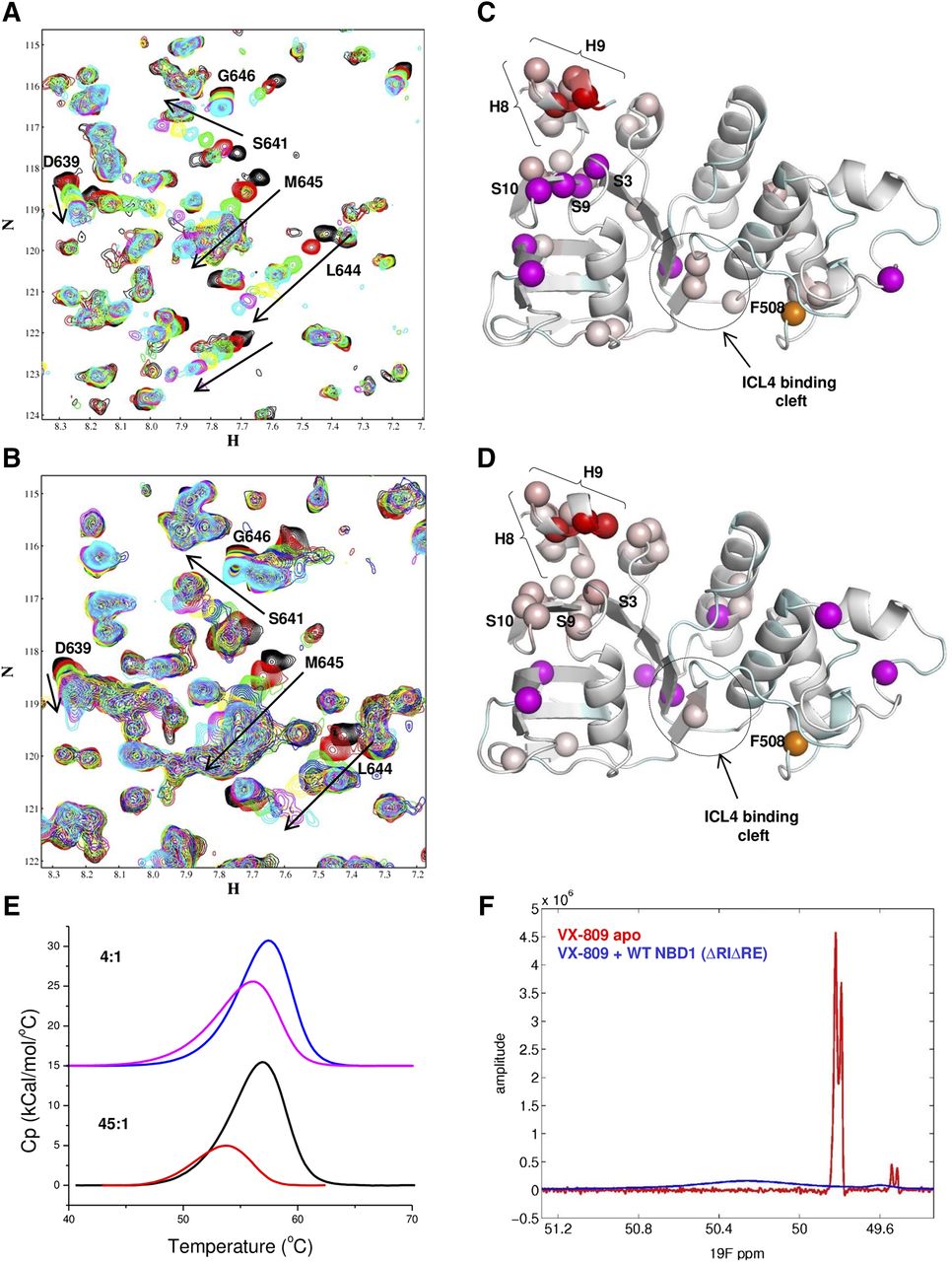
- Fig. 1.
VX-809 reduces helicity of H8 and H9 in the C-terminus of WT and F508del NBD1 ∆RI∆RE. (A) and (B) Overlays of 15N-1H correlation spectra at 500 MHz for 250 μm WT and F508del NBD1 ΔRI ΔRE, respectively, in the absence (black; background) and presence of titration points of VX-809 (1:1, red; 2:1, green; 3:1, cyan; 4:1, yellow; 5:1, magenta; 6:1, cyan). Arrows in (A) and (B) are paired with the assigned peak (where known) and indicate the subset of peaks that shift upon addition of compound and show the direction of chemical shift changes in the titration. (C) and (D) Ribbon diagrams of WT and F508del NBD1 ∆RI ∆RE (PDB ID: 2PZE), respectively, mapping chemical shift changes shown in (A) and (B). Unassigned residues are shown in cyan, and assigned residues that do not shift upon compound addition are shown in light gray. The N atoms for residues that show chemical shift changes are shown as spheres colored with a linear gradient from light pink to red, where light pink corresponds to the smallest shifts (beginning at 10 Hz), and red to the largest shifts. Magenta spheres represent N atoms for residues whose chemical shift changes upon the addition of VX-809 yet are not included in the linear gradient as their post-VX-809–shifted position cannot be known with certainty. F508 is shown as an orange sphere, and the CL4 binding cleft is shown as indicated. (E) DSC traces for WT NBD1 ΔRI ΔRE at concentrations of 4:1 (blue/magenta) and 45:1 (black/orange) compound/protein ratio. (VX-809 concentrations are apparent only due to limited solubility.) The blue and black lines represent the absence of compound, and the magenta and orange lines represent the addition of compound. (F) Overlay of 1D-fluorine NMR spectra of VX-809 with (blue) and without (red) WT NBD1 ΔRI ΔRE. Spectral processing was the same in each case except that line broadening was set to 2 and 0.2, respectively, due to the huge difference in the observed line broadening.
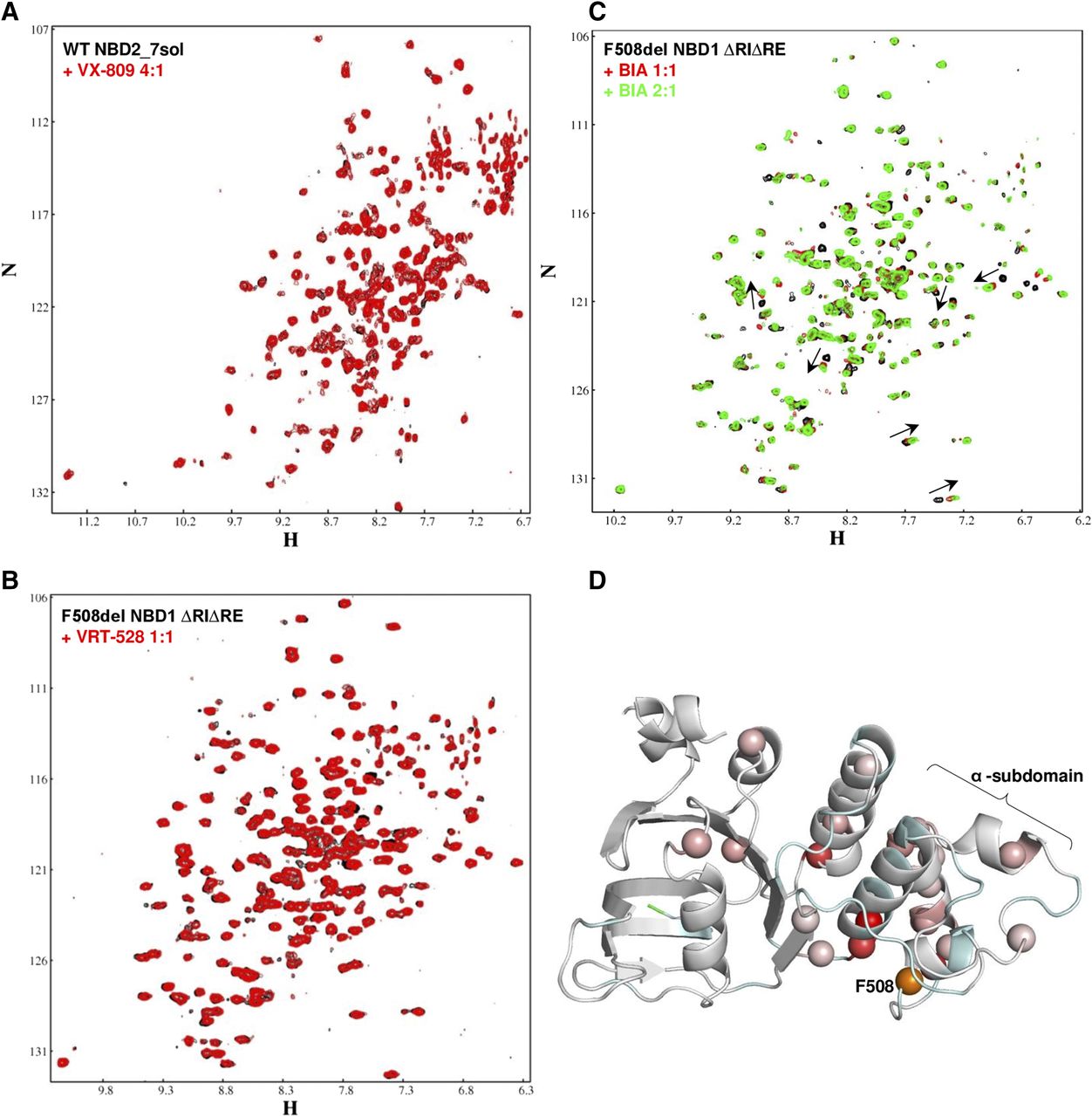
- Fig. 2.
NBD2, VRT-528, and BIA as controls for VX-809 binding. Overlays of 15N-1H correlation spectra at 500 MHz in the absence (black, background) and presence (red, foreground) of VX-809 for 250 μm NBD2 (A) or of VRT-528 (B) and BIA (C) for F508del NBD1 ΔRI ΔRE. Note the absence of chemical shift changes in (A) and (B) and the subset of peak shift changes in (C) (arrows) that differs from those seen upon a VX-809 titration in NBD1. (D) Ribbon diagrams of WT NBD1 ∆RI ∆RE (PDB ID: 2PZE) mapping the chemical shift changes shown in (C). Coloring is as described in the legend of Fig. 1.
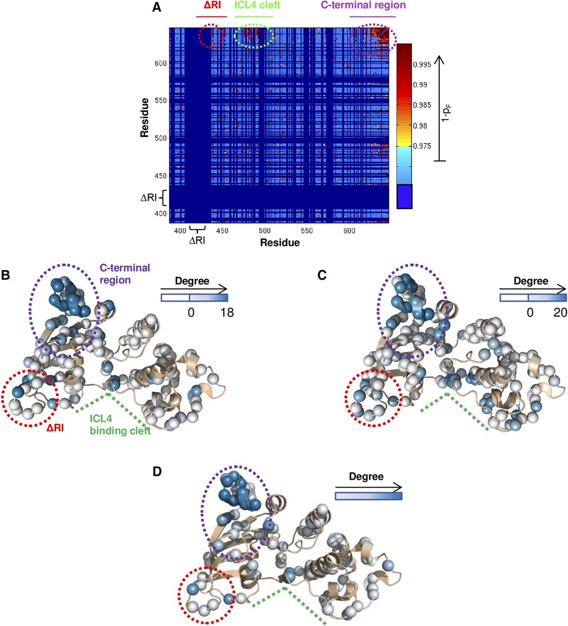
- Fig. 3.
(A) Inter-residue correlation contact map for VX-809:NBD1 titration. Pairs of residues with significant correlations (1-PF ≥ 0.975) are shown in a yellow-to-red heat map, with red being the most significant. Light blue indicates nonsignificant correlations, and dark blue marks the residue pairs that were not analyzed due to the lack of assignment or deletion from the construct (∆RI). The inter-residue correlations among the RI deletion site (red circle), CL4 binding cleft (green circle), and the C-terminal β-strands/helices region (purple circle) are indicated. VX-809:NBD1 (B) and CL4:NBD1 (C) degree values that are above the threshold have been mapped onto the NBD1 structure (PDB ID: 2PZE). Similar network theory analysis of the CL4:NBD1 data found that a threshold at degree >20 eliminated 98% of the noise. Analyzed residues are indicated as spheres with above threshold degree values colored by a white-to-blue gradient and residues below threshold are shown as white. Unassigned residues are shown as tan ribbons. VX-809:NBD1 and CL4:NBD1 data have similar patterns of mapped degree values. (D) The similarity between the sets of residues probed by VX-809 and CL4 was found to be statistically significant, implying that the ligands probe the same allosteric network. Two hundred fifty inter-residue correlations are found in common for both sets of binding data. The allosteric network coupling the C-terminal region and the CL4 binding cleft is shown by mapping these common correlations onto the NBD1 structure as degree values.
- Fig. 4.
Ribbon diagrams of the S3, S9, and S10 region of NBD1 with VX-809 docked. NBD1 and VX-809 are shown in green and violet, respectively. Nitrogen, oxygen, and fluorine atoms are shown in blue, red, and cyan, respectively. VX-809 docked in two main orientations. The VX-809 conformers in the dominant docking orientation, which was present in six of nine of the lowest energy conformers, are shown in (A). The remaining three low-energy VX-809 conformers in the secondary docking orientation are shown in (B).
- Fig. 5.
H620D abrogates binding of VX-809 to WT NBD1 ∆RI∆RE. Overlay of 15N-1H correlation spectra at 500 MHz for 250 μm WT NBD1 ΔRI ΔRE (black, background) and H620D NBD1 ΔRI ΔRE (red, foreground) (A); 250 μm H620D NBD1 ΔRI ΔRE in the absence (black; background) and presence of titration points (1:1, red; 2:1, blue; 3:1, green) of VX-809 (B) and CFFT-001 (C). Note the absence of chemical shift changes. (D) Ribbon diagram of WT NBD1 ΔRI ΔRE (PDB ID: 2PZE) where the N atom of the H620 is shown as a yellow sphere. Coloring is as described in Supplemental Fig. 1B. (E) HEK-293 cells expressing F508del-CFTR or F508del-H620D-CFTR were treated with 1 μm VX-809 or CFFT-001 and were incubated for 24 hours at 30°C. CFTR correction by VX-809 and CFFT-001, as shown by Band C, is reduced by the H620D mutation. (F) Quantitative analysis (n = 6) of HEK-293 cells expressing F508del-CFTR (○) or F508del-H620D-CFTR (●) and incubated with increasing concentrations of VX-809 demonstrate the suppression, but not complete abrogation, of correction.


Additional Files
Data Supplement
- Supplemental Data -
Supplemental figures 1-5.
- Supplemental Data -


