


Article Figures & Data
Figures
- Fig. 1.
Effect of LEF treatment on the plasma concentrations of MTX and 7OH-MTX. (A and B) Concentration-time curves of MTX (A) and 7OH MTX (B). Male C57BL/6 mice were intraperitoneally administered LEF (40 mg/kg) or vehicle (PEG400) for 3 consecutive days, and then all mice were administered MTX (50 mg/kg, i.v.). Blood samples were collected and analyzed as described in the Materials and Methods. The data are representative of two independent experiments, and each point indicates the mean ± S.D. (n = 5).
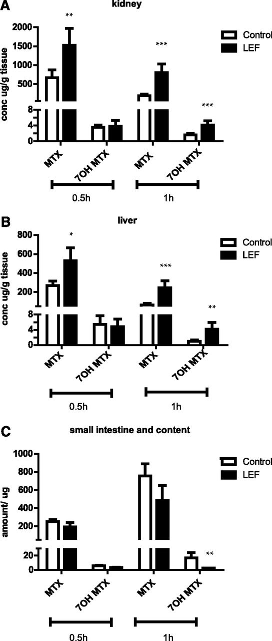
- Fig. 2.
Effect of LEF treatment on the tissue distributions of MTX and 7OH-MTX. (A–C) The concentrations (conc) of MTX and 7OH MTX in the kidney (A), liver (B), and small intestine (C) were assessed at 30 minutes and 1 hour. Male C57BL/6 mice were intraperitoneally administered LEF (40 mg/kg) or vehicle (PEG400) for 3 consecutive days, and then all mice were administered MTX (50 mg/kg, i.v.). At 30 minutes and 1 hour after the last dose, the mice were euthanized and tissue samples were collected and analyzed as described in the Materials and Methods. The data are representative of two independent experiments, and each point indicates the mean ± S.D. (n = 5). The raw data were log-transformed and the significant difference analysis was done on the transformed data. Differences between two groups were analyzed using the t test (*P < 0.05; **P < 0.01; ***P < 0.001).
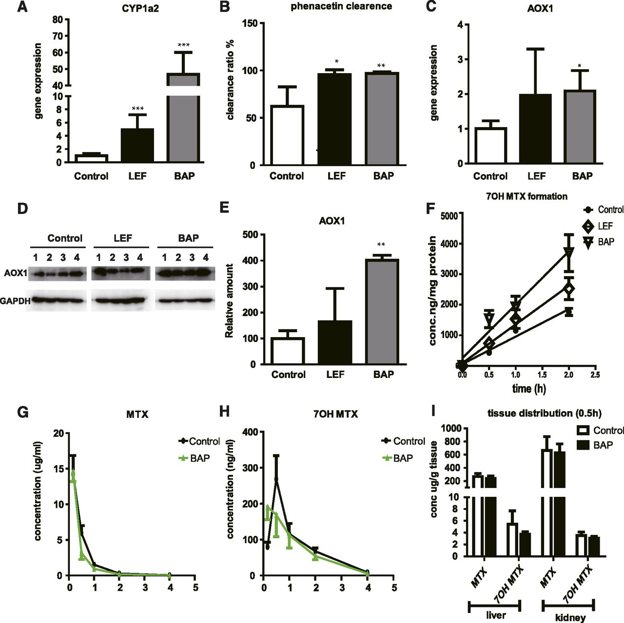
- Fig. 3.
Effect of LEF treatment on the expression and activity of liver CYP1a2 and AOX. (A and B) Effect of LEF on the gene expression of CYP1a2 (A) and the activity of CYP1a2 (B) in liver microsomes. Phenacetin was chosen as a probe substrate of CYP1a2. (C–E) Effect of LEF on AOX1 gene expression (C) and AOX1 protein expression (D and E). (F) Effect of LEF on AOX activity in the cytoplasm of liver cells. 7OH MTX was chosen as a probe product of AOX. (G and H) Effect of BAP on the plasma concentration of MTX (G) and 7OH MTX (H). (I) Effect of BAP on the liver and kidney concentrations of MTX and 7OH MTX at 30 minutes after the last dose. (A–F) Male C57BL/6 mice were intraperitoneally administered LEF (40 mg/kg, i.p.), BAP (12.5 mg/kg, i.p.), or vehicle (PEG400) for 3 consecutive days. The mice were then euthanized, and liver samples were collected and processed as described in the Materials and Methods. BAP was employed as a positive control. Gene expression and Western blotting data were normalized against the endogenous control GAPDH. The data are representative of three independent experiments, and each point indicates the mean ± S.D. (n = 4). The raw data were log-transformed and the significant difference analysis was done on the transformed data. Differences between the three groups were analyzed using one-way analysis of variance (*P < 0.05; **P < 0.01; ***P < 0.001). (G–I) Male C57BL6 mice were intraperitoneally administered BAP (12.5 mg/kg, i.p.) or vehicle (PEG400) for 3 consecutive days, and then all of the mice were administered MTX (50 mg/kg, i.v.). Blood and tissue samples were collected and the concentration of MTX/7OH MTX were measured. The data are representative of two independent experiments, and each point indicates the mean ± S.D. (n = 5).
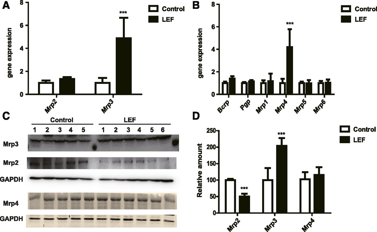
- Fig. 4.
Effect of LEF treatment on the mRNA and protein expression of liver efflux transporters. (A and B) Gene expression of Mrp2 and Mrp3 (A) and other transporters (Bcrp, P-gp, Mrp1, Mrp4, and Mrp5) (B). (C and D) Protein expression of Mrp2, Mrp3, and Mrp4. Male C57BL/6 mice were intraperitoneally administered LEF (40 mg/kg, i.p.) or vehicle (PEG400) for 3 consecutive days, and then the mice were euthanized. Liver samples were collected and processed as described in the Materials and Methods. Gene expression and Western blotting data were normalized against the endogenous control GAPDH. The data are representative of three independent experiments, and each point indicates the mean ± S.D. (control, n = 5; LEF, n = 6). The raw data were log-transformed and the significant difference analysis was done on the transformed data. Differences between two groups were analyzed using the t test (***P < 0.001). Bcrp, breast cancer resistance protein; P-gp, P-glycoprotein.
- Fig. 5.
Effect of LEF treatment on plasma and tissue concentrations of 7OH-MTX after 7OH MTX administration. (A–D) The concentration of 7OH-MTX in the plasma (A) liver (B), kidney (C), and small intestine (D) at 30 minutes and 1 hour after the last dose. Male C57BL/6 mice were intraperitoneally administered LEF (40 mg/kg) or vehicle (PEG400) for 3 consecutive days, and then the mice were administered 7OH MTX (10 mg/kg, i.v.) Blood and tissue samples were collected and analyzed as described in the Materials and Methods. The data are representative of two independent experiments, and each point indicates the mean ± S.D. (n = 5). The raw data were log-transformed and the significant difference analysis was done on the transformed data. Differences between two groups were analyzed using the t test (***P < 0.001).
- Fig. 6.
Effect of LEF treatment on liver nuclear receptors. (A–D) The gene expression of nuclear receptors (Car, Fxr, Nrf2, Pxr, and PPARα) (A), downstream target gene expression (CYP2b10, Cyp7a1, HO-1, CYP3a11, CYP4a10, and CYP 4a14) (B), protein expression of Cyp4a10 and Cyp4a14 (proteomics data) (C), and the PPARα reporter luciferase assay (D). (A and B) Male C57BL/6 mice were administered LEF (40 mg/kg) or vehicle (PEG400) for 3 consecutive days, and then the mice were euthanized. Liver samples were collected and processed as previously described in the Materials and Methods. Gene expression was normalized against the endogenous control GAPDH. The data are representative of two independent experiments, and each point indicates the mean ± S.D. (n = 5). (C) Proteomics data showing the relative fold change from the control group (PEG400). Each point indicates the mean ± S.D (n = 5). (D) Cos-7 cells were transiently cotransfected with 200 ng plasmid containing the luciferase gene and 100 ng GAL4-PPARα LBD expression plasmid, and then the cells were treated with GFT-505 (a positive control) and LEF at the indicated concentrations for 24 hours. Luciferase activity was determined and normalized to Renilla luciferase activity. Each condition was performed with n = 3 for each experiment. As a control, the activity was measured in the presence of vehicle (DMSO). The data are representative of two independent experiments. Each point indicates the mean ± S.D. The raw data were log-transformed and the significant difference analysis was done on the transformed data. Differences between two groups were analyzed using the t test (*P < 0.05; **P < 0.01; ***P < 0.001). DMSO, dimethylsulfoxide; HO, heme oxygenase; Pxr, pregnane X receptor.
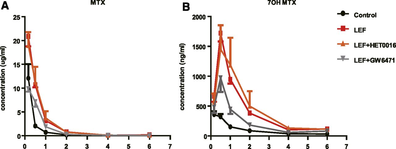
- Fig. 7.
Effect of LEF, LEF plus HET0016, and LEF plus GW6471 treatment on the plasma concentrations of MTX and 7OH MTX. (A and B)The plasma concentration of MTX (A) and 7OH-MTX (B). Male C57BL/6 mice were administered LEF (40 mg/kg plus 5% DMSO, i.p.), LEF plus HET0016 (LEF, 40 mg/kg; HET0016, 5 mg/kg, i.p.), LEF plus GW6471 (LEF, 40 mg/kg; GW6471, 5 mg/kg, i.p.), or vehicle (equal volume of PEG400 plus 5% DMSO) for 3 consecutive days, and then the mice were given a tail vein injection of MTX (50 mg/kg). Blood samples were collected and analyzed as described in the Materials and Methods. The data are representative of two independent experiments, and each point indicates the mean ± S.D. (n = 5).
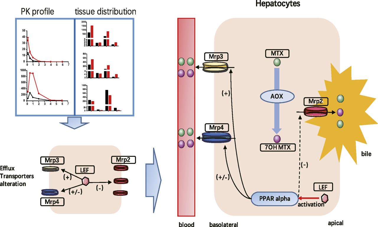
- Fig. 8.
Model of how LEF increases exposure to MTX and 7OH MTX via PPARα activation and Mrp2/3 alterations in the mouse liver. MTX transforms to 7OH MTX through AOX activity. Both MTX and 7OH MTX are substrates of Mrp2/3. LEF downregulated Mrp2 and upregulated Mrp3 (both gene and protein levels) and Mrp4 (only gene level). Through Mrp2 downregulation, the biliary efflux of MTX/7OH MTX was damaged. Through Mpr3 (major) and Mrp4 (minor) upregulation, the concentrations of MTX and 7OH MTX in the plasma and kidneys were increased. Because the upregulation of Mrp3 did not completely compensate for the downregulation of Mrp2, LEF increased MTX/7OH MTX accumulation in the liver.


Tables
Gene Forward Reverse GAPDH aactttggcattgtggaagg acacattgggggtaggaaca AOX1 actgcgtgggccatcttgtctg ttcctggccgcctatgtgtat Mrp1 tgcgcttcccactcaacatcc cgggccaggctcacacg Mrp2 gtgtggattcccttgggcttt cacaacgaacacctgcttgg Mrp3 gcagagacaggcaatgtgaa gaaagctgacagcatgacca Mrp4 catcgcggtaaccgtcctc ccgcagttttactccgcag Mrp5 gtctcctttcctctcccacatcac cttgatgcagcctccagatagc Mrp6 agtcgggaacctgctgaacc cacgcgctccacggctaccats Bcrp aaatggagcacctcaacctg cccatcacaacgtcatcttg P-gp cagcagtcagtgtgcttacaa atggctcttttatcggcctca CYP4a10 tgagggagagctggaaaaga ctgttggtgatcagggtgtg CYP4a14 tggggagatcagatccaaag gacagagtccgccatgattt ACOX1 caggaagagcaaggaagtgg cctttctggctgatcccata CPT1 ccaggctacagtgggacatt gaacttgcccatgtccttgt Nrf2 ctttagtcagcgacagaaggac aggcatcttgtttgggaatgtg Fxr gcttgatgtgctacaaaagctg cgtggtgatggttgaatgtcc Pxr gatggaggtcttcaaatctgcc cagccggacattgcgtttc Car ttcaagcctccggcctatct tgatctgttgcaccataaacgtg ACOX1, peroxisomal acyl-coenzyme A oxidase 1; Bcrp, breast cancer resistance protein; CPT1, carnitine palmitoyltransferase I; P-gp, P-glycoprotein; Pxr, pregnane X receptor.
- TABLE 2
PK parameters of MTX and 7OH MTX
PK parameters are presented as the mean ± S.D. (n = 5). The raw data were log-transformed and the significant difference analysis was done on the transformed data.
PK Parameter MTX 7OH MTX Control LEF Control LEF t1/2 (h) 0.53 ± 0.11 0.41 ± 0.02 0.91 ± 0.29 1.26 ± 0.68 Tmax (h) NA NA 0.80 ± 0.67 0.75 ± 0.27 Cmax (μg/ml) NA NA 0.271 ± 0.143 1.075 ± 0.320** AUC (μg/ml*h) 9.56 ± 3.39 25.8 ± 6.5* 0.344 ± 0.099 1.558 ± 0.247** CL (l/h per kilogram) 5.64 ± 1.54 2.05 ± 0.51* NA NA MRT0–t (h) 0.42 ± 0.05 0.49 ± 0.07 1.26 ± 0.26 1.22 ± 0.16 See the illustration in Fig. 1 for details. CL, clearance; MRT0–t, mean residence time from time zero extrapolated until the end of the dosing interval; NA, not applicable; t1/2, elimination half-life; Tmax, time to reach Cmax.
↵* P < 0.01; **P < 0.001 (differences between two groups were analyzed using the t test).
- TABLE 3
PK parameters of 7OH MTX
PK parameters are presented as the mean ± S.D. (n = 5). The raw data were log-transformed and the significant difference analysis was done on the transformed data.
PK Parameter Control LEF 7OH MTX t1/2 (h) 0.91 ± 0.29 0.99 ± 0.19 AUC (μg/ml*h) 13.1 ± 4.6 44.2 ± 25.6* CL (l/h per kilogram) 0.83 ± 0.27 0.28 ± 0.13 MRT0–t (h) 0.82 ± 0.16 1.28 ± 0.18 See the illustration in Fig. 5 for details. CL, clearance; MRT0–t, mean residence time from time zero extrapolated until the end of the dosing interval; NA, not applicable; t1/2, elimination half-life.
↵* P < 0.01 (differences between four groups were analyzed using one-way analysis of variance).
- TABLE 4
PK parameters of MTX and 7OH MTX
PK parameters are presented as the mean ± S.D. (n = 5). The raw data were log-transformed and the significant difference analysis was done on the transformed data.
PK Parameter MTX 7OH MTX Control LEF LEF + HET0016 LEF + GW6471 Control LEF LEF + HET0016 LEF + GW6471 t1/2 (h) 1.49 ± 0.66 0.89 ± 0.13 0.77 ± 0.08 0.77 ± 0.11 2.50 ± 0.53 1.57 ± 0.41 1.97 ± 1.02 1.91 ± 0.74 Tmax (h) NA NA NA NA 0.25 ± 0.17 0.50 ± 0.00 0.60 ± 0.22 0.50 ± 0.00 Cmax (μg/ml) NA NA NA NA 0.384 ± 0.080 1.721 ± 0.343*** 1.479 ± 0.948** 0.972 ± 0.411 AUC (μg/ml*h) 7.50 ± 3.30 16.2 ± 2.6* 15.9 ± 7.3* 8.52 ± 1.52 0.576 ± 0.055 2.488 ± 0.407*** 2.765 ± 2.158*** 1.274 ± 0.333 CL (l/h per kilogram) 7.48 ± 3.04 3.11 ± 0.59** 3.53 ± 4.27 4.62 ± 5.61 NA NA NA NA MRT0–t (h) 0.46 ± 0.13 0.62 ± 0.02 0.59 ± 0.12 0.61 ± 0.02 1.67 ± 0.19 1.49 ± 0.25 1.68 ± 0.29 1.45 ± 0.36 See the illustration in Fig. 7 for details. CL, clearance; MRT0–t, mean residence time from time zero extrapolated until the end of the dosing interval; NA, not applicable; t1/2, elimination half-life; Tmax, time to reach Cmax.
↵* P < 0.05; **P < 0.01; ***P < 0.001 (differences between four groups were analyzed using one-way analysis of variance).
Data Supplement
- Supplemental Data -
Supplemental Figure 1 - Effect of BAP treatment on the mRNA expression of liver efflux transporters and on the PK profile of 7OH MTX
Supplemental Figure 2 - Effect of LEF treatment on mRNA expression of PPAR α downstream probe genes
Supplemental Figure 3 - Effect of LEF treatment on liver reactive oxygen species (ROS) content and bile acid components of the plasma and liver
Supplemental Table 1 - Effect of LEF and TER treatment on protein change in human hepatocytes
Supplemental Methods
References
- Supplemental Data -


