

Abstract
Nociceptin/orphanin FQ (N/OFQ) and nocistatin (NST) are two recently identified neuropeptides with opposing effects on several CNS functions, including spinal nociception. The cellular mechanisms that underlie this antagonism are not known. Here, we have investigated the effects of both peptides on synaptic transmission mediated by the three fast neurotransmitters l-glutamate, glycine, and GABA in the superficial layers of the rat spinal cord horn, which constitute the first important site of integration of nociceptive information in the pain pathway. NST selectively reduced transmitter release from inhibitory interneurons via a presynaptic Bordetella pertussis toxin-sensitive mechanism but left excitatory glutamatergic transmission unaffected. In contrast, N/OFQ only inhibited excitatory transmission. In the rat formalin test, an animal model of tonic pain in which N/OFQ exerts antinociceptive activity, NST induced profound hyperalgesia after intrathecal application. Similar to glycine and GABAA receptor antagonists, NST had no significant effects in the rat tail-flick test, a model of acute thermal pain. Our results provide a cellular basis for the antagonism of N/OFQ and NST and suggest the existence of a so far unidentified membrane receptor for NST. In addition, they support a role of NST as an endogenous inhibitor of glycinergic and GABAergic neurotransmission in the sensory part of the spinal cord and as a mediator of spinal hyperalgesia.
Hyperalgesia, i.e., an increased sensitivity of noxious stimuli, and allodynia, i.e., a painful sensation of otherwise innocuous stimuli, often accompany chronic pain states seen after prolonged tissue damage. Nociceptin (Meunier et al., 1995), also called orphanin FQ (Reinscheid et al., 1995), and nocistatin (NST) (Okuda-Ashitaka et al., 1998) are two recently identified neuropeptides that have been implicated in the development and/or modulation of hyperalgesia and allodynia. Nociceptin/orphanin FQ (N/OFQ) is an endogenous ligand of the opioid receptor-like 1 receptor (Mollereau et al., 1994), which is now also called N/OFQ receptor. This receptor shares ∼60% homology with classical opioid receptors and is negatively coupled to adenylate cyclase via inhibitory G-proteins, but unlike the μ, κ, and δ opioid receptors, it does not bind classical opioids.
Like other neuropeptides and peptide hormones, N/OFQ is proteolytically released from a larger precursor protein, called preproN/OFQ (Saito et al., 1995; Houtani et al., 1996; Nothacker et al., 1996; Pan et al., 1996). In addition to the cleavage sites necessary for release of N/OFQ, preproN/OFQ contains several other potential cleavage sites, i.e., pairs of the basic amino acids lysine and arginine. Depending on the species, preproN/OFQ contains three or four putative neuropeptides in addition to N/OFQ. At least for one of these peptides, the bovine 17 amino acid peptide b-PNP-3, or bovine nocistatin (bNST), biological activity has been demonstrated (Okuda-Ashitaka et al., 1998). bNST has been detected in the CSF of bovines and, when applied to mice intrathecally, reversed N/OFQ- or prostaglandin E2 (PGE2)-induced hyperalgesia and allodynia via a so far unknown mechanism. Biological activity has also been demonstrated for the mouse (Okuda-Ashitaka et al., 1998) and meanwhile for the human homolog of bNST (Minami et al., 1998). These results suggest that preproN/OFQ contains at least two biologically active peptides, which appear to affect spinal nociception and possibly other CNS functions in opposite directions. So far, nothing is known about the cellular mechanisms that underlie these opposing effects. Here, we have investigated the effects of N/OFQ and NST on excitatory and inhibitory neurotransmission in the superficial layers of the rat spinal cord dorsal horn, which constitutes the first site of synaptic integration of nociceptive information (Yaksh and Malmberg, 1994). We demonstrate that NST reduces inhibitory glycinergic and GABAergic synaptic transmission but leaves excitatory glutamatergic transmission unaffected, whereas N/OFQ only interferes with glutamatergic transmission.
MATERIALS AND METHODS
Slice preparation and electrophysiological recordings. Ten- to 16-d-old Sprague Dawley rats of either sex were killed under ether narcosis by decapitation. Transverse slices (250-μm-thick) of the lumbar spinal cord were prepared as described previously (Liebel et al., 1997). Whole-cell patch-clamp recordings were performed from neurons identified under visual control using the infrared gradient contrast technique coupled to a video microscopy system (Dodt and Zieglgänsberger, 1994). Slices were completely submerged and continuously superfused with external solution, which contained (in mm): 125 NaCl, 26 NaHCO3, 1.25 NaH2PO4, 2.5 KCl, 2 CaCl2, 1 MgCl2, and 10 glucose, pH 7.30 (315 mOsm/l), bubbled with 95% O2–5% CO2. Patch pipettes (4–5 MΩ) were filled with internal solution containing (in mm): 130 K-gluconate, 20 KCl, 2 MgCl2, 0.05 EGTA, 3 Na-ATP, 0.1 Na-GTP, and 10 Na-HEPES, pH 7.30. QX-314 (5 mm) was added to the internal solution to block voltage-activated sodium currents. EPSCs and IPSCs were evoked at a frequency of 0.1–0.07 Hz and recorded at a holding potential of −80 mV at room temperature. Short hyperpolarizing voltage steps to −90 mV were applied every minute to monitor input and access resistance. EPSCs were elicited by ipsilateral extracellular electrical stimulation (100 μsec, 3–10 V) of the dorsal root entry zone using a glass electrode filled with 1 mNaCl. To record IPSCs, the stimulation electrode was placed ∼50 μm away from the recorded neuron. Peptide or drug-containing solutions were applied by bath perfusion at a rate of 1–2 ml/min. Percent inhibition of PSCs by the neuropeptides was determined from the average amplitude of 10 consecutive PSCs evoked immediately before application of the peptides and when a steady state of inhibition was reached, usually ∼3 min after application. In some experiments, a cocktail of protease inhibitors (Complete mini, EDTA-free; Roche Diagnostics, Mannheim, Germany) was added to the external solution. Spontaneously occurring mIPSCs were recorded in the presence of tetrodotoxin (TTX) (1 μm). Amplitude and frequency distributions were analyzed using a custom-made Igor macro (Liebel et al., 1997).
Pertussis toxin treatment. Treatment with pertussis toxin (PTX) was performed similar to the method described by Meis and Pape (1998). In brief, slices prepared as described above were incubated in 35 mm tissue culture dishes filled with 2 ml of Eagle's basal medium supplemented with 50% HBSS, 50% horse serum, 2 mm glutamine, and 0.65%d-glucose, in a standard tissue culture incubator at 34°C, 5% CO2–95% air for 14–18 hr. PTX (500 ng/ml) (Calbiochem, La Jolla, CA) or vehicle (NaCl) was added to PTX-treated and control slices, respectively. Apart from that, incubation conditions of PTX-treated slices and control slices were identical. PTX-treated slices and control slices were recorded alternately.
Behavioral testing. The pronociceptive and/or antinociceptive effects of rat NST consisting of the 17 C-terminal amino acids (rNST1–17) were analyzed in the rat formalin test (Dubuisson and Dennis, 1977) and the tail-flick test. Experiments were performed at room temperature. Sprague Dawley rats weighing 350–400 gm were anesthetized with ketamine (100 mg/kg, i.p.) and xylacine (5 mg/kg, i.p.) and implanted with polyethylene catheters (inner diameter of 0.28 mm, outer diameter of 0.61 mm), which were extended from the cisterna to the rostral edge of the lumbar enlargement. Only rats with unimpaired motor function were used. Formalin tests and tail-flick tests were performed 6–10 d after implantation. Rats were randomly assigned to the different treatment groups consisting of four to six rats each. rNST or vehicle (0.9% NaCl) were delivered to the spinal cord via the catheters in a total volume of 10 μl.
In the formalin tests, formalin (50 μl, 5%) was injected subcutaneously into the dorsal surface of the left hind paw 10 min after intrathecal injection of rNST or vehicle. Flinches of the injected paw were counted at 1 min intervals for 60 min starting 10 min after intrathecal injection. Tail-flick latencies (TFL) were determined using an electronically controlled algesiometer (Ugo Basile, Comerio-Varese, Italy). During measurements of TFL, rats were kept in a plastic restrainer. TFL were determined 20, 40, and 60 min before intrathecal injection and every 10 min for 1 hr after intrathecal injection. Cutoff time was set to 15 sec to avoid unnecessary tissue damage. After the tests, rats were killed by CO2inhalation, and proper position of the catheter tip was visually verified after laminectomy and methylene blue injection.
All behavioral tests and the killing of the animals were performed in accordance with the institutional guidelines of the University of Erlangen-Nürnberg and with the Society for Neuroscience.
Peptides. rNST1–35 (rat preproN/OFQ 98–132) was from Phoenix Pharmaceuticals (Mountain View, CA); its C-terminal 17 amino acid peptide (rNST1–17, rat preproN/OFQ 116–132) was obtained from Dr. M. Herkert (Institut für Biochemie, Universität Erlangen, Erlangen, Germany) and from Research Genetics (Huntsville, AL); bNST was from Tocris Cookson (Bristol, UK); and N/OFQ was purchased from Dr. M. Herkert and from Tocris Cookson. Peptides (purity >95%) were dissolved in external recording solution and stored in aliquots (1 mm) at −20°C. Fresh dilutions were made with standard external solution on every experimental day.
RESULTS
The rat homolog of bNST is a 35 amino acid neuropeptide, of which the C-terminal eight amino acids are conserved among mice and rats and are crucial for biological activity (Okuda-Ashitaka et al., 1998). We have used a peptide consisting of the 17 C-terminal amino acids (rNST1–17) to investigate the effects of rNST on neurotransmission in the spinal cord mediated by the three major fast neurotransmittersl-glutamate, glycine, and GABA. PSCs were evoked by extracellular electrical stimulation and recorded in the whole-cell configuration of the patch-clamp technique from neurons in the superficial layers (substantia gelatinosa) of the spinal cord dorsal horn in which thin and unmyelinated (nociceptive) nerve fibers terminate (Willis et al., 1995). IPSCs were recorded in isolation after blockade of EPSCs with 6-cyano-7-nitroquinoxaline-2,3-dione (CNQX) (10 μm) and d(−)-2-amino-5-phosphonovaleric acid (d-APV) (50 μm). IPSCs typically reversed polarity near −30 mV, which was close to the chloride equilibrium potential of the recording solutions used. IPSCs could almost completely be blocked by a combination of bicuculline and strychnine, indicating that they were mediated by glycine and/or GABAA receptors.
At saturating concentrations (≥1 μm), rNST1–17 reversibly decreased the amplitudes of IPSCs by ∼50% (Fig.1A) but had no effect on excitatory synaptic transmission (compare with Fig. 5). Inhibition occurred in a concentration-dependent manner with half-maximum effective concentration in the submicromolar range. Experiments performed with the complete rat homolog of NST (rNST1–35) and with bNST yielded similar results. Both peptides reduced IPSCs with the same efficacy as rNST1–17 but were slightly less potent (Fig.1B). Several experiments were performed in the presence of protease inhibitors (for details, see Materials and Methods) to exclude the possibility that significant amounts of the peptide were degraded by proteases in the spinal cord tissue. Inhibition of IPSCs was compared in the presence and absence of protease inhibitors at the lowest effective concentration of rNST1–17 (100 nm) at which the effect of protease inhibition should be most striking. However, inhibition was nearly identical under both conditions (6.2 ± 4.6 vs 5.4 ± 5.0%; number of cells, n = 5 each). This result also argues against the possibility that the effects observed were mediated by degradation products of rNST1–17 rather than by rNST1–17 itself.

Effects of NST on inhibitory synaptic transmission in rat dorsal horn neurons. A, Averages of 10 consecutively recorded IPSCs under control condition (control), in the presence of rNST1–17 (rNST; 10 μm), and after removal of rNST1–17 (wash). B, Concentration–response curves of rNST1–17 (solid line) and rNST1–35 and bNST (both dashed lines). Ordinate, Effect of NST on IPSC amplitudes expressed as relative remaining amplitude. Abscissa, NST concentration on a logarithmic scale. Data points were fitted to the following equation: y =ymax − [(ymax −ymin)/(1 + (IC50/C)nH)].ymax is the normalized IPSC amplitude under control conditions, ymin is the relative IPSC amplitude in the presence of saturating concentrations of NST,C is the NST concentration, IC50 is the half-maximum effective concentration, and nHis the Hill coefficient. rNST1–17 (●): average maximum inhibition, 45.7 ± 3.3%; IC50, 486 ± 129 nm;nH, 1.44 ± 0.59. rNST1–35 (▪): IC50, 863 ± 242 nm. bNST (■): IC50, 979 ± 347 nm. Inhibition was statistically significant (p = 0.05) for all concentrations of rNST1–17 (≥500 nm) (ANOVA followed by a Bonferronipost hoc test). n, Number of cells was 5–20 for each peptide and each concentration.
Most neuropeptides exert their action via the activation of seven transmembrane receptors coupled to GTP binding proteins (G-proteins) (Strand, 1999). We tested for a possible involvement of G-proteins of the Gi/Go type using PTX, a selective inhibitor of these G-proteins (Hille, 1994). Overnight incubation of the slices with PTX (500 ng/ml) had no effect on baseline synaptic transmission. In the absence of rNST1–17, IPSC amplitudes were similar in control slices and PTX-treated slices (409 ± 130 pA, mean ± SEM,n = 6; vs 461 ± 198 pA, n = 4). However, the inhibitory effect of rNST1–17 was completely prevented by PTX treatment. The average reduction of IPSC amplitudes by rNST1–17 (10 μm) was 5.6 ± 3.7% (n = 6) in PTX-treated slices compared with 32.5 ± 8.6% (n = 4) in control slices (Fig.2).

Involvement of PTX-sensitive G-proteins.A, Averages of 10 consecutive IPSCs recorded under control conditions or in the presence of rNST1–17 (10 μm). Incubation with PTX (500 ng/ml, 14–18 hr) completely suppressed the effect of rNST1–17 (right), whereas incubation with vehicle had no effect (left).B, Normalized IPSC amplitudes versus time from six PTX-treated (●) and four control (○) slices.
Because inhibitory neurotransmission in the rat spinal cord is mediated by both glycine and GABA (Grudt and Henderson, 1998), we have investigated the effects of rNST1–17 on the GABAergic and glycinergic transmission separately. As shown in Figure3, A and B, rNST1–17 suppressed glycinergic and GABAergic components of IPSCs almost equally (glycinergic component, 48.8 ± 6.2%,n = 14; GABAergic component, 49.9 ± 6.9%,n = 8).
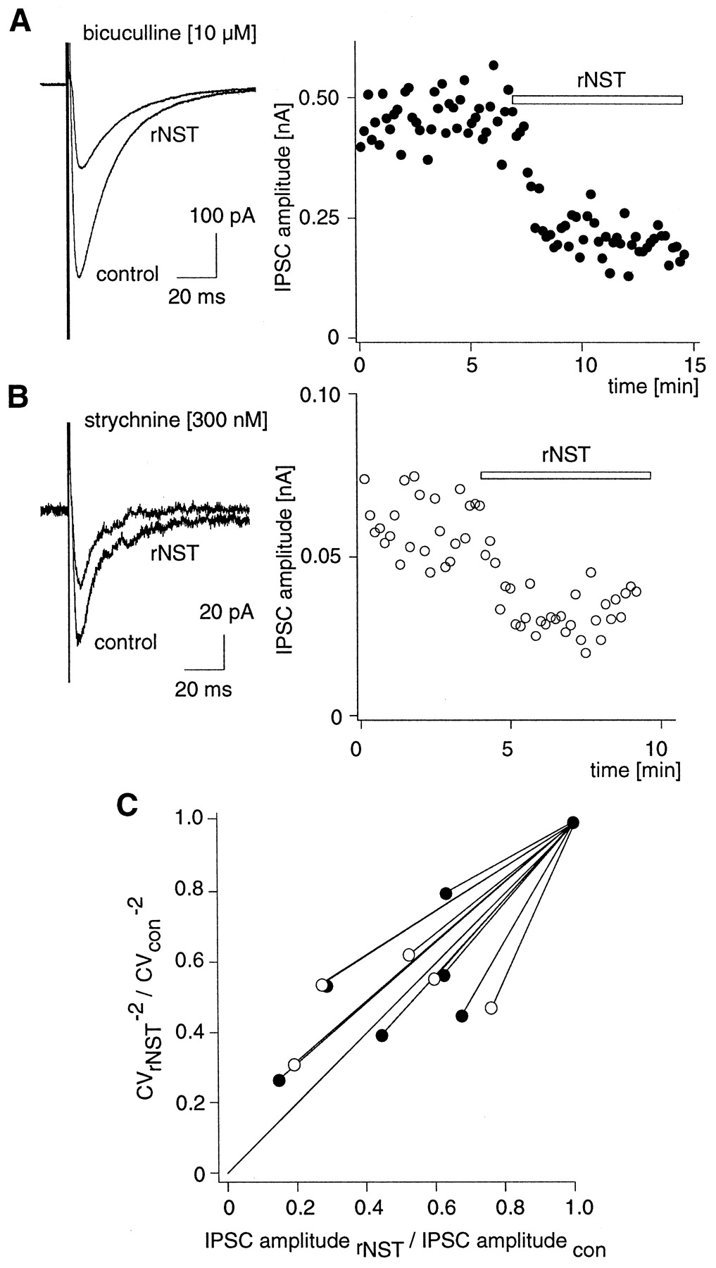
Variation analysis indicates a presynaptic site of rNST action. A, B, Changes in the coefficient of variation of IPSC amplitudes were analyzed for the glycinergic (A) and GABAergic (B) component of inhibitory synaptic transmission. The GABAergic and glycinergic IPSC components were isolated with strychnine (300 nm) and bicuculline (10 μm), respectively. Left, Current traces averaged from 20 consecutive stimulations under control conditions (control) and in the presence of rNST1–17 (10 μm). At least 20 IPSCs were recorded under control conditions and after reaching a steady-state degree of inhibition.Right, Complete time course of representative experiments. C, The coefficient of variation to the (−2) power of the IPSC amplitudes in the presence of rNST1–17 (rNST; 10 μm) (CV−2 rNST ) was plotted against the average amplitude in the presence of rNST1–17 (IPSC amplituderNST) both normalized to the respective control values (CV−2con andIPSC amplitudecon). ●, Glycinergic component; ○, GABAergic component. Data points for both components were close to the identity line, indicting a presynaptic site of action.
To determine the site of action of rNST1–17, we have used two different methods. First, changes in the coefficient of variation of IPSC amplitudes were analyzed (Malinow and Tsien, 1990; Jonas et al., 1998). A plot of the coefficient of variation to the (−2) power versus the average IPSC amplitude both normalized to the respective control value shows that the data points were all close to unity, which indicates a presynaptic rather than a postsynaptic site of action (Fig.3B).
Additional evidence against a postsynaptic site of action was obtained from the analysis of spontaneously occurring miniature IPSCs (mIPSCs), which were recorded in the presence of TTX (1 μm). rNST1–17 had no effect on the amplitude distribution of these mIPSCs, indicating that it did not act postsynaptically by modulating GABAA or glycine receptor function (Fig.4). This is in line with our observation that currents elicited by exogenous application of GABA or glycine were not affected by rNST1–17 (10 μm; data not shown). In this context, it is interesting to note that no change in input resistance was observed when rNST1–17 (10 μm) was applied (ΔRmem, 0.8 ± 6.4%;n = 28), which also argues against the possibility that rNST1–17 opens postsynaptic G-protein-activated potassium channels in spinal cord dorsal horn neurons. In contrast to what one would expect for a presynaptic site of action, no decrease in the frequency of mIPSCs was found. Instead, mIPSC frequency slightly increased in five of seven recordings (Fig. 4E). This increase was statistically not significant (p = 0.242; paired Student's t test) and did not correlate with the application of rNST1–17. It might rather reflect a slight gradual increase in mIPSC frequency, which was sometimes observed in our recordings.

rNST has no effect on spontaneously occurring miniature IPSCs. A, Spontaneously occurring mIPSCs recorded in the presence of TTX (1 μm) at a holding potential of −80 mV. B, Normalized amplitude histograms of mIPCs recorded under control conditions (left) and in the presence of rNST (right) obtained from the same cell. Left trace in each histogram shows the amplitude distribution of the current noise. Insets show mIPSC averaged from 25 consecutive events. C, Changes in the median mIPSC amplitude in eight neurons. D, Normalized cumulative amplitude histograms derived from eight experiments.E, Changes in the average frequency of mIPSCs.
We have next compared the effects of rNST1–17 on EPSCs and IPSCs with those of N/OFQ. IPSCs were isolated as described above. EPSCs were recorded in the presence of bicuculline (10 μm) and strychnine (2 μm). They were almost completely blocked by CNQX (10 μm) and d-APV (50 μm), indicating that they were mediated by ionotropic glutamate receptors. In these experiments, all neurons tested were exposed to both peptides consecutively (Fig. 5). Again, average IPSC amplitudes were significantly reduced to 59.5 ± 2.7% by rNST1–17 (10 μm) but remained unaffected (98.5 ± 1.9%; n = 10) after application of N/OFQ (10 μm) (Fig. 5A). In contrast, EPSCs were insensitive to application of rNST1–17 at concentrations up to 10 μm. The average EPSC amplitude was 102.3 ± 1.2% of the control amplitude (n = 14) (Fig.5B). However, a reversible decrease in the amplitudes of EPSCs to 57 ± 3.2% (n = 14) was observed after application of N/OFQ (10 μm). Thus, rNST1–17 turned out to be a specific inhibitor of inhibitory synaptic transmission, whereas N/OFQ selectively interfered with excitatory transmission.

Inhibition of EPSCs and IPSCs by N/OFQ and rNST. A, B, IPSCs (A) and EPSCs (B) were recorded in the presence of CNQX (10 μm) andd-APV (20 μm) or of bicuculline (10 μm) and strychnine (2 μm), respectively.Top, Current traces are averages of 10 consecutive traces each, recorded under control conditions, in the presence of N/OFQ or in the presence of rNST1–17 (both 10 μm) and after removal of the peptides (wash).Middle, Complete time course of representative experiments. Bottom, Changes in the average IPSC and EPSC amplitudes in individual cells (A,n = 9; B, n = 14) during application of N/OFQ and rNST1–17 (both 10 μm) and after removal of the peptides, all normalized to the respective control amplitudes. Bars represent the average PSC amplitudes (blue, N/OFQ; red, r-NST1–17). Note that the order of N/OFQ and rNST1–17 application is different in A and B.
To test for the relevance of these actions for spinal nociception, we investigated the effects of rNST1–17 in the rat formalin test and the tail-flick test, which are differentially sensitive to changes in the strength of inhibitory synaptic transmission (Yaksh and Malmberg, 1994). In the rat formalin test, a typical biphasic reaction was observed in both control and rNST1–17-treated rats. When applied to the subarachnoid space of the lumbar spinal cord via a surgically implanted catheter (i.e., intrathecally), rNST1–17 dose-dependently increased the number of flinches during all three phases of the test at doses ranging from 1 pmol/rat to 10 nmol/rat (Fig.6A). Statistically significant hyperalgesia was achieved at a dose of 10 nmol/rat for all phases. At lower doses, the effect was most prominent during phase IIa (Fig. 6B). In the tail-flick test, an animal model of acute thermal pain, only a small and statistically not significant decrease in TFL was found after intrathecal injection of rNST1–17 (Fig. 6C).

Effects of rNST in the rat formalin test and the tail-flick test. A, B, Rat formalin test. Flinches were counted starting 10 min after intrathecal administration of rNST in 1 min intervals over a period of 60 min. A, Number of flinched per minute (mean ± SEM). ●, Vehicle; ○, rNST1–17, 10 nmol/rat; ■, rNST1–17, 0.1 nmol/rat; ▵, rNST1–17 0.001 nmol/rat. B, Bars show number of flinches per minute (mean ± SEM) averaged during phase I (0–10 min), phase IIa (20–39 min), and phase IIb (40–60 min) under control conditions and after application of different doses of rNST. Statistically significant mean difference versus respective control (*p ≤ 0.05; **p ≤ 0.01;n = 4–6; ANOVA followed by Scheffépost hoc test). Experiments with rNST1–35 (10 nmol/rat) yielded similar results. C, Tail-flick latencies (mean ± SEM) were determined under control conditions (baseline, averaged from measurements taken 20, 40, and 60 min before intrathecal injection) and every 10 min after intrathecal injection. ●, Vehicle; ○, rNST1–17, 10 nmol/rat; ■, rNST1–17, 1 nmol/rat; ▵, rNST1–17, 0.1 nmol/rat (differences between the treatment groups were not significant at all time points; n = 5–6; ANOVA).
DISCUSSION
The recently discovered neuropeptide NST has originally been described as a functional antagonist of N/OFQ- and PGE2-induced hyperalgesia (Okuda-Ashitaka et al., 1998). Our results indicate that NST has per se biological activity. It specifically suppresses transmitter release from inhibitory (GABAergic and glycinergic) interneurons in the rat spinal cord dorsal horn. Thereby, it serves as a functional antagonist of N/OFQ, which inhibits excitatory (glutamatergic) synaptic transmission. Because neurons in the substantia gelatinosa do not represent a homogeneous group, recordings were probably made from different cell types that cannot be easily distinguished on electrophysiological or morphological criteria in a vital unstained slice preparation. Despite this heterogeneity, the effects of NST and N/OFQ on transmitter release were very reliably observed, suggesting that they represent a widespread phenomenon in the substantia gelatinosa.
Our results suggest a so far unknown membrane receptor to which NST binds in the spinal cord dorsal horn. The sensitivity to PTX of rNST1–17-mediated suppression of inhibitory neurotransmission indicates that this putative NST receptor couples to GTP binding proteins of the Gi/Go type and presumably belongs to the large group of membrane receptors with seven transmembrane domains. The presynaptic nature of the suppression of inhibitory neurotransmission by NST and the lack of impairment of motor function in the behavioral pharmacology tests suggest that, in the spinal cord, the putative NST receptor is preferentially expressed in dorsal horn inhibitory interneurons.
Well established mechanisms involved in the modulation of transmitter release include the activation or facilitation of potassium channels and the inhibition of voltage-gated Ca2+channels. In most CNS regions, including the spinal cord, action potential-evoked Ca2+ influx into presynaptic nerve terminals and transmitter release are mediated primarily by N- and/or P/Q-type Ca2+channels (Takahashi and Momiyama, 1993). NST may thus reduce action potential-triggered GABA and glycine release via inhibition of N- and P/Q-type Ca2+ channels, which are well known targets of a variety of G-protein-coupled receptors (Zamponi and Snutch, 1998). These Ca2+ channels are closed at the resting potential of the cell and hence are of only minor relevance for spontaneous transmitter release (Bao et al., 1998). This may explain why rNST1–17 reduced action potential-evoked GABA and glycine release but did not decrease the frequency of mIPSCs. Alternative mechanisms by which NST might suppress inhibitory synaptic transmission include the opening G-protein-activated potassium channels or an interaction with the vesicle fusion apparatus (Jarolimek and Misgeld, 1997). However, these possibilities appear less likely. Activation of potassium channels should change membrane resistance, which remained constant when rNST1–17 was applied, and a direct interference with the release process should decrease spontaneous transmitter release, which also not observed in our experiments.
Much attention has been focused on glutamatergic synaptic transmission in the spinal cord dorsal horn (for review, see Yaksh et al., 1999). Facilitation of glutamatergic transmission has been proposed as a mechanism of activity-dependent generation of chronic of pain (Zieglgänsberger and Tölle, 1993), and its inhibition is generally accepted as an important target for the analgesic action of opioid peptides. Less is known about the functional significance of endogenous modulators of glycinergic and GABAergic synaptic transmission for spinal nociception. Although the predominant effects of blockers of inhibitory neurotransmission, e.g., of strychnine and picrotoxin, are convulsions, there is considerable evidence that glycine and/or GABA are also involved in sensory information processing. Glycine and GABAAreceptor antagonists increase the size of receptive fields of dorsal horn neurons (Zieglgänsberger and Herz, 1971). Strychnine intoxication in humans (Arena, 1979) and strychnine- or bicuculline-treatment of mice (Yaksh and Rudy, 1977) are characterized by a hypersensitivity to mechanical stimulation. The spastic mouse mutant, which has a strongly reduced number of functional glycine receptors (Becker et al., 1986), is particularly sensitive to mechanical stimulation (White and Heller, 1982). The encoding of low-threshold mechanical stimulation as innocuous is thought to depend on the presence of tonic inhibition by glycinergic and GABAergic interneurons (Yaksh and Malmberg, 1994). Suppression of inhibitory interneurons, which are located between primary afferent low-threshold mechanoreceptors and centrally projecting wide dynamic range neurons (Carlton and Hayes, 1990; Hayes and Carlton, 1992), would then result in pain or pain-related reactions to otherwise innocuous stimuli (Yaksh and Malmberg, 1994). Indeed, NST augmented nociceptive behavior in the rat formalin test (this report) and facilitated nociceptive flexor reflexes (Xu et al., 1999) but had only little effect on acute thermal pain (this report; Yamamoto and Sakashita, 1999). The effects of NST in the different pain models are therefore similar to those of postsynaptic blockers of GABAA and glycine receptors (for review, see Yaksh and Malmberg, 1994). When injected intrathecally, bicuculline (Kaneko and Hammond, 1997) and strychnine (Yaksh and Malmberg, 1994) increase nociceptive behavior in the formalin test but induce only modest changes in tail-flick latencies (Yaksh and Malmberg, 1994).
On the other hand, GABA released from local interneurons in the substantia gelatinosa can depolarize primary afferent nerve terminals (Thompson and Wall, 1996) and thereby augment hyperalgesia and inflammation (Sluka et al., 1993). A preferential reduction of GABA release from these interneurons might explain the anti-allodynic or analgesic effects seen by others after injection of NST (Okuda-Ashitaka et al., 1998; Yamamoto and Sakashita, 1999).
Antinociceptive effects of N/OFQ have been reported repeatedly after intrathecal injection in both tonic pain models, e.g., the rat formalin test (Erb et al., 1997; Yamamoto et al., 1997), and acute pain models, such as the tail-flick test (Xu et al., 1996; Tian et al., 1997). In this respect, the pharmacological profile of N/OFQ resembles that ofl-glutamate receptor antagonists. NMDA receptor blockers reduce nociceptive behavior in the rat formalin test (Coderre and Melzack, 1992; Yamamoto and Yaksh, 1992; Chaplan et al., 1997), and inhibition of AMPA receptors has been demonstrated to increase latencies in the tail-flick test (Näsström et al., 1992;Lutfy et al., 1997). Inhibition of l-glutamate release by N/OFQ in the spinal cord dorsal horn in which l-glutamate is the dominant fast excitatory neurotransmitter might thus very well underlie the antinociceptive effects of spinal N/OFQ. On the other hand, extrapolation of cellular data obtained from young rats to nociceptive behavior in adult animals must be done with caution. There is considerable evidence that significant changes occur in the primary afferent input to substantia gelatinosa neurons during postnatal development. Within the first 8 weeks after birth, low-threshold mechanoreceptors (Aβ fibers) retract from the substantia gelatinosa, and Aδ and C fiber input dominates in mature animals (Fitzgerald et al., 1994; Park et al., 1999).
The physiological role of N/OFQ has been addressed with the use of mutant mice lacking the N/OFQ receptor (Nishi et al., 1997). Although these animals showed several behavioral abnormalities, no alteration in nociceptive thresholds was detected. It is, however, interesting to note that these mice differed from those with a disrupted preproN/OFQ gene in as much as the latter mice exhibited increased nociceptive thresholds (Köster et al., 1999). This difference might be explained by the lack not only of N/OFQ but also of other preproN/OFQ-derived neuropeptides, including NST.
N/OFQ and NST are derived from the same precursor peptide, preproN/OFQ, which in the spinal cord dorsal horn is mainly expressed in local interneurons (Riedl et al., 1996; Neal et al., 1999). Because both N/OFQ (Liebel et al., 1997) and NST act at least in part via a presynaptic site, our results suggest that N/OFQ and NST are released from such local interneurons onto the presynaptic terminals of excitatory and inhibitory neurons. It is at present not known under what physiological or pathophysiological conditions these peptides are released and whether they are always coreleased. There is evidence from other neuropeptide or hormone precursors, such as proopiomelanocortin, that post-translational modifications, peptide sorting, and secretion can occur in a peptide-specific manner (Strand, 1999). Such post-translational modifications can be tissue-specific and may be under control of certain stimuli. A group of enzymes called sortases can intracellularly bind certain hormones (e.g., prolactin, insulin, and human growth factor) and prevent them thereby from storage and secretion (Chung et al., 1989). Different peptides may be transported to different secretory vesicles enabling differential and controlled release (Fumagalli and Zanini, 1985). It appears therefore possible that, under certain conditions, the production and/or release of N/OFQ and NST are differentially regulated.
In summary, we have shown that NST inhibits GABAergic and glycinergic neurotransmission in the spinal cord dorsal horn via a presynaptic mechanism involving PTX-sensitive G-proteins. This effect provides a cellular mechanism for the hyperalgesic action of this peptide observed after spinal application. In concert with N/OFQ, NST presents as a modulatory machinery capable of tuning the spinal nociceptive system to states of both increased and decreased sensitivity to painful stimuli. Inhibition by NST of synaptic release of GABA and glycine in other areas of the CNS, including the hippocampus, may account for other effects of NST, including those on learning and memory (Nicol et al., 1998; Hiramatsu and Inoue, 1999).
Footnotes
This work was supported by grants from the Deutsche Forschungsgemeinschaft Ze 377/4–1 and SFB 353/A8 to H.U.Z. We thank Dr. Kay Brune for critical reading of this manuscript, Angelien Heister and Dr. Matthias Herkert for peptide synthesis, and Susanne Gabriel, Tanja Mittmann, and Claudia Labahn for excellent technical assistance.
Correspondence should be addressed to Dr. H. U. Zeilhofer at his present address: Institute of Pharmacology and Toxicology, Winterthurerstrasse 190, CH-8057 Zürich, Switzerland. E-mail:zeilhofe{at}pharma.unizh.ch.


