


Visual Overview

Abstract
Over the past decade the kinetics of ligand binding to a receptor have received increasing interest. The concept of drug-target residence time is becoming an invaluable parameter for drug optimization. It holds great promise for drug development, and its optimization is thought to reduce off-target effects. The success of long-acting drugs like tiotropium support this hypothesis. Nonetheless, we know surprisingly little about the dynamics and the molecular detail of the drug binding process. Because protein dynamics and adaptation during the binding event will change the conformation of the protein, ligand binding will not be the static process that is often described. This can cause problems because simple mathematical models often fail to adequately describe the dynamics of the binding process. In this minireview we will discuss the current situation with an emphasis on G-protein–coupled receptors. These are important membrane protein drug targets that undergo conformational changes upon agonist binding to communicate signaling information across the plasma membrane of cells.
Introduction
G-protein–coupled receptors (GPCRs) represent attractive pharmacological targets. A long tradition of research in this field has led to the development of several successful drug classes that make up almost 30% of all marketed drugs. Such drugs can interfere with a given GPCR by binding to the receptor and preventing the binding of the endogenous ligand (in which case they are known as antagonists) or they can mimic the endogenous ligand and stimulate a functional response (in which case they are agonists). Such simple views are currently still found in many pharmacological textbooks, and although this simplicity helps to teach beginners the basic principles of receptor pharmacology, we know that this view is overly simplified. Within the past 20 years or so, we have witnessed a dramatic increase in our knowledge of how GPCRs function. We have learned that GPCRs can undergo different conformational changes when different ligands bind to the same receptor (Nygaard et al., 2013). We have seen a tremendous community-wide effort on GPCR crystallization achieve substantial success, and to date more than 100 X-ray structures of 28 different GPCRs have become available to the public (Shonberg et al., 2015). It is likely that many more are available within commercial research groups. We also learned recently that receptor internalization does not necessarily stop a GPCR from being able to continuously signal from the inside of a cell (Irannejad et al., 2013). Nonetheless, our current knowledge of the earliest steps involved in ligand binding appears to be very rudimentary (Pan et al., 2013), especially compared with other aspects of GPCR biology. Ligand binding to a given receptor protein is a dynamic process and is not distinct in its general rules from enzymes, ligand-gated ion channels, or other GPCRs (Colquhoun, 1998, 2006). Although we can learn a lot from equilibrium binding assays and determine ligand binding affinities, the underlying constant flux in ligand binding (on-rate) and unbinding (off-rate) of a ligand has been largely ignored, although this can have a significant influence in vivo, where equilibrium conditions are rarely achieved.
In this short minireview, we briefly introduce the term ligand residence time and briefly discuss the currently available assays that are used to study ligand residence time for GPCRs. We will also critically discuss some known shortcomings and limitations of such assays. Furthermore, we will discuss recent technical advances that might provide insight into the molecular determinants of ligand binding and contribute to a more systematic evaluation of ligand residence time in the future. Finally, we will outline the potential influence of different ligand residence times on GPCR signaling and name successful examples where optimization of ligand residence time has improved drug performance in patients.
The Concept of Ligand Residence Time at GPCRs
Within the last 10 years several excellent reviews have appeared on the general topic of drug-target residence time, each covering the topic from a different perspective (Copeland et al., 2006; Tummino and Copeland, 2008; Lu and Tonge, 2010; Dahl and Akerud, 2013, Vauquelin and Charlton, 2013; Guo et al., 2014 to name a few), and have highlighted this parameter for drug discovery. To keep this minireview focused, we will refer the reader to those articles or the recently published book Thermodynamics and Kinetics of Drug Binding (Keserü and Swinney, 2015) for an in-depth discussion, and we will focus on the concept of residence time at GPCRs.
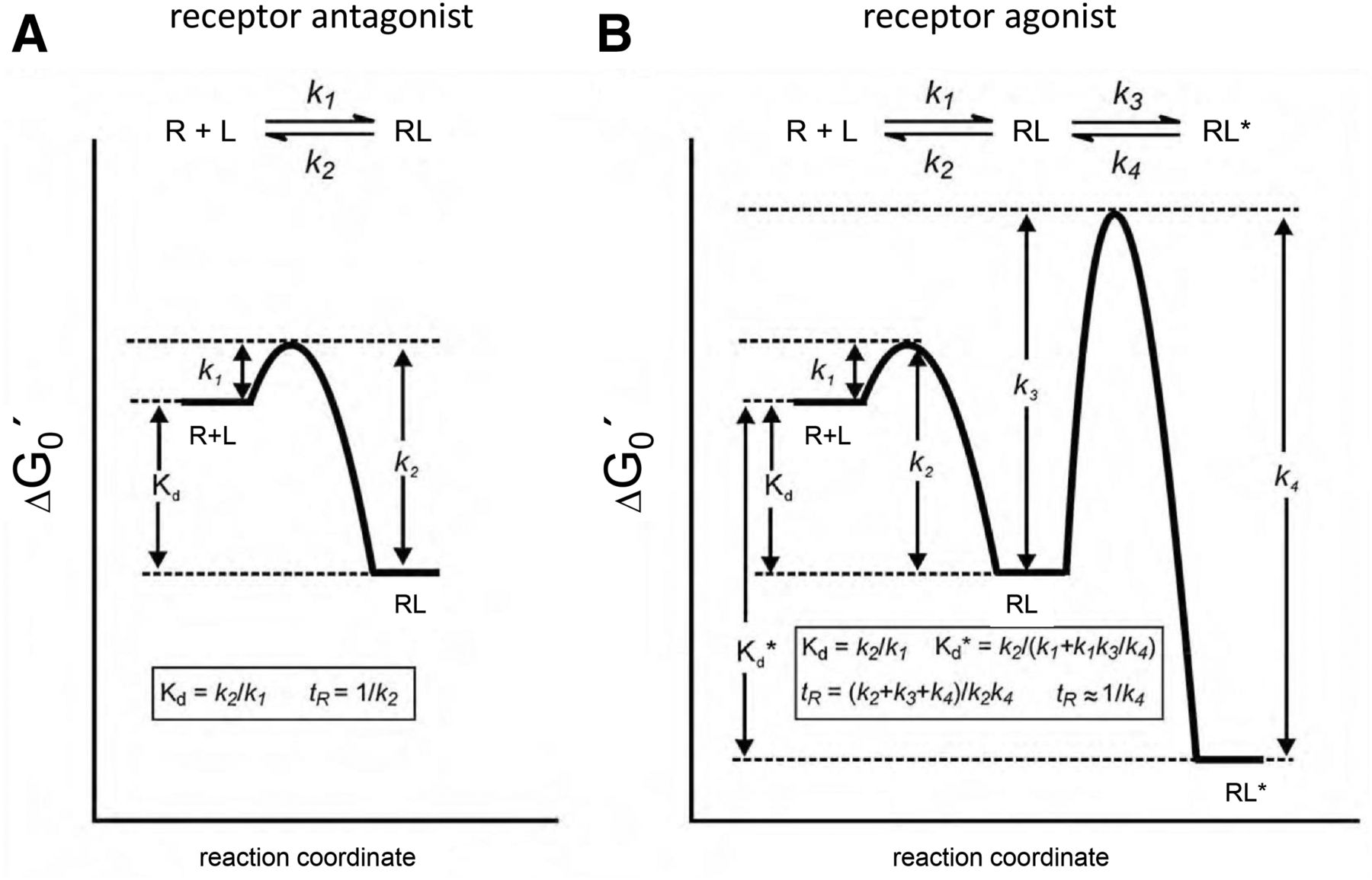
The signal transduction cascade that is mediated by a GPCR is initiated by the binding of agonist to the receptor. The newly formed agonist-receptor complex generates a signal in the given cell, and the lifetime of this complex has a big impact on the efficiency of signal transduction. As a consequence, drug-target residence time has become an important parameter for drug discovery, alongside classic affinity parameters such as IC50 and Ki values (Copeland et al., 2006). In an in vivo system, the residence time becomes crucial if the pharmacokinetic drug elimination is faster than its dissociation from the receptor complex (Dahl and Akerud, 2013). In this case the residence time directly depends on the dissociation rate of the drug from its complex with the receptor. The detection of dissociation rates initially looks straightforward, because most often ligand receptor interactions are illustrated in terms of structurally static binding and dissociation events. This is schematically depicted in Fig. 1A for the case of an antagonist binding to a GPCR, assuming there is no conformational change occurring. Because the ligand dissociation rate constant (k2, also termed koff) is inversely proportional to drug residence time (1/koff), the residence time can be experimentally determined by measuring ligand dissociation rate constants (Copeland, 2011). However, it has become evident that such descriptions are inadequate to explain the impact of conformational dynamics on this process (Copeland, 2011). It was recently shown that the dynamic of a protein can greatly influence ligand dissociation (Teague, 2003; Carroll et al., 2012), or in other words conformational adaptation of the receptor can greatly influence the residence time of a ligand on its receptor or a drug on its target. This is schematically depicted in Fig. 1B for the case of an agonist of a GPCR. If the dissociation rate constant k4 is small compared with the dissociation rate constant k2 of the inactive receptor complex, then the active complex will be stable and the ligand residence time will be determined largely by k4. In case of a GPCR, receptor activation generates a high-affinity agonist complex. This phenomenon can sometimes be observed in binding experiments as a high-affinity state, which can be eliminated by the addition of guanosine triphosphate. The active receptor complex can thus extend significantly the drug’s residence time (Copeland et al., 2006), and this situation becomes more complex if, in addition to an orthosteric ligand, allosteric regulators are also present (May et al., 2011; Corriden et al., 2014; Christopoulos, 2014).

Free-energy plot for ligand (L)-receptor (R) interaction. (A) Shows a simple binding mechanism in case of an antagonist binding to a GPCR without conformational change (one-step binding mechanism). In this simple case, Kd is the dissociation constant given by k1 and k2 as the forward and reverse rate constants, respectively. (B) Shows a more complex two-step induced fit binding mechanism. In this case, k1 and k2 represent the rate constants for formation of the initial RL complex, whereas k3 and k4 are the rate constants for the isomerization step, leading to the final active receptor complex RL*. The case shown chooses k3 and k4 to be small. Hence, formation and breakdown of RL* is correspondingly slow. Kd is the dissociation constant of RL, whereas Kd* represents the dissociation constant of RL* and determines the true affinity of the ligand to the receptor. (Modified from Lu and Tonge, 2010.)
State of the Art: Currently Used Assays
Radioligand Binding Assays.
As stated above, the ligand dissociation rate constant (koff) is inversely proportional to drug residence time (1/koff) and can be experimentally determined by measuring ligand dissociation rate (Copeland, 2011). The determination of the residence time of a drug in complex with a receptor became possible only after the development of radioligand binding assays (Paton and Rang, 1965). Until recently, this was the major method available to assess ligand binding directly and is still the most frequently used assay format (see Table 1). This approach allows the direct detection of on- and off-rates for a high-affinity radioligand. This method, however, predominantly gives information about the labeled ligand itself, although this has been invaluable in the study allosteric regulatory effects (De Amici et al., 2010). The technique has several limitations, because the radioligand binding assay requires separation of the bound ligand from the free ligand fraction, and the binding itself may have several steps. If we are interested in a nonlabeled competing ligand, then the situation is more complex. In such cases, only a fraction of the receptor-ligand complexes might be detected if the radioligand and test compound do not bind to the same receptor conformational state. Different assay formats for competition binding are available that allow radioligand and competitor kinetic binding constants (e.g., kon and koff) to be determined (Guo et al., 2014). The relative strengths and weaknesses of each procedure have been described previously (Guo et al., 2014). Nonetheless, if the compound of interest itself is not labeled, only indirect information of its residence time will be acquired. In addition, nonhomogeneity of this assay system complicates the interpretation of the results obtained. Further problems might arise if the radiolabeled ligand cannot easily be removed during the assay. In these cases the phenomenon of rebinding can occur, and this can complicate the determination of koff values (Vauquelin and Charlton, 2010). Furthermore, such assays currently ignore conformational dynamics and, hence, are largely unsuitable for GPCR receptor agonists that induce a conformational change in the receptor. This problem also extends to data fitting procedures, because often researchers use very simple kinetic models to fit the data that do not account for such details (Motulsky and Mahan, 1984). Furthermore, radioligand binding assays are often conducted on ice and in nonphysiologic buffer conditions, and it has been shown that 10-fold shorter residence times of tiotropium at the M3 acetylcholine receptor are obtained under physiologic conditions (Sykes et al., 2012).
Advantages and disadvantages of different methods for kinetic binding experiments
Label-free approaches (see next section) have also been used to study ligand protein interaction kinetics using purified proteins and immobilization strategies. This has the advantage of using known and well characterized proteins but comes at the cost of the need for detergents and nonnative membranes. For the β2-adrenergic receptor, the local membrane environment was demonstrated recently to have a significant influence on receptor-ligand interactions, and it has been recommended that residence time measurements should be conducted using conditions that are as close as possible to the natural conditions, for example using whole cell experiments or even tissue slices (Sykes et al., 2014). Notwithstanding the difficulties mentioned above, the concept of residence time optimization of GPCR ligands has led to the development of antagonist drugs with long residence times such as tiotropium (Tautermann et al., 2013). Even for agonists, a positive correlation between agonist efficacy and residence time has been observed in the case of the M3 acetylcholine receptor (Sykes et al., 2009) and at the adenosine A2A receptor (Guo et al., 2012), although no such correlation was observed for the adenosine A1 receptor (Louvel et al., 2014).
Surface Plasmon Resonance Analysis
An alternative biophysical approach that is frequently used to determine kinetic ligand binding in drug discovery is represented by surface plasmon resonance (SPR) analysis (see Table 1). This method can be considered as a label-free method with respect to the ligand. To generate a plasmon, polarized light is directed via a prism onto a gold-coated glass surface on which the sample is bound. The refractive index of the medium near the gold surface is a major parameter that influences the critical angle of the polarized light. If the refractive index changes, for example during the formation of the ligand-receptor complex, a signal will be detected due to a shift in the critical angle. This relationship is used to analyze the dynamics of ligand binding. Because of recent technical advancements, this technique is now capable of detecting the binding of molecules as small as 200 Da (Aristotelous et al., 2015) and is now well suited to investigate GPCR ligands. The application of this approach to GPCRs has recently been reviewed (Aristotelous et al., 2015). Currently six GPCRs have been investigated using this approach (rhodopsin, CXCR4, CCR5, adenosine A2A receptor, β1- and β2-adrenergic receptors; Aristotelous et al., 2015). One major drawback is the need to use purified proteins, and the purification often limits the application of this approach. Furthermore, the required immobilization of the protein on the SPR chip can potentially block the accessibility of the intra- or extracellular side of the receptor. However, because of the label-free approach with respect to the ligand, both orthosteric and allosteric ligands can be investigated. This approach has been used to investigate the ligand binding pocket of a stabilized version of the adenosine A2A receptor (Zhukov et al., 2011) and to perform a fragment screening at the β1-adrenergic receptor (Christopher et al., 2013) that identified novel lead structures for this receptor. Both studies demonstrate the powerful potential of this approach. The influence of lipid composition upon assay performance was recently investigated in a comparative study of the adenosine A2A receptor employing four different reconstitution approaches (Bocquet et al., 2015). When the receptor was reconstituted in lipid nanodiscs, protein stability was enhanced and the kinetic data obtained were more similar to native receptors compared with those solubilized in detergents. Similar results were obtained for the CXCR4 receptor when the receptor was embedded in lipoparticles largely consisting of native cell membrane (Heym et al., 2015). In combination their studies demonstrate the influence of native membrane composition upon protein performance. Very recently, the application of SPR was extended to investigate binding kinetics to whole cells using a Herceptin (Genentech Inc., San Francisco, CA)-Her2 combination where the mass increase was detectable in whole cells (Wang et al., 2014).
Novel Approaches and Promising Developments
Quartz Crystal Microbalance.
A very recent development to study the kinetics of ligand binding to whole cells is provided by quartz crystal microbalance technology. This approach uses changes in the frequency of a quartz crystal resonator to provide information on mass changes. If a quartz crystal is placed in between two electrodes in a sandwich like arrangement and an alternating electric potential is applied over the crystal, the crystal will start to vibrate. At a given frequency, resonance occurs and this forms the basis of quartz crystal microbalance technology (Aastrup, 2013). The resonance frequency depends on the mass of the total system, and thus, if cells are placed on the crystal, ligand binding will alter the resonance frequency that forms the basis of signal detection. The principal was originally discovered in 1959, but it is only recently that commercial devices have become available. One major advantage of this technique is the ability to use two flow chambers, in which transfected or nontransfected cells can be compared in a paralleled fashion, to provide an indication of receptor-specific binding (Wright et al., 2014).
Binding of Fluorescent Ligands by Fluorescence Intensity
Alternatives to radioligand binding opened up when novel fluorescence methods for the characterization of ligand binding to GPCRs were implemented. Fluorescent ligands have been used to characterize GPCRs for almost four decades (Melamed et al., 1976; Atlas and Levitzki, 1977). In these studies staining patterns were evaluated by fluorescence microscopy, which gave valuable information about receptor localization at the subcellular level but did not add information about ligand binding properties. Several attempts to distinguish bound fluorescent ligands from free ligand and to quantify their signal after separation were done, but these attempts turned out to be difficult (Sridharan et al., 2014). If the binding to the receptor changes the fluorescence emission spectrum or fluorescence intensity of the ligand, this alteration can be used for quantification of the receptor-bound ligands. However, because of cellular autofluorescence and high level of nonspecific signals, a wide use of this method was prevented (Sridharan et al., 2014). However, the use of red fluorescent dyes such as BODIPY-630/650 has enabled quantitative evaluation of ligand-receptor interactions in the case of a number of GPCRs, including the β1-adrenoceptor and the adenosine A1 and A3 receptors (May et al., 2011; Stoddart et al., 2012; Hill et al., 2014; Gherbi et al., 2014, 2015).
Binding Determined by Fluorescence Anisotropy
An alternative approach is offered by the detection of changes of fluorescence anisotropy (see Table 1). Binding of labeled ligand to the receptor restricts its freedom to rotate within the lifetime of the activated fluorophore. Therefore the portion of polarized light emitted by the ligand increases. In this case one does not have to physically separate bound ligand from unbound, and one can monitor the binding as process in real time. This method has been used to characterize ligand binding to receptors of hormones like endothelin (Junge et al., 2010) and melanocortin (Veiksina et al., 2010). However, it has also been demonstrated for GPCRs of small molecules like acetylcholine for mACh receptors (Huwiler et al., 2010) and serotonin (Tõntson et al., 2014). However, the ratiometric nature of this assay format generates certain limitations for itself—the changes in anisotropy can only be detected if the ratio of bound to free fluorescent ligand has been significantly altered (Nosjean et al., 2006). This is only the case when the concentrations of receptor and ligand are in the same order and at the level of the dissociation constant of the interaction. Such a high level of receptor binding sites, to allow reliable measurements, is usually difficult to achieve, and one also has to be aware of substantial background autofluorescence. One possible solution, other than the use of purified proteins, has been shown by the use of budding baculoviruses, which display GPCRs on their surfaces at such high density that these assays became suitable (Veiksina et al., 2014).
Binding Determined by Fluorescence Correlation Spectroscopy
Similar information about fluorescent ligand binding can be obtained if changes in the particle number and mobility of fluorescently labeled species are detected with fluorescence correlation spectroscopy. This technique measures fluctuations in the fluorescence intensity of fluorescently labeled particles diffusing through a small illuminated detection volume. This allows free ligands to be distinguished from slowly diffusing receptor-bound ligands without their physical separation (Briddon and Hill, 2007). The technique works best at low fluorescent particle numbers and can therefore be used to monitor binding to endogenously expressed receptors (Briddon and Hill, 2007). Furthermore, low concentrations of fluorescent agonists and antagonists can be used to detect active (R*) and inactive (R) receptor conformations (Cordeaux et al., 2008; Corriden et al., 2014). One major advantage of this method is that the actual ligand amounts can be measured. It can be used at the single cell level and even at the level of single molecules (compare Table 1). This approach has already been used for the characterization of ligand binding to different GPCRs, including adenosine A1 and A3 (Middleton et al., 2007; Cordeaux et al., 2008; Corriden et al., 2014) and adrenergic receptors (Prenner et al., 2007).
Binding Determined by Resonance Energy Transfer Techniques
Fluorescence resonance energy transfer (FRET)–based methods have been widely acknowledged for studies of GPCRs. This has been mainly used for the characterization of signaling and oligomerization (Lohse et al., 2012; van Unen et al., 2015). Monitoring direct ligand binding by FRET at GPCRs usually requires that the receptor is labeled on the extracellular side with a fluorophore. This can be achieved by fusing a fluorescence protein (Castro et al., 2005; Fernandez-Duenas et al., 2012) or a SNAP tag (previously known as genetically modified AGT: O6-alkylguanine-DNA alkyltransferase) to the N terminus of the receptor (Lohse et al., 2012). More recently, a bioluminescent protein (NanoLuc) was fused to the N terminus of GPCRs to allow bioluminescence resonance energy transfer to a fluorescent ligand bound to the target GPCR (Stoddart et al., 2015).
Indirectly, ligand binding can also be monitored by a GPCR-based FRET sensor, which allows the study of receptor activation to be monitored by FRET and report upon ligand binding by a conformational change that alters the observed FRET signal (Lohse et al., 2012). With such approaches, kinetic differences in on-rates for ligand binding were observed at the α2a-adrenergic receptor for ligands with different efficacies (Nikolaev et al., 2006). Very recently, this approach was used to study dynamic conformational changes at the M3 acetylcholine receptor and a constitutively active receptor. Agonists exhibited a higher affinity at the constitutively active receptor with unaltered ligand on-rates. The major difference observed at both receptor variants was a 10-fold increase in receptor deactivation time for the constitutively active receptor. This indicated that the observed higher ligand affinity would solely be due to a decrease in ligand off-rates and hence increase in ligand residence time (Hoffmann et al., 2012). Such approaches allow protein dynamics to be taken into account but currently do not allow ligand binding to be observed directly. When concentration-dependent receptor activation was analyzed under nonequilibrium conditions at different time points and in the time range of seconds, it was observed that concentration-effect curves were shifted to higher affinity in a time-dependent manner (Ambrosio and Lohse, 2012). This phenomenon has also been described in a recent simulation of ligand binding and was predicted to result in a kinetic discrimination between different receptors (Ventura et al., 2014). Earlier work by the group of Jennifer J. Linderman has simulated the impact of different ligand off-rates on receptor signaling and receptor desensitization (Woolf and Linderman, 2003). It was also proposed that these processes would be differentially affected by different ligand off-rates and could even be used to design biased agonism to a certain degree.
Examples for Biologic Discrimination by Different Ligand Residence Times
Muscarinic receptor antagonists employed as bronchodilators in the treatment of chronic obstructive pulmonary disease constitute perhaps the best example of drugs for which optimization of binding kinetic parameters is critical for their in vivo profile. Acetylcholine promotes bronchoconstriction and mucus secretion via stimulation of muscarinic receptors present in the airways. Although blocking M1/M3 receptor subtypes would counteract airway limitation in chronic obstructive pulmonary disease patients, blocking presynaptic M2 autoreceptors would be detrimental for this purpose and systemic M2 antagonism would increase the risk of tachycardia as side effect. The difficulties in finding muscarinic receptor subtype-selective ligands were successfully overcome by the development of ipratropium, a short-acting muscarinic antagonist, and long-acting muscarinic antagonists (LAMAs) tiotropium (Disse et al., 1993) and the novel aclidinium (Gavaldà et al., 2009) and glycopyrronium (Casarosa et al., 2009) that are particularly indicated for maintenance therapy. All these drugs dissociate more rapidly from M2 than from M3 receptors. Apart from the advantageous kinetic subtype selectivity of these drugs, the duration of action of the LAMAs was suggested to be primarily related to their long residence time at M3 receptors (Disse et al., 1993; Casarosa et al., 2009). Hence, it was shown that the duration of the bronchodilator action in vivo of different muscarinic antagonists resembles their residence times at M3 receptors determined in vitro under nonphysiologic conditions (Gavaldà et al., 2014). However, other factors, particularly rebinding of the dissociated drug to receptors in the effect compartment, seem likely to contribute to the long duration of action of LAMAs in vivo (Sykes et al., 2012).
The histamine H1 receptor (H1R) increases vascular permeability and smooth muscle contraction during allergic responses. The first generation antagonist mepyramine (pyrilamine) competitively antagonizes histamine-induced increase in intracellular [Ca2+] and guinea pig ileum contraction, resulting in a right shift of the histamine concentration response curves without affecting the maximal response (Anthes et al., 2002; Slack et al., 2011b). In contrast, other antihistamines such as azelastine, desloratidine (Aerius; Merck Sharp & Dohme, Whitehouse Station, NJ), GSK1004723 [4-[(4-chlorophenyl)methyl]-2-({(2R)-1-[4-(4-{[3- (hexahydro-1H-azepin-1-yl)propyl]oxy}phenyl)butyl]-2-pyrro lidinyl}methyl)-1(2H)-phthalazinone], and ceterizine (Zyrtec; UCB, Brussels, Belgium) inhibited histamine-induced Ca2+ signaling and/or smooth muscle cell contraction in an apparently noncompetitive manner, resulting in an attenuated maximal response (Anthes et al., 2002; Slack et al., 2011a,b). Indeed, these insurmountable antagonists dissociated at least 70-fold more slowly from the H1R compared with mepyramine (Gillard et al., 2002; Anthes et al., 2002; Gillard and Chatelain, 2006; Slack et al., 2011b).
Interestingly, the two enantiomers of cetirizine display a 25-fold difference in affinity for the H1R, which results from different dissociation rate constants. Levocetirizine (Xyzal; UCB) dissociates from the H1R with a half-time of 142 minutes, whereas (S)-cetirizine has a dissociation half-time of 6 minutes (Gillard et al., 2002). The long residence time of levocetirizine on the H1R has been related to the interaction of its carboxylic moiety with lysine 191(5.39) in transmembrane helix 5 (Wieland et al., 1999; Gillard et al., 2002). Substitution of lysine 191 with alanine on the receptor side or the carboxyl with methyl ester or hydroxyl moieties on the ligand side significantly accelerated the dissociation rate (Gillard et al., 2002). Although for several of these ligands the long duration of action of in vitro and in vivo preparations has been linked to the long residence time; for azelastine, retention in the airway epithelium has also been suggested to be implicated (Slack et al., 2011a).
In addition to slow dissociation from the H1R, GSK1004723 is also reported as an insurmountable antagonist on the histamine H3 receptor (H3R) with slow dissociation kinetics at the human H3R (Slack, et al., 2011b). So far, no data are available for other H3R ligands, making a direct link with functional effects difficult. The H4R antagonist JNJ7777120 [1-[(5-chloro-1H-indol-2-yl)carbonyl]-4-methylpiperazine] shows best efficacy in in vivo models compared with other classes of H4R antagonists, despite its relatively short half-life time in the circulation. Indeed, JNJ7777120 has a longer target residence time on the H4R as compared with other tested antagonists (Smits et al., 2012; Andaloussi et al., 2013).
Dissociation rates might also be related to qualitative differences in the cellular effects of drugs belonging to the same class. Endothelin receptors mediate calcium signals elicited by endothelin-1 in pulmonary artery smooth muscle cells (PASMC). These signals are described by a first rapid transient peak response followed by a sustained and lower magnitude plateau in intracellular calcium concentration. Sustained Ca2+ signals in PASMC have been related to sustained pulmonary vasoconstriction and pulmonary vascular remodeling in pulmonary arterial hypertension through PASMC contraction and proliferation (Kuhr et al., 2012). Functional studies in PASMC indicate that the slowly dissociating endothelin receptor antagonist macitentan is differentiated from the competitive antagonists bosentan or ambrisentan through its insurmountable antagonism of the sustained Ca2+ signal elicited by endothelin-1, at least under nonequilibrium conditions. This difference among drugs was not revealed when the first fast Ca2+ peak in response to ET-1 was considered (Gatfield et al., 2012). It is conceivable that this qualitative difference in the modulation of a complex cellular response by these drugs might result in a better control of pathologic processes involving PASMC by the novel drug macitentan.
Along the same lines, there is growing evidence for the relevance of kinetics for the cellular responses elicited by GPCRs upon interaction with different ligands. Second or third wave signals can occur (Lohse and Calebiro, 2013), which in some cases include nonclassic signals dependent on β-arrestins such as the regulation of mitogen-activated protein kinases (Shukla et al., 2011). There is also the potential for sustained second messenger signals from internalized receptors (Calebiro et al., 2009; Roed et al., 2015), which are internalized in functional complexes together with their cognate G-proteins (Calebiro et al., 2009; Irannejad et al., 2013). These events are being resolved by performing dynamic measurements of receptor activation and cellular signaling in live cells with different biosensors and, in some cases, by following the intracellular fate of the cointernalized ligand (Calebiro et al., 2009; Roed et al., 2014). In this context, different receptor conformations resulting from the interaction of structurally different ligands might account for ligand functional bias, but it is not clear to what extent ligand-receptor binding/unbinding kinetics also might play a role. Some examples of functional bias on GPCRs include that of the PTH1R, for which peptide ligands with different patterns of biased signaling have been described (Gesty-Palmer et al., 2009; Cupp et al., 2013). Human parathyroid hormone (PTH) and PTH-related protein (PTHrP), the two endogenous agonists of PTH1R, elicit different effects on the renal synthesis of 1,25(OH)2 vitamin D and, therefore, on hypercalcemia in humans in continuous infusion (Horwitz et al., 2005), a dose regimen that discards differences in pharmacokinetics as the only explanation for the discordant effects. The fully active portions of these peptides, PTH-(1-34) and PTHrP-(1-36), associate and dissociate from the receptor with different kinetics, as determined by radioligand binding assays (Dean et al., 2008) and by FRET approaches using fluorescent-labeled peptides and receptor tagged with green fluorescent protein (Castro et al., 2005; Ferrandon et al., 2009). Furthermore, the slow dissociating PTH, in contrast to the fast dissociating PTHrP, cointernalized with the receptor and Gs proteins and elicited a sustained intracellular cAMP signal (Ferrandon et al., 2009). Although the impact of ligand binding kinetics of PTH1R on the distinct cellular responses promoted by different peptides is not known, these observations suggest that certain ligands might show a stable binding to a conformational state of the receptor capable of generating a prolonged cAMP signal by isomerization to a different active conformation without dissociation of the bound agonist (Vilardaga et al., 2014).
In the case of the glucagon-like peptide (GLP)-1 receptor (GLP-1R), a therapeutic target in type 2 diabetes, it was found that it internalizes rapidly and with similar kinetics upon activation with its endogenous ligand GLP-1 or with the two stable GLP-1 analogs exendin-4 (exenatide) and liraglutide (Roed et al., 2014). However, upon interaction with GLP-1, the receptor underwent recycling with 2–3 times faster kinetics than obtained with the two stable agonists. This observation corresponded with a longer colocalization of GLP-1R and the internalized ligand in early recycling endosomes in the case of exendin-4 and liraglutide compared with GLP-1 (Roed et al., 2014). Recent evidence indicates the requirement of internalized GLP-1R/GLP-1 complexes in endosomes for endosomal cAMP signaling and regulation of insulin secretion by GLP-1 in pancreatic β-cells (Kuna et al., 2013). It was suggested that the acidic environment in the endosomal compartment could facilitate the dissociation of cointernalized receptor-ligand complexes (Lu and Willars, 2013). In this context, the work of Roed et al. (2014) points to the fact that ligands with different on/off binding kinetics might be able to differentially promote receptor internalization (as proposed by Woolf and Linderman, 2003), postendocytic sorting, and/or recycling and thus displaying a “kinetic functional selectivity.” This paradigm might be interpreted as a further enrichment of previous biased signaling found for this receptor in yeast, where exenatide displayed a significant bias for the Gi pathway (Weston et al., 2014). Therefore, it would be of interest to know the dissociation rates of cointernalized ligands from their receptors in intracellular compartments.
In conclusion, we think that it has become clear that studying residence time will add significant information to our understanding of ligand binding at GPCRs or any other proteins. Successful examples like tiotropium for drug optimization exist that demonstrate the potential to improve target selectivity by kinetic optimization. Nonetheless, we think the currently used assays, particularly at GPCRs, fail to take into account the conformational dynamics of GPCRs, especially for agonists. Therefore, we need to develop novel assay formats that take into account the conformational changes upon ligand binding. Such assays should close the gap by ideally detecting binding and conformational changes in parallel and at the same time as has been shown for ion channels. This will be technically challenging, but fluorescence technologies might be helpful in this respect, but all cautions discussed need to be appropriately addressed. However, we think it will be worth the effort if better medication can be designed.
Author Contributions
Wrote or contributed to the writing of the manuscript: Hoffmann, Castro, Rinken, Leurs, Hill, Vischer.
Footnotes
- Received April 28, 2015.
- Accepted July 7, 2015.
This work was supported by the Deutsche Forschungsgemeinschaft, Transregio 166 (Project C2) to C.H., the Spanish Ministry of Economy and Competitiveness [SAF2014-57138-C2-1-R] to M.C., the Estonian Ministry of Education and Science (IUT 20-17) to A.R., the Innovative Medicines Initiative Grant K4DD “Kinetics for Drug Discovery” to R.L. and S.J.H., the UK Medical Research Council grant [G0800006] to S.J.H., and the “TOPPUNT grant of the Netherlands Organization of Scientific Research-Chemical Sciences” to R.L. and H.F.V.
The authors declare no conflict of interest.
Abbreviations
- FRET
- fluorescence resonance energy transfer
- GLP
- glucagon-like peptide
- GPCR
- G-protein–coupled receptor
- GSK1004723
- 4-[(4-chlorophenyl)methyl]-2-({(2R)-1-[4-(4-{[3-(hexahydro-1H-azepin-1-yl)propyl]oxy}phenyl)butyl]-2-pyrro lidinyl}methyl)-1(2H)-phthalazinone
- JNJ7777120
- 1-[(5-chloro-1H-indol-2-yl)carbonyl]-4-methylpiperazine
- LAMA
- long-acting muscarinic antagonist
- PASMC
- pulmonary artery smooth muscle cells
- PTH
- parathyroid hormone
- SPR
- surface plasmon resonance
- Copyright © 2015 by The American Society for Pharmacology and Experimental Therapeutics
References
In this issue


Jump to section
- Article
- Visual Overview
- Abstract
- Introduction
- The Concept of Ligand Residence Time at GPCRs
- State of the Art: Currently Used Assays
- Surface Plasmon Resonance Analysis
- Novel Approaches and Promising Developments
- Binding of Fluorescent Ligands by Fluorescence Intensity
- Binding Determined by Fluorescence Anisotropy
- Binding Determined by Fluorescence Correlation Spectroscopy
- Binding Determined by Resonance Energy Transfer Techniques
- Examples for Biologic Discrimination by Different Ligand Residence Times
- Author Contributions
- Footnotes
- Abbreviations
- References
- Figures & Data
- Info & Metrics
- eLetters