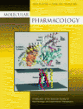
Cover Caption
About the cover: Model of the receptor-G protein complex. The G protein heterotrimer is an αt/αi1 chimera complexed with βtγt (Lambright et al., 1996). The receptor model is the MII rhodopsin model (Pogozheva et al., 1997). Positioning of the heterotrimer relative to the receptor is based on the model in Bourne (1997). The GTPase domain of the α-subunit is light blue. The helical domain of the α-subunit is pink. The GDP is yellow. Switch I is dark blue, switch II is green, and switch III is magenta. The α2/β4 loop is yellow, the α3/β5 loop is orange, and the α4/β6 loop is red. The carboxyl terminus (labeled C) of the α-subunit is magenta. Selected regions of secondary structure in the α-subunit, including the amino-terminal α-helix (αN), are indicated. The β-strands of the β-subunit are orange, and the amino-terminal helix and connecting loops are yellow. the γ-subunit is white. Receptor helices are numbered, and those connected to each other by an intracellular loop are the same color. These figures were drawn using MidasPlus, developed by the Computer Graphics Laboratory at the University of California at San Francisco. From Grishna G and Berlot CH (2000) A surface-exposed region of Gsα in which substitutions decrease receptor-mediated activation and increase receptor affinity. Mol Pharmacol57:1081–1092.


