Cover Caption
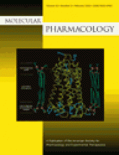
About the cover: Three-dimensional molecular representation of the distribution of positively charge residues in rhodopsin. The Cα trace of rhodopsin (yellow) is viewed perpendicular to the transmembrane domain axis, with the extracellular side on the top and the intracellular side on the bottom. The side chains of Arg and Lys are shownin blue sticks. To represent the membrane bilayer, several phospholipid molecules (phosphatidylcholines in the extracellular leaflet and negatively charged phosphatidylserines in the cytoplasmic leaflet) are shown. Mol Pharmacol 60:1–19.


