Cover Caption
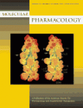
About the cover: DAT TM3 electrostatic potential models. A, dot surface displays of electrostatic potentials in WT (left) and F154A (right) TM3 helices. Each TM3 was built as an -helix and energy-minimized. The 21 amino acids of TM3 cross the plasma membrane from the lower (N-terminal, cytoplasmic) side to the upper (C-terminal, extracellular) side. F154A is indicated by arrows. Dot surfaces were displayed using Sybyl 6.6 "vdW Dot Surface" with red indicating electrostatic potential measures of "V" > 24.9; orange, yellow or green 3.3 > V > 0, cyan 0 > V > 3.3 and blue, purple or white V < 24.9. "V" is defined as described previously [Weiner PA, Langridge R, Blaney JM, Scharfer R, and Kollman PK (1982) Electrostatic potential molecular surfaces. Proc Natl Acad Sci USA 79:3754-3758] at an arbitrary distance of 1.4 Å. At the helical side important for cocaine binding, there are seven residues; three (42%) are decreased and another three increased in electrostatic potentials. At the side for dopamine uptake, there are eight residues; seven of them (87%) are decreased and none of them is increased in electrostatic potentials. Indicated are amino acid residues visible on this graph to the reader and whose electrostatic potentials are reduced by the F154A substitution. Italic lables denote residues hiding from the reader (located at the opposite side of the domain). #, increased electrostatic potentials for asparagine 157 (above the arrow) and phenylalanine 150 (below the arrow). Scale bar, 5 Å. Mol Pharmacol 61:885–981.


